Review
. 2017 Jan 24;11(1):69-83.
doi: 10.1021/acsnano.6b07607. Epub 2017 Jan 9.

Re-Engineering Extracellular Vesicles as Smart Nanoscale Therapeutics

Affiliations
Review

Re-Engineering Extracellular Vesicles as Smart Nanoscale Therapeutics

James P K Armstrong et al. ACS Nano. .

Abstract

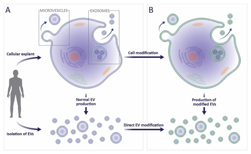
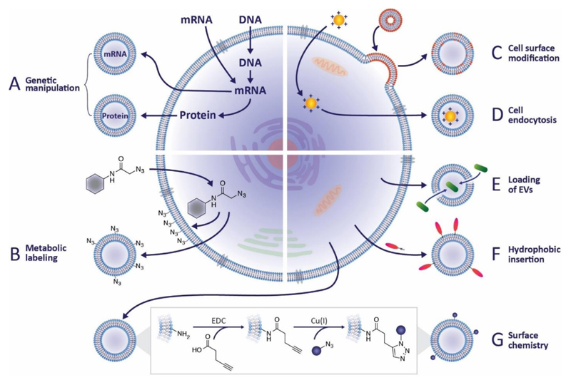
In the past decade, extracellular vesicles (EVs) have emerged as a key cell-free strategy for the treatment of a range of pathologies, including cancer, myocardial infarction, and inflammatory diseases. Indeed, the field is rapidly transitioning from promising in vitro reports toward in vivo animal models and early clinical studies. These investigations exploit the high physicochemical stability and biocompatibility of EVs as well as their innate capacity to communicate with cells via signal transduction and membrane fusion. This review focuses on methods in which EVs can be chemically or biologically modified to broaden, alter, or enhance their therapeutic capability. We examine two broad strategies, which have been used to introduce a wide range of nanoparticles, reporter systems, targeting peptides, pharmaceutics, and functional RNA molecules. First, we explore how EVs can be modified by manipulating their parent cells, either through genetic or metabolic engineering or by introducing exogenous material that is subsequently incorporated into secreted EVs. Second, we consider how EVs can be directly functionalized using strategies such as hydrophobic insertion, covalent surface chemistry, and membrane permeabilization. We discuss the historical context of each specific technology, present prominent examples, and evaluate the complexities, potential pitfalls, and opportunities presented by different re-engineering strategies.

Keywords: cell-free therapy; drug loading; exosomes; extracellular vesicles; functionalization; genetic manipulation; membrane modification; microvesicles.


Figures

Figure 1
Figure 1. Clinical and pre-clinical studies using EVs derived from mesenchymal stem cells.
(a) Exosomes are well known to be effective in myocardial tissue repair after ischemia-reperfusion injury; in this mouse model, infarct size (IS, stained white) as a proportion of area-at-risk (AAR, stained red) was reduced from 39 ± 2% to 21 ± 2% (t = 1 day). Images were adapted from Arslan et al. and reproduced with permission from Elsevier. (b) Exosomes were used to promote wound healing in a rat model of skin deep second degree burn injury. Immunostaining of CK19 expression (red) along with Hoechst stain (blue) showed re-epithelization at the wound area for rats treated with exosomes (t = 2 weeks, scale bars = 200 μm). Images were reproduced under creative commons licence from Zhang B et al. (2015) Stem Cells doi:10.1002/stem.1771. (c) Exosomes were used in a clinical study to reduce pro-inflammatory cytokine response and alleviate the symptoms of therapy-refractive graft-versus-host disease. Images were adapted from Kordelas et al. and reproduced with permission from Nature Publishing Group. (d) Exosomes have also been used to enhance in vivo cartilage repair in 1 mm deep osteochondral defects created on the trochlear grooves of distal femurs of adult rats (t = 6 weeks). Images reproduced under creative commons licence from Zhang S et al. (2016) Osteoarthritis and Cartilage doi:10.1016/j.joca.2016.06.022. (e) Microvesicles have been shown to provide protection against tubular injury in an acute kidney injury mouse model. Here, cisplatin was used to induce intra-tubular casts (asterisks) and tubular necrosis (arrows), which was alleviated with multiple injections of microvesicles (t = 4 days, magnification = 200X). Images were reproduced under open access from Bruno S et al. (2012) PLoS ONE 7(3) doi:10.1371/journal.pone.0033115.
Figure 2
Figure 2. The isolation, secretion and modification of EVs.
(a) EVs can either be isolated directly from bodily fluids or indirectly from in vitro cultured cells. As part of normal exocytosis, cells will shed microvesicles from the cytoplasmic membrane, and release exosomes from multivesicular bodies. (b) Cell manipulation can indirectly lead to modified exosomes and microvesicles, alternatively, the EVs themselves can be directly functionalized or loaded.
Figure 3
Figure 3. Strategies for EV modification.
(a) Genetic engineering can be used to introduce coding and non-coding oligonucleotides into cells. There it can be packaged into EVs to promote gene expression or regulate transcription in recipient cells. Alternatively, transgenic proteins can be incorporated into EVs, for instance, as fluorescent reporters or targeting moieties. (b) Metabolic labeling, in which metabolite analogues are incorporated into cell biosynthesis, has been widely used to introduce non-native moieties into cells. This approach can be used to introduce functional groups, such as azides, to EVs, which allows subsequent bio-orthogonal reactions to be performed. (c) Exogenous material may be introduced to EVs via liposomes or micelles that fuse with cytoplasmic membranes. (d) Alternatively, the process of packaging endocytosed material into EVs as part of normal membrane turnover and exocytosis can be hijacked to introduce exogenous species to EVs. (e) A direct EV modification strategy is to permeabilize the vesicle membrane to allow the active loading of molecules into the EV interior, an approach that has been exploited for drug delivery. (f) A similar approach uses lipophilic or amphiphilic molecules that can insert into the EV membrane via hydrophobic interactions with the phospholipid bilayer. (g) Chemical reactions may also be performed directly on the vesicle membrane, for instance, carbodiimides can be used to modify native amines in order to present azide groups for click chemistry reactions.
Figure 4
Figure 4. Examples and applications of re-engineered EVs.
(a) A quantitative comparison of different strategies for loading porphyrin drugs into EVs showed that active loading strategies resulted in higher loading, with saponin treatment and hypotonic dialysis offering the greatest efficiency. Image reproduced from Fuhrmann et al. (b) Iron oxide nanoparticles exposed to macrophages can be passively packaged into EVs, as shown by these electron micrographs. Image reproduced from Silva et al. (c) Suetsugu et al. generated a mouse breast cancer cell line (MMT) expressing a CD63-GFP hybrid that was packaged into EVs and used to visualize intercellular vesicle transfer. Reproduced with permission from Elsevier. (d) Takahashi et al. used a truncated lactadherin fused with Gaussia luciferase to produce artificially chemiluminescent EVs. This allowed EVs to be traced after systemic administration into a mouse model using chemiluminescent imaging, an analysis that revealed rapid clearance from the blood circulation. Reproduced with kind permission from Elsevier. (e) Qi et al. used superparamagnetic nanoparticles functionalized with transferrin, which allowed them to bind to receptors present on the surface of blood-derived EVs. These responsive EVs were used in combination with an external magnetic field (MF) to enhance delivery to a tumor site, as shown in this ex vivo near infrared fluorescence image.

References

    1. Raposo G, Stoorvogel W. Extracellular Vesicles: Exosomes, Microvesicles, and Friends. J Cell Biol. 2013;200:373–383. - PMC - PubMed
    1. Gould SJ, Raposo G. As We Wait: Coping with an Imperfect Nomenclature for Extracellular Vesicles. J Extracell Vesicles. 2013;2:3–5. - PMC - PubMed
    1. Théry C, Zitvogel L, Amigorena S. Exosomes: Composition, Biogenesis and Function. Nat Rev Immunol. 2002;2:569–579. - PubMed
    1. Gyorgy B, Szabo RG, Pasztoi M, Pal Z, Misjak P, Aradi B, Laszlo V, Pallinger E, Pap E, Kittel A, Nagy G, et al. Membrane Vesicles, Current State-of-the-Art: Emerging Role of Extracellular Vesicles. Cell Mol Life Sci. 2011;68:2667–2688. - PMC - PubMed
    1. El Andaloussi S, Mäger I, Breakefield XO, Wood MJA. Extracellular Vesicles: Biology and Emerging Therapeutic Opportunities. Nat Rev Drug Discov. 2013;12:347–357. - PubMed

Publication types

MeSH terms