. 2017 Mar;187(3):528-542.
doi: 10.1016/j.ajpath.2016.11.012. Epub 2017 Jan 6.

Modulation of Intersectin-1s Lung Expression Induces Obliterative Remodeling and Severe Plexiform Arteriopathy in the Murine Pulmonary Vascular Bed

Affiliations

Modulation of Intersectin-1s Lung Expression Induces Obliterative Remodeling and Severe Plexiform Arteriopathy in the Murine Pulmonary Vascular Bed

Monal Patel et al. Am J Pathol. 2017 Mar.

Abstract

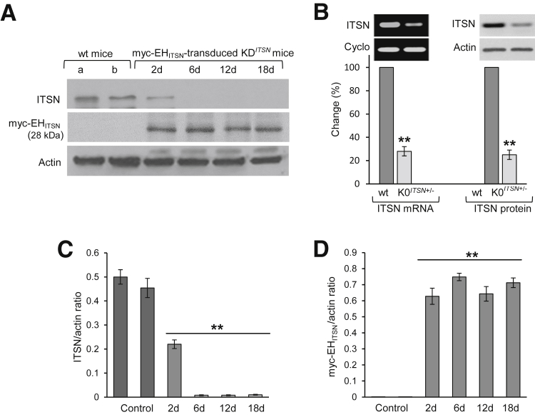
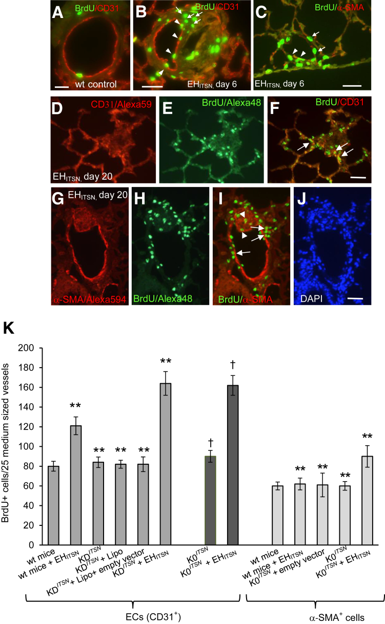
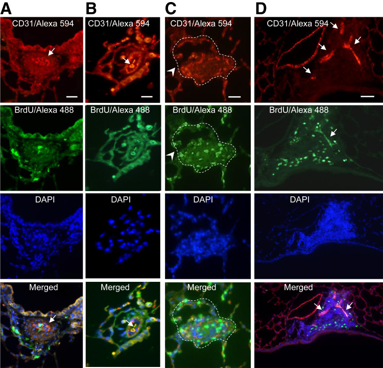
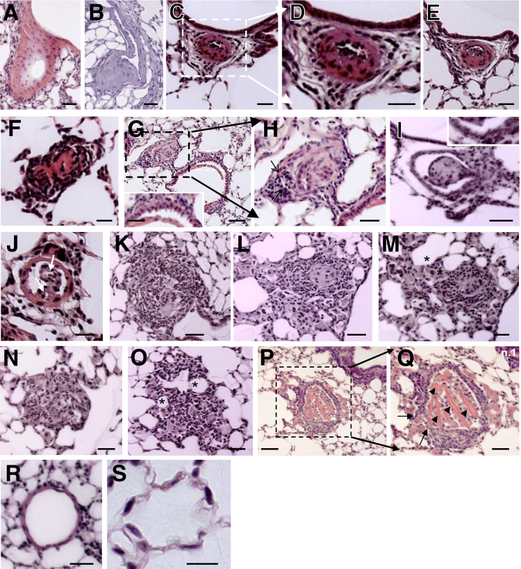
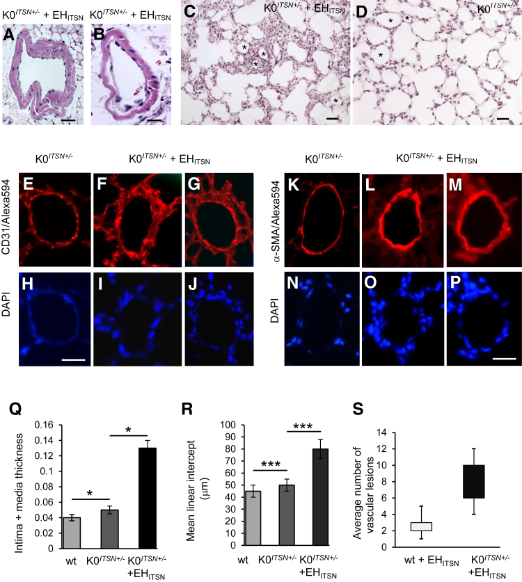
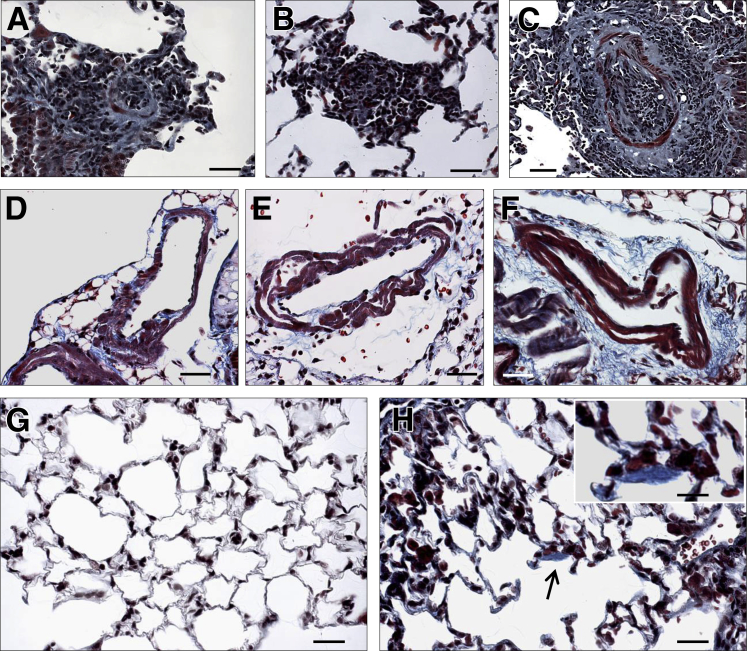
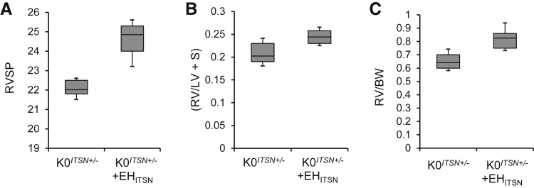
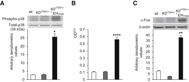
Murine models of pulmonary arterial hypertension (PAH) that recapitulate the plexiform and obliterative arteriopathy seen in PAH patients and help in defining the molecular mechanisms involved are missing. Herein, we investigated whether intersectin-1s (ITSN) deficiency and prolonged lung expression of an ITSN fragment with endothelial cell (EC) proliferative potential (EHITSN), present in the lungs of PAH animal models and human patients, induce formation of plexiform/obliterative lesions and defined the molecular mechanisms involved. ITSN-deficient mice (knockout/heterozygous and knockdown) were subjected to targeted lung delivery of EHITSN via liposomes for 20 days. Immunohistochemistry and histological and morphometric analyses revealed a twofold increase in proliferative ECs and a 1.35-fold increase in proliferative α-smooth muscle actin-positive cells in the lungs of ITSN-deficient mice, transduced with the EHITSN relative to wild-type littermates. Treated mice developed severe medial wall hypertrophy, intima proliferation, and various forms of obliterative and plexiform-like lesions in pulmonary arteries, similar to PAH patients. Hemodynamic measurements indicated modest increases in the right ventricular systolic pressure and right ventricle hypertrophy. Transcriptional and protein assays of lung tissue indicated p38MAPK-dependent activation of Elk-1 transcription factor and increased expression of c-Fos gene. This unique murine model of PAH-like plexiform/obliterative arteriopathy induced via a two-hit pathophysiological mechanism without hypoxia provides novel druggable targets to ameliorate and, perhaps, reverse the EC plexiform phenotype in severe human PAH.


Figures

Figure 1
Figure 1
Repeated delivery of lipoplexes containing the ITSN-1–specific siRNA and myc-EHITSN plasmid to murine lungs produces efficient long-term ITSN down-regulation and EHITSN protein expression; genotyping of K0ITSN mice. A: Lung lysates (70 μg per lane) of KDITSN transduced with the myc-EHITSN lipoplexes (days 2, 6, 12, and 18 of treatment) were subjected to Western blotting (WB) using ITSN antibody (Ab) and myc Ab. Actin was used as loading control. Wild-type (wt) mice (lane a), mice injected with empty liposomes (lane b, top panel) or empty vector (lane b, bottom panel) were used as controls. B: Conventional RT-PCR on lung samples from wt and K0ITSN+/− mice was used to analyze ITSN-1s mRNA levels, relative to internal control, cyclophilin (cyclo, left panels). ITSN protein levels were analyzed in lung lysates (70 μg total protein per lane) of wt and K0ITSN+/− mice by WB using ITSN Ab. Actin was used as loading control (right panels). All mice used in this study were genotyped. C and D: Densitometric analyses of ITSNand myc-EHITSN expression, respectively, in control mice and KDITSN mice transduced with the myc-EHITSN. Data are shown as ITSN/actin (C) or myc-EHITSN/actin (D) ratio. Data are expressed as means ± SEM (BD). n = 3 (A, C, and D, mice per group); n = 3 (A, BD, different experiments). ∗∗P < 0.01 versus wt mice.
Figure 2
Figure 2
Myc-EHITSN expression in K0ITSN+/− murine lungs triggers proliferation of endothelial cell (EC) and α-smooth muscle actin (α-SMA)+ lung resident cells. A–C: Wild-type (wt) (A) and myc-EHITSN-transduced mice, day 6 of treatment, were subjected to bromodeoxyuridine (BrdU) assay, followed by BrdU–fluorescein isothiocyanate and CD31 Alexa Fluor 594 (B) or α-SMA Alexa Fluor 594 (C) antibody immunohistochemistry for positive identification of ECs and myofibroblasts, smooth muscle cells, and undifferentiated cells, respectively. Representative micrograph illustrates the BrdU/CD31 colocalization (B, arrowheads) as well as significant proliferation of other lung resident cells (B, arrows). C: α-SMA+/BrdU colocalization (arrowheads) within the wall of a pulmonary arteriole or their close proximity (arrows) was also detected. D–I: Representative CD31/BrdU (D–F) and α-SMA+/BrdU (G–I) immunohistochemistry of lung sections of myc-EHITSN-transduced K0ITSN+/− mouse lung sections, day 20 of treatment, illustrates clusters of proliferative cells [ECs and α-SMA+ cells (F and I, arrows, respectively)], as well as other lung resident cells (frequently detected in vascular lesions that obliterate the vessel lumina; I, arrowheads). J: DAPI staining illustrates the hypercellularity associated with the remodeled vessel. K: Quantification of the number of BrdU+ ECs and BrdU+/α-SMAs+ in the mid-sized lung vessels of controls versus EHITSN-treated mice. All data shown are representative of four different experiments with three mice for the control groups and five mice per experimental condition. Wt mice include CD1 as well as 129SV/J genetic background mice. No significant differences were noticed in proliferation of α-SMAs+ between wt mice, untreated KDITSN, and K0ITSN+/− mice. n = 3 mice per group in three independent experiments (A–C); n = 12 (J); n = 4 different experiments (K); n = 3 mice for the control groups (K); n = 5 mice per experimental condition (K). ∗∗P < 0.01 versus wt mice; P < 0.05 versus K0ITSN+/− mice. Scale bars: 20 μm (A–C); 25 μm (D–F); 40 μm (G–J).
Figure 3
Figure 3
Myc-EHITSN expression in K0ITSN+/− murine lungs triggers severe vasculopathy. Representative double CD31/Alexa Fluor 594 antibody– bromodeoxyuridine (BrdU)/Alexa Fluor 488 immunohistochemistry shows BrdU+ endothelial cells (ECs) within complex lesions in myc-EHITSN-transduced K0ITSN+/− mice lungs. A and D: DAPI staining of the nuclei documents the hypercellularity of intimal cells. The merged images illustrate the significant BrdU/CD31 colocalization within the vascular lesions (A, B, and D, arrows), the severe narrowing of the vessel lumen (arrow, merged image, D) because of concentric medial fibroproliferation, and the presence of slit-like or irregular-shaped CD31+ channels (arrows, merged images, B and D,); these complex lesions containing proliferative cells, many of them ECs, partially obliterate the lumen of a pulmonary artery with partial destruction of the vessel wall (C, arrowheads). The dotted lines in C mark the perimeter of the vessel wall. Data are representative of four different experiments. n = 3 mice for the control groups and per experimental condition. Scale bars: 50 μm (A and D); 25 μm (B and C).
Figure 4
Figure 4
Myc-EHITSN expression in K0ITSN+/− mouse lungs leads to vascular remodeling and formation of vascular lesions, including pulmonary vascular occlusion and plexiform-like lesions. Representative hematoxylin and eosin (H&E) staining of paraffin-embedded lung tissue sections of myc-EHITSN-transduced K0ITSN+/− mice shows various forms of vascular remodeling. A and B: Media fibroproliferation with vessel wall thickening. C–F, D, and H: Concentric intimal proliferation with severe obliteration of the vessel lumen (arrows). G: Higher magnification of the lesion in C. The proliferative endothelial cells were located in a hobnail pattern in a vessel, most likely affected by a lesion in other plan (G, inset). The inset in G shows the area indicated by a white dashed box in the main image. H: Higher magnification of the black dashed box in G. I and J: Stalk-like lesions. Proliferative endothelial cells arranged in a hobnail pattern (I, inset). K–O: Complex plexiform-like lesions. Arrows indicate two irregular slit-like channels separated by large hyperchromatic cells. M and O: Frequently, the plexiform lesions feed into dilated thin-walled vessels (asterisks). P and Q: A transversal section through a complex plexiform lesion (P and Q) illustrates the obliteration of the vessel lumen, partial destruction of the vessel wall (Q, arrows), and multiple endothelial channels (Q, arrowheads). Q: Higher magnification of the black dashed box in P. R and S: Untreated K0ITSN+/− mice lung used as control did not show vascular remodeling. Three independent experiments were performed with three mice for the control groups and five mice for each experimental condition; 8 to 10 H&E-stained slides per mouse were used. Scale bars: 40 μm (AB, I, K, M, and Q); 20 μm (C, D, F, and J); 35 μm (E); 30 μm (H, N, O, and G, inset); 60 μm (G, main image, and P); 50 μm (L and R); 10 μm (S).
Figure 5
Figure 5
Histological and morphometry analyses revealed the severity of vascular remodeling in the lungs of myc-EHITSN-transduced K0ITSN+/− mice. A–C: Representative hematoxylin and eosin staining illustrates medial proliferation (A and B) and alveolar septa thickening (C) in myc-EHITSN-transduced K0ITSN+/− mice. D: Lung morphology of untreated K0ITSN+/− mice. Several vascular profiles in EHITSN-treated (C) and untreated K0ITSN+/− (D) mice are marked by asterisks. Representative CD31/DAPI (E–J) and α-smooth muscle actin (α-SMA)/DAPI (K–P) of lung arterioles in untreated mice versus K0ITSN+/− mice transduced with the myc-EHITSN. Q: Intima plus media thickening in K0ITSN+/− plus myc-EHITSN versus controls (wt and K0ITSN+/− mice). A, B, Q, and R: The mean linear intercept was increased in the myc-EHITSN-transduced K0ITSN+/− mice compared to untreated mice (wt and K0ITSN+/−). Quantification of affected vessels was performed on small- and medium-sized blood vessels (20 mm ≥ diameter ≤ 100 mm), as above, using three sections per mouse, three mice in the control group and five mice in the experimental group, with the experiments performed at least three times with reproducible results (A, B, Q). S: Average number of profiles of vascular lesions per section in myc-EHITSN-treated mice. n = 3 mice for the control group (C, D, E–P, R, S); n = 5 mice for the experimental group (C, D, E–P, R, S); n = 3 independent experiments (C, D, E–P, R, S). Data are expressed as means ± SEM (QS). P < 0.05, ∗∗∗P < 0.001. Scale bars: 20 μm (A, C, and D); 10 μm (B); 35 μm (E–J); 40 μm (K–P).
Figure 6
Figure 6
Collagen deposition within plexiform lesion, perivascular spaces, and alveolar septa, in EHITSN-transduced K0ITSN+/− mouse lungs. Representative Masson's trichrome staining of EHITSN-transduced K0ITSN+/− mouse lung sections illustrates collagen deposition within vascular obliterative lesions in pulmonary vessels with a diameter between 20 and 60 μm (A–C), within the layers of muscularized vessel walls (D–F), and in the interstitial space (F). Limited collagen deposition was seen in the alveolar septa of both untreated K0ITSN+/− mice (G) and EHITSN-treated K0ITSN+/− mice (H). Occasionally, however, focal collagen deposition was seen in the alveolar wall of EHITSN-treated K0ITSN+/− mice (arrow and inset in H). n = 3 mice for the control groups (G); n = 5 mice for the experimental group (A–F and H); n = 6 Masson's trichrome stained slides per mouse. Scale bars: 30 μm (A–H); 15 μm (inset).
Figure 7
Figure 7
Myc-EHITSN-transduced K0ITSN+/− mice show modest increase in right ventricular systolic pressure (RVSP) and right ventricle (RV) hypertrophy. Treatment of K0ITSN+/− mice with myc-EHITSN induces modest increases in the RVSP (P < 0.049) (A), RV/left ventricle (LV) + septum (S) weight ratio (B), and RV weight/body weight (BW) (P < 0.05) (C). Lines within the boxes show medians; boxes show 25th and 75th percentiles of the data, respectively. Data are expressed as medians ± SEM. n = 3 independent experiments were performed; n = 5 mice per experimental condition.
Figure 8
Figure 8
Myc-EHITSN expression leads to activation of p38MAPK as well as downstream Elk-1 transcription factor and c-fos expression in K0ITSN+/− mouse lungs. A: Lung lysates of untreated wt mice (white bars), K0ITSN+/− mice (grey bars), and myc-EHITSN-transduced K0ITSN+/− mice (black bars) were assessed for p38MAPK phosphorylation. Total p38 kinase was used as a loading control. Densitometry values are representative for three independent experiments. B: Nuclear extracts prepared from controls and treated mice were assayed for Elk-1 activity. Data are representative of three independent experiments performed in triplicate. C: Western blotting of nuclear extracts (45 μg per lane) from controls and myc-EHITSN-transduced K0ITSN+/− mice using c-Fos antibody and consequent densitometry analysis. Data were normalized to β-actin. Data represent three different experiments. Data are expressed as means ± SEM (A–C). n = 3 mice per group (A–C). P < 0.05, ∗∗P < 0.01, and ∗∗∗∗P < 0.005 versus wt mice.

Similar articles

Cited by

References

    1. Tuder R.M., Cool C.D., Yeager M., Taraseviciene-Stewart L., Bull T.M., Voelkel N.F. The pathobiology of pulmonary hypertension: endothelium. Clin Chest Med. 2001;22:405–418. - PubMed
    1. Tuder R.M., Marecki J.C., Richter A., Fijalkowska I., Flores S. Pathology of pulmonary hypertension. Clin Chest Med. 2007;28:23–42. vii. - PMC - PubMed
    1. Voelkel N.F., Cool C. Pathology of pulmonary hypertension. Cardiol Clin. 2004;22:343–351. v. - PubMed
    1. Tuder R.M., Abman S.H., Braun T., Capron F., Stevens T., Thistlethwaite P.A., Haworth S.G. Development and pathology of pulmonary hypertension. J Am Coll Cardiol. 2009;54:S3–S9. - PubMed
    1. Rabinovitch M. Molecular pathogenesis of pulmonary arterial hypertension. J Clin Invest. 2008;118:2372–2379. - PMC - PubMed

MeSH terms