. 2017 Jan 10;14(1):1.
doi: 10.1186/s12989-016-0182-0.

In vitro biological responses to nanofibrillated cellulose by human dermal, lung and immune cells: surface chemistry aspect

Affiliations

In vitro biological responses to nanofibrillated cellulose by human dermal, lung and immune cells: surface chemistry aspect

Viviana R Lopes et al. Part Fibre Toxicol. .

Abstract

Background: Nanocellulose, and particularly nanofibrillated cellulose (NFC), has been proposed for a diversity of applications in industry and in the biomedical field. Its unique physicochemical and structural features distinguish nanocellulose from traditional materials and enable its use as an advance nanomaterial. However, its nanoscale features may induce unknown biological responses. Limited studies with NFC are available and the biological impacts of its use have not been thoroughly explored. This study assesses the in vitro biological responses elicited by wood-derived NFC gels, when human dermal fibroblasts, lung MRC-5 cells and THP-1 macrophage cells are exposed to the nanomaterial. Furthermore, whether the presence of surface charged groups (i.e. carboxymethyl and hydroxypropyltrimethylammonium groups) on NFC can induce distinct biological responses is investigated.

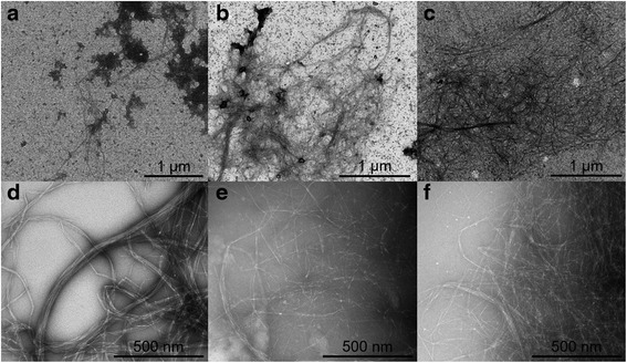
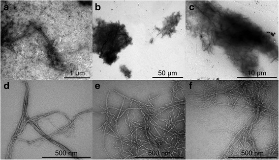
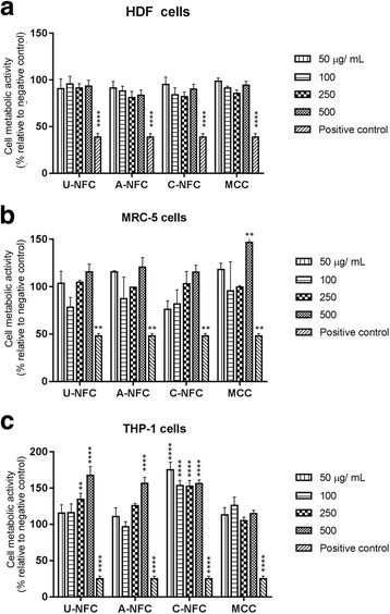
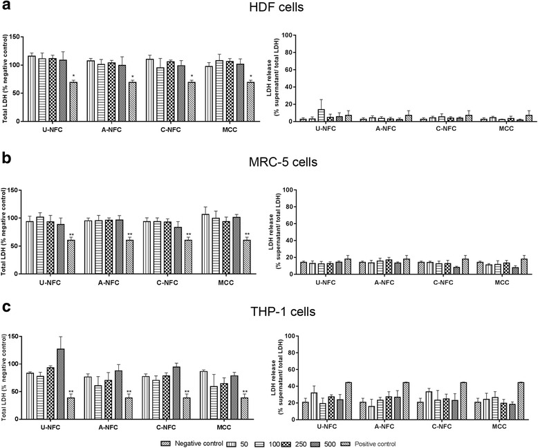
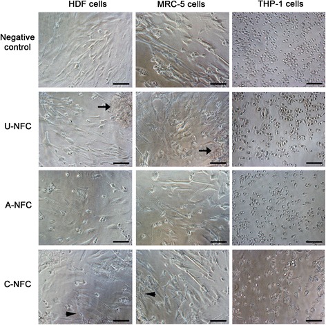
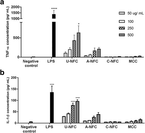
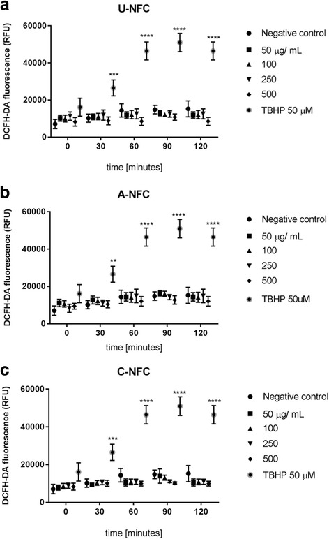
Results: The introduction of surface charged groups resulted in individual nanofibrils, while fibril aggregates predominated in the unmodified NFC gel suspensions as observed by transmission electron microscopy. In the presence of proteins, the surface modified NFCs formed compact agglomerates while the agglomeration pattern of the unmodified NFC was similar in the presence of proteins and in physiological buffer. Unmodified and modified NFC gels did not induce cytotoxicity in human dermal fibroblasts, lung and macrophage cells. No significant ROS production by THP-1 macrophages was found and no cellular uptake was observed. However, an inflammatory response was detected when THP-1 macrophages were treated with unmodified NFC as assessed by an increase in TNF-α and IL1-β levels, an effect that was absent when surface charged groups were introduced into NFC.

Conclusions: Taken together, the data presented here show the absence of cytotoxic effects associated with the exposure to unmodified, carboxymethylated and hydroxypropyltrimethylammonium-modified NFCs. Unmodified NFC presented a pro-inflammatory effect which can be further moderated by introducing surface modifications such as carboxymethyl and hydroxypropyltrimethylammonium groups into the nanofibrils. The present findings suggest that the inflammatory response to NFC might be driven by the material surface chemistry, and thus open up for the possibility of designing safe nanocellulose materials.

Keywords: Cytotoxicity; Inflammation; Nanocellulose; Surface chemistry.


Figures

Fig. 1
Fig. 1
Morphology of NFC suspended in phosphate buffer. Transmission electron microscopy images of U-NFC (a and d), A-NFC (b and e) and C-NFC (c and f)
Fig. 2
Fig. 2
Morphology of NFC suspended in cell culture medium. Transmission electron microscopy images of U-NFC (a and d), A-NFC (b and e) and C-NFC (c and f)
Fig. 3
Fig. 3
Metabolic activity of cells after NFC exposure. Cell viability of NFC-treated cells was assesed by evaluating cell metabolic activity using the alamar blue assay. a HDF cells, b MRC-5 cells and c THP-1 macrophages exposed to increasing doses of NFC (50–500 μg/mL) for 24 h. MCC is a food grade nanocellulose used as a reference material. The positive control was DMSO (5% v/v in cell culture media) an inducer of cytotoxic effects. Data are expressed as percentage relative to the negative control (untreated cells) and presented as mean ± SEM of three independent experiments. Significant results as compared to the negative control are marked with asterisks (** p < 0.01 and **** p < 0.0001)
Fig. 4
Fig. 4
Lactate dehydrogenase (LDH) activity of cells after NFC exposure. Cytotoxicity of NFC-treated cells was evaluated by measuring total and extracellular LDH activity. a HDF cells, b MRC-5 cells and c THP-1 macrophages were treated with a range of NFC concentration from 50 to 500 μg/mL during 24 h. MCC is a food grade nanocellulose used as a reference material. The positive control was DMSO (5% v/v in cell culture media) an inducer of cytotoxicity. Data are presented as mean ± SEM of three independent experiments. Significant results as compared to the negative control are marked with asterisks (* p < 0.05 and ** p < 0.01)
Fig. 5
Fig. 5
Cell Morphology after NFC exposure. Morphology of HDF, MRC-5 and THP-1 macrophages after direct contact with NFC. Top images show HDF, MRC-5 and THP-1 cells untreated (negative control). For all other conditions, HDF, MRC-5 and THP-1 cells were treated with the highest concentration of U-NFC, A-NFC and C-NFC (500 μg/mL) for 24 h. Black arrows indicate agglomerates of fibres. Images of cells treated with the positive control (DMSO 5%) are given in Additional file 1: Figure S1. Scale bars represent 100 μm
Fig. 6
Fig. 6
Cytokine production after NFC exposure. a TNF-α and b IL1-β concentration in the supernatants of THP-1 macrophages exposed to increasing doses of NFC (50–500 μg/mL) for 24 h. For the U-NFC sample cytokine secretion was assessed in the presence of PMB in order to supress the contribution of endotoxin contamination to the secreted cytokine levels. MCC is a commercial grade nanocellulose used as a reference material. Negative control represents untreated cells. Cells treated with LPS (1 ng/mL), an inducer of cytokine production, represent the positive control of the assay. The data are presented as mean ± SEM of three independent experiments. Significant results as compared to the negative control are marked with asterisks (*p < 0.05 and **** p < 0.0001)
Fig. 7
Fig. 7
Cellular reactive oxygen species (ROS) production after NFC exposure. Kinetic study of ROS production of THP-1 macrophages treated with increasing doses (50–500 μg/mL) of (a) U-NFC, (b) A- NFC and (c) C-NFC. ROS assessed with the ROS-specific fluorescent probe DCFDA-DA every 30 min during 120 min. Negative control represents untreated cells. Tert-butyl hydroperoxide (TBHP), an inducer of oxidative stress, represents the positive control. Data are expressed as relative fluorescence units (RFU) and presented as the mean ± SEM of three independent experiments

References

    1. Azizi Samir MAS, Alloin F, Dufresne A. Review of recent research into cellulosic whiskers, their properties and their application in nanocomposite field. Biomacromolecules. 2005;6(2):612–626. doi: 10.1021/bm0493685. - DOI - PubMed
    1. Eichhorn SJ, Dufresne A, Aranguren M, Marcovich NE, Capadona JR, Rowan SJ, et al. Review: Current international research into cellulose nanofibres and nanocomposites. J Mater Sci. 2010;45:1. doi: 10.1007/s10853-009-3874-0. - DOI - PMC - PubMed
    1. Moon RJ, Martini A, Nairn J, Simonsen J, Youngblood J. Cellulose nanomaterials review: Structure, properties and nanocomposites. Chem Soc Rev. 2011;40:7. doi: 10.1039/c0cs00108b. - DOI - PubMed
    1. Dufresne A. Nanocellulose: a new ageless bionanomaterial. Mater Today. 2013;16(6):220–227. doi: 10.1016/j.mattod.2013.06.004. - DOI
    1. Klemm D, Kramer F, Moritz S, Lindström T, Ankerfors M, Gray D, et al. Nanocelluloses: A New Family of Nature-Based Materials. Angew Chem Int Ed Engl. 2011;50:24. doi: 10.1002/anie.201001273. - DOI - PubMed

Publication types