. 2017 Jan 9;36(1):8.
doi: 10.1186/s13046-016-0478-9.

FGF19/FGFR4 signaling contributes to the resistance of hepatocellular carcinoma to sorafenib

Affiliations

FGF19/FGFR4 signaling contributes to the resistance of hepatocellular carcinoma to sorafenib

Lixia Gao et al. J Exp Clin Cancer Res. .

Abstract

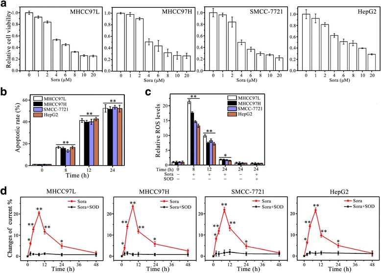
Background: Sorafenib, a multi-kinase inhibitor, is used as a standard therapy for advanced hepatocellular carcinoma (HCC). However, complete remission has not been achieved and the molecular basis of HCC resistance to sorafenib remains largely unknown. Previous studies have shown that fibroblast growth factor 19 (FGF19) expression correlates with tumor progression and poor prognosis of HCC. Here, we demonstrate the novel role of FGF19 in HCC resistance to sorafenib therapy.

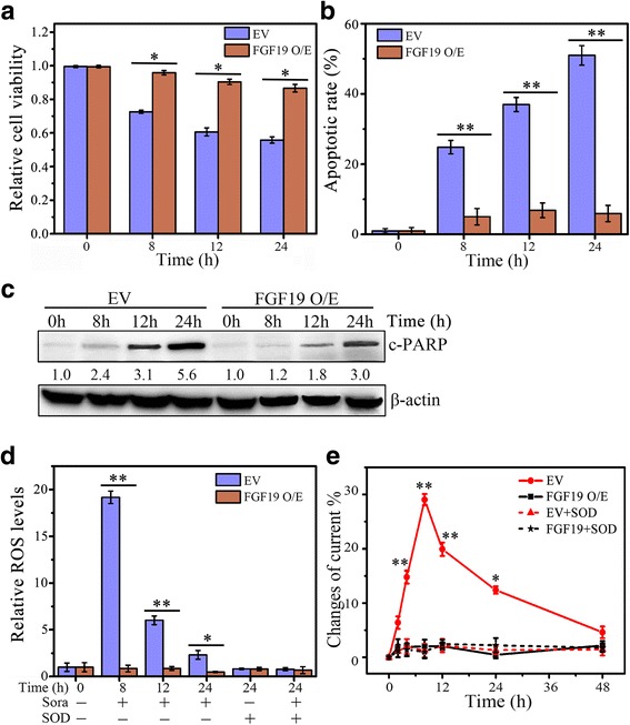
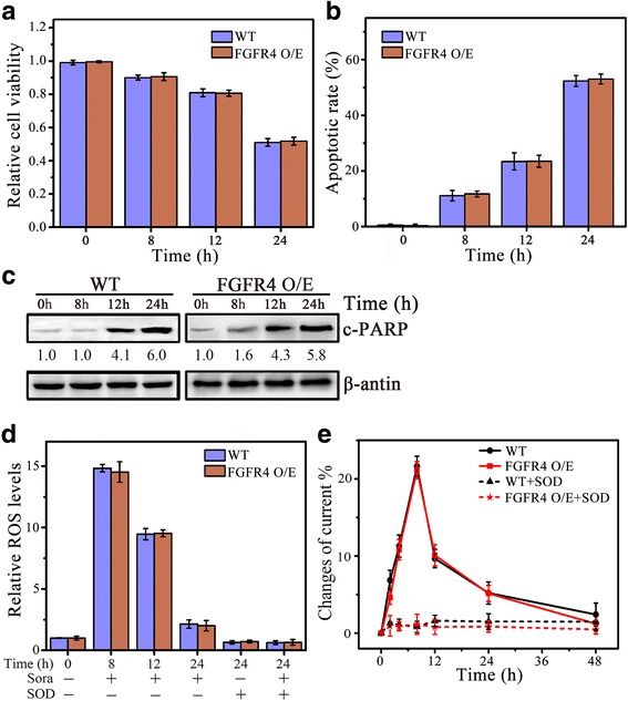
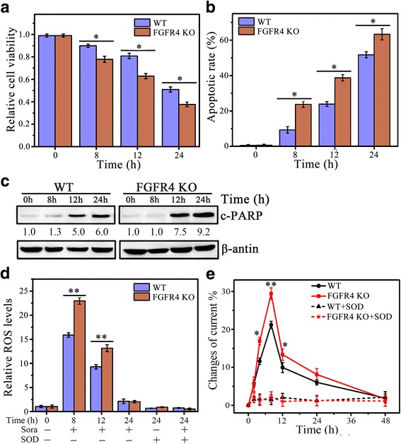
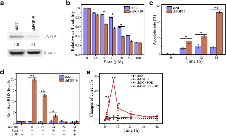
Methods: FGF19 Knockdown cells were achieved by lentiviral-mediated interference, and FGFR4 knockout cells were achieved by CRISPR-Cas9. Protein levels of FGF19, FGFR4 and c-PARP in various HCC cell lines were measured by Western blotting analysis. Cell viability was determined by MTS assay, apoptosis was determined by DAPI nuclear staining and Western blot of c-PRAP, and ROS generation was determined by DCFH-DA staining and electrochemical biosensor.

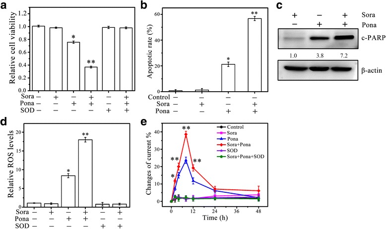
Results: We showed that FGF19, when overexpressed, inhibited the effect of sorafenib on ROS generation and apoptosis in HCC. In contrast, loss of FGF19 or its receptor FGFR4 led to a remarkable increase in sorafenib-induced ROS generation and apoptosis. In addition, knockdown of FGF19 in sorafenib-resistant HCC cells significantly enhanced the sensitivity to sorafenib. Importantly, targeting FGF19/FGFR4 axis by ponatinib, a third-generation inhibitor of chronic myeloid leukemia, overcomes HCC resistance of sorafenib by enhancing ROS-associated apoptosis in sorafenib-treated HCC.

Conclusion: Our results provide the first evidence that inhibition of FGF19/FGFR4 signaling significantly overcomes sorafenib resistance in HCC. Co-treatment of ponatinib and sorafinib may represent an effective therapeutic approach for eradicating HCC.

Keywords: Drug resistance; FGF19; FGFR4; Hepatocellular carcinoma; Sorafenib; Synergistic effect.


Figures

Fig. 1
Fig. 1
Sorafenib induces apoptosis and ROS generation in HCC cells. a The effect of sorafenib (Sora) on cell viability. The HCC cell lines (MHCC97L, MHCC97H, HepG2 and SMCC-7721) were treated with indicated concentrations of Sora for 24 h, and cell viability was determined by MTS assays as previously described. b–d The effect of Sora on cell apoptosis and ROS generation. The HCC cell lines were treated with Sora (4 μM for MHCC97L, MHCC97H and SMCC-7721, and 6 μM for HepG2) over a series of time points. Apoptosis was determined by DAPI staining (b); ROS generation was determined by DCFH-DA staining (c); and O2 •− generation was determined by electrochemical biosensor (d). SOD: superoxide dismutase. * p < 0.05; ** p < 0.01
Fig. 2
Fig. 2
FGF19 overexpression protects HCC cells against sorafenib. ae The effect of FGF19 overexpression on Sora-induced HCC cell apoptosis and ROS generation. MHCC97L cells expressing pcDNA3.1-FGF19 (FGF19 O/E) or empty vector (EV) were treated with 4 μM of Sora over a series of time points before analysis. Cell viability was determined by MTS assays (a); cell apoptosis was determined by DAPI staining (b) and Western blot of cleaved PARP (c-PARP) (c); ROS generation was determined by DCFH-DA staining (d); and O2 •− generation was determined by electrochemical biosensor (e). In (c), expression levels were normalized against actin and reported relative to controls (fold changes shown below each lane). * p < 0.05; ** p < 0.01
Fig. 3
Fig. 3
Loss of FGF19 expression enhances sorafenib-induced apoptosis associated with ROS. ae The effect of FGF19 depletion on Sora-induced HCC cell apoptosis and ROS generation. MHCC97H cells expressing FGF19 shRNA (shFGF19) or control shRNA (shNC) were treated with 4 μM of Sora over a series of time points before analysis. Cell viability was determined by MTS assays (a); apoptosis was determined by DAPI staining (b) and Western blot of c-PRAP (c); ROS generation was determined by DCFH-DA staining (d); and O2 •− generation was determined by electrochemical biosensor (e). In c, expression levels were normalized against actin and reported relative to controls (fold changes shown below each lane). * p < 0.05; ** p < 0.01
Fig. 4
Fig. 4
Knockdown of FGFR4 promotes ROS-associated HCC cell apoptosis by sorafenib. ae The effect of FGFR4 knockout on Sora-induced HCC cell apoptosis and ROS generation. FGFR4 knockout MHCC97L cells by CRISPR sgRNA targeting exonic regions (FGFR4 KO) were treated with 4 μM of Sora over a series of time points before analysis. Cell viability was determined by MTS assays (a); Apoptosis was determined by DAPI staining (b) and Western blot of c-PARP (c); ROS generation was determined by DCFH-DA staining (d); and O2 •− generation was determined by electrochemical biosensor (e). In c, expression levels were normalized against actin and reported relative to controls (fold changes shown below each lane). * p < 0.05; ** p < 0.01
Fig. 5
Fig. 5
FGF19 Knockdown of in sorafenib-resistant HCC cells increases the sensitivity to sorafenib. (a) The knockdown effect of FGF19 in Sora-resistant MHCC97H (MHCC97H Sora-R) cells. (b-e) The effect of FGF19 knockdown on Sora-induced apoptosis in MHCC97H Sora-R cells. FGF19 was knocked down in MHCC97H Sora-R cells by lentiviral shRNA. FGF19 knockdown cells (shFGF19) and the control cells (shNC) were treated with different doses of Sora for 24 hours, and cell viability was determined by MTS assays (b). FGF19 knockdown cells (shFGF19) and the control cells (shNC) were treated with 20 μM of Sora over a series of time points before analysis. Apoptosis was determined by DAPI staining (c); ROS generation was determined by DCFH-DA staining (d); and O2 •− generation was determined by electrochemical biosensor (e). In a, expression levels were normalized against actin and reported relative to controls (fold changes shown below each lane). * p < 0.05; ** p < 0.01
Fig. 6
Fig. 6
Ponatinib facilitates sorafenib-resistant HCC cells to death. a-e The effect of ponatinib (Pona) on Sora-induced ROS generation and apoptosis in Sora-resistant HCC cells. Sora-resistant MHCC97H (MHCC97H Sora-R) cells were treated with 20 µM of Sora and 10 µM of Pona singly or in combination. Cell viability was determined by MTS assays (a); apoptosis was determined by DAPI staining (b) and Western blot (c); ROS generation was determined by DCFH-DA staining (d); and O2•− generation was determined by electrochemical biosensor (e). In c, expression levels were normalized against actin and reported relative to controls (fold changes shown below each lane). * p<0.05; ** p<0.01

Similar articles

Cited by

References

    1. Njei B, Rotman Y, Ditah I, Lim JK. Emerging trends in hepatocellular carcinoma incidence and mortality. Hepatology. 2015;61:191–99. doi: 10.1002/hep.27388. - DOI - PMC - PubMed
    1. Salem R, Gilbertsen M, Butt Z, Memon K, Vouche M, Hickey R, et al. Increased quality of life among hepatocellular carcinoma patients treated with radioembolization, compared with chemoembolization. Clin Gastroenterol H. 2013;11:1358–65. doi: 10.1016/j.cgh.2013.04.028. - DOI - PubMed
    1. Memon K, Kulik L, Lewandowski RJ, Mulcahy MF, Benson AB, Ganger D, et al. Radioembolization for hepatocellular carcinoma with portal vein thrombosis: impact of liver function on systemic treatment options at disease progression. J Hepatol. 2013;58:73–80. doi: 10.1016/j.jhep.2012.09.003. - DOI - PMC - PubMed
    1. Chang TS, Huang Y-H. Role of SENP1 in HBx-induced cell migration and stemness-related properties in hepatocellular carcinoma. Cancer Res. 2016;76:1720–20. doi: 10.1158/1538-7445.AM2016-1720. - DOI
    1. Salvi A, Conde I, Abeni E, Arici B, Grossi I, Specchia C, et al. Effects of miR-193a and sorafenib on hepatocellular carcinoma cells. Mol Cancer. 2013;12:162. doi: 10.1186/1476-4598-12-162. - DOI - PMC - PubMed

MeSH terms