. 2017 Jan;113(1):40-51.
doi: 10.1093/cvr/cvw229. Epub 2016 Nov 7.

Loss of Axin2 results in impaired heart valve maturation and subsequent myxomatous valve disease

Affiliations

Loss of Axin2 results in impaired heart valve maturation and subsequent myxomatous valve disease

Alexia Hulin et al. Cardiovasc Res. 2017 Jan.

Abstract

Aims: Myxomatous valve disease (MVD) is the most common aetiology of primary mitral regurgitation. Recent studies suggest that defects in heart valve development can lead to heart valve disease in adults. Wnt/β-catenin signalling is active during heart valve development and has been reported in human MVD. The consequences of increased Wnt/β-catenin signalling due to Axin2 deficiency in postnatal valve remodelling and pathogenesis of MVD were determined.

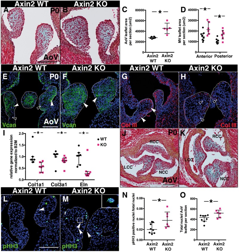
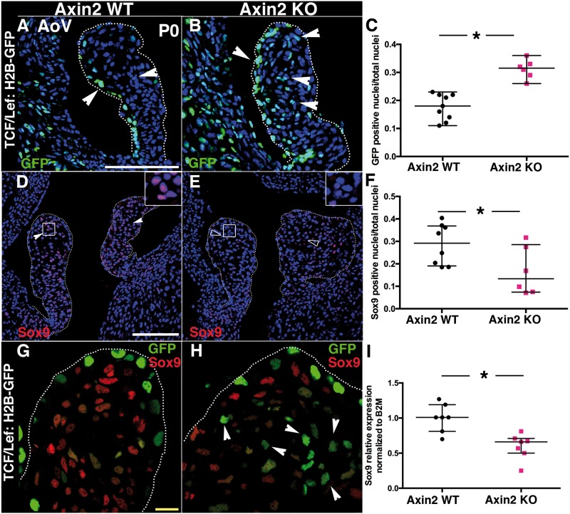
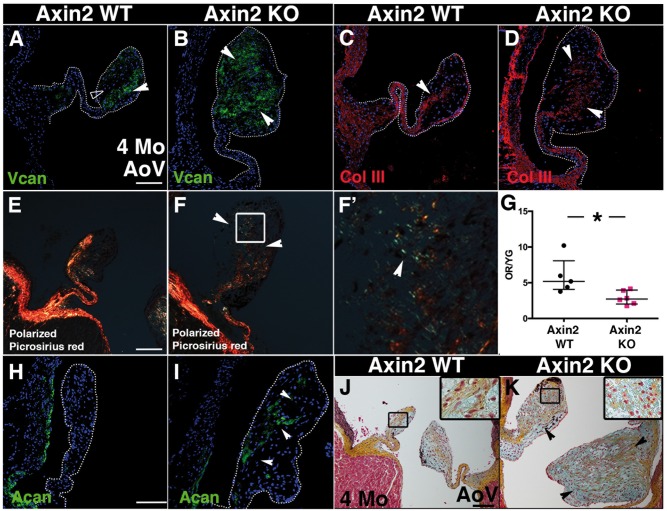
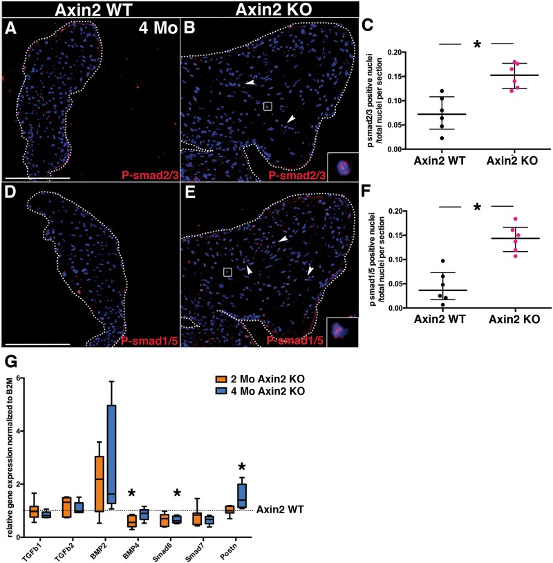
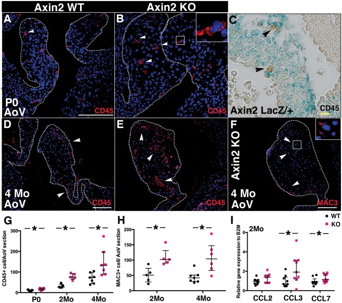
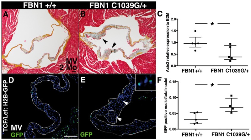
Methods and results: To investigate the role of Wnt/β-catenin signalling, we analysed heart valves from mice deficient in Axin2 (KO), a negative regulator of Wnt/β-catenin signalling. Axin2 KO mice display enlarged mitral and aortic valves (AoV) after birth with increased Wnt/β-catenin signalling and cell proliferation, whereas Sox9 expression and collagen deposition are decreased. At 2 months in Axin2 KO mice, the valve extracellular matrix (ECM) is stratified but distal AoV leaflets remain thickened and develop aortic insufficiency. Progressive myxomatous degeneration is apparent at 4 months with extensive ECM remodelling and focal aggrecan-rich areas, along with increased BMP signalling. Infiltration of inflammatory cells is also observed in Axin2 KO AoV prior to ECM remodelling. Overall, these features are consistent with the progression of human MVD. Finally, Axin2 expression is decreased and Wnt/β-catenin signalling is increased in myxomatous mitral valves in a murine model of Marfan syndrome, supporting the importance of Wnt/β-catenin signalling in the development of MVD.

Conclusions: Altogether, these data indicate that Axin2 limits Wnt/β-catenin signalling after birth and allows proper heart valve maturation. Moreover, dysregulation of Wnt/β-catenin signalling resulting from loss of Axin2 leads to progressive MVD.

Keywords: Axin2 • Heart valve development • Myxomatous valve disease • ECM remodelling • Wnt/β-catenin signalling.


Figures

Figure 1
Figure 1
Axin2 KO mice display immature thickened aortic valves (AoV) and mitral valves (MV). (A and B) Representative Movat’s Pentachrome staining performed on P0 heart sections from Axin2 WT and KO mice. (C) AoV leaflet area per section was measured (WT n = 6, KO n = 5). (D) Anterior and posterior MV leaflet areas per section were measured (WT n = 8 and KO n = 6). (E and F) Representative versican (vcan) staining performed on Axin2 WT and KO P0 heart sections. (G and H) Representative collagen type III (col III) staining performed on P0 heart sections from Axin2 WT and Axin2 KO mice. (I) Collagen 1a1, collagen 3a1 and elastin expression were measured by qPCR in P0 Axin2 KO AoV, normalized to B2M and compared to Axin2 WT (WT n = 7, KO n = 7). (J and K) Representative Movat’s Pentachrome staining performed on cross sections of P0 hearts from Axin2 WT and KO mice (RCC, LCC and NCC for right, left and non-coronary cusps). (L and M) Representative pHH3 staining of P0 heart sections from Axin2 WT and Axin2 KO mice. Inset displays nuclear pHH3 staining. (N) The number of pHH3-positive nuclei/total nuclei was quantified in AoV leaflet sections (WT n = 8, KO n = 6). (O) The total number of nuclei per AoV leaflets section was counted (WT n = 8 and KO n = 6). Data are reported as dot plots and interquartile ranges, where each dot represents a mouse (C, D and N) or biological sample (I) and the horizontal bar indicates the median. *P < 0.05 determined with Mann–Whitney U test. Scale bar =100 μm.
Figure 2
Figure 2
Increased Wnt/β-catenin signalling in P0 Axin2 KO mice correlates with decreased Sox9 expression. (A and B) Wnt/β-catenin signalling was detected at P0 with GFP staining in TCF/Lef:H2B-GFP/Axin2 WT and TCF/Lef:H2B-GFP/Axin2 KO mice. (C) The number of GFP-positive cells/total nuclei was quantified (WT n = 9, KO n = 6). (D and E) Representative Sox9 immunofluorescence of P0 heart sections from Axin2 KO and WT mice. (Solid arrowheads indicate Sox9 expression). (F) The number of Sox9-positive cells/total nuclei was quantified in AoV leaflets section (WT n = 8, KO n = 6). (G and H) Co-staining of Sox9 and GFP in TCF/Lef:H2B-GFP/Axin2 WT and TCF/Lef:H2B-GFP/Axin2 KO). (I) Sox9 expression was measured by qPCR in P0 Axin2 KO AoV, normalized to B2M and compared to Axin2 WT set to 1 (WT n = 7, KO n = 7). Data are reported as dot plots and interquartile ranges, where each dot represents a mouse (C and F) or biological sample (I) and the horizontal bar indicates the median. *P < 0.05 determined with Mann–Whitney U test. Scale bar =100 μm. Yellow scale bar =10 μm.
Figure 3
Figure 3
Aortic valve regurgitation occurs in a subset of 2-month-old Axin2 KO mice. (A and B) Representative Movat’s pentachrome staining performed on cross sections of 2-month-old Axin2 WT and KO heart sections (RCC, LCC and NCC for right, left and non-coronary cusps). (C) AoV area per section was measured (WT n = 6 and KO n = 6). (D) The total number of nuclei per AoV leaflets section was counted (WT n = 5 and KO n = 5). (E and F) Representative pulsed wave Doppler tracings of Axin2 WT (E) and KO (F) mice show normal systolic and diastolic flow patterns across the aortic valve. (G) Representative pulsed wave Doppler tracing of Axin2 KO with holodiastolic flow reversal (arrowhead), indicative of aortic insufficiency (AI) (H). (I) Representative color Doppler images obtained in the apical 4-chamber view. (H) Diastolic inflow in WT mouse across the tricuspid and mitral valves is shown as a red signal. (I) Diastolic inflow is abnormal in an Axin2 KO mouse. The transducer was angled slightly anteriorly to demonstrate the left ventricular outflow track and the red jet of diastolic aortic insufficiency (AI) is indicated. TV, tricuspid valve; MV, mitral valve; AI, aortic insufficiency. (J) Hemodynamic evaluation in Axin2 KO mice with AI compared to Axin2 KO without AI and Axin2 WT mice. Measurements were performed on WT n = 14 and KO n = 12. Data are reported as dot plots and interquartile ranges, where each dot represents a mouse and the horizontal bar indicates the median. *P < 0.05 determined with Mann–Whitney U test. Scale bar =100 μm.
Figure 4
Figure 4
4-month-old Axin2 KO AoV display myxomatous valve disease features. (A and B) Representative versican staining performed on heart sections from 4-month-old Axin2 WT and KO mice. (C and D) Representative collagen type III staining performed on heart sections from 4-month-old Axin2 WT and KO mice. (E and F) Representation picrosirius red staining under polarized light (F′ higher magnification). (G) The ratio of orange red (OR) per yellow green (YG) under polarized light was quantified (WT n = 5 and KO n = 6). (H and I) Representative aggrecan immunofluorescence of heart sections from 4-month-old Axin2 WT and KO mice (WT n = 6 and KO n = 6). (J and K) Representative Movat’s Pentachrome staining performed on P0 heart sections from Axin2 WT and KO mice. Data are reported as dot plots and interquartile ranges, where each dot represents a mouse and the horizontal bar indicates the median. *P < 0.05 determined with Mann–Whitney U test. Scale bar =100 μm.
Figure 5
Figure 5
Phosphorylation of smad2/3 and smad1/5 is increased in 4-month-old Axin2-deficient AoV leaflets. (A and B) Representative p-smad2/3 immunofluorescence performed on heart sections from 4-month-old Axin2 WT and KO mice. (C) The number of p-smad2/3-positive cells/total nuclei was quantified in AoV leaflets (WT n = 6 and KO n = 6). (D and E) Representative p-smad1/5 immunofluorescence performed on heart sections from 4-month-old Axin2 WT and KO mice. (inset indicate positive nuclei). (F) The number of p-smad1/5-positive cells/total nuclei was quantified in AoV leaflets (WT n = 6 and KO n = 6). Data are reported as dot plots and interquartile ranges, where each dot represents a mouse and the horizontal bar indicates the median. (G) TGF-β1, TGF-β2, BMP2, BMP4, smad6, smad7 and periostin (postn) expression were measured by qPCR in 2-month-old (WT n = 7, KO n = 7) and 4-month-old (WT n = 6, KO n = 5) Axin2 KO AoV, normalized to B2M and compared to Axin2 WT (set as 1, dotted line). Data are represented as box and whiskers. *P < 0.05 determined with Mann–Whitney U test. Scale bar =100 μm.
Figure 6
Figure 6
Increased inflammatory cell infiltration in Axin2 KO AoV. (A and B) Representative CD45 immunofluorescence performed on P0 heart sections from Axin2 WT and KO mice. (C) CD45 immunostaining performed on X-gal stained P0 valve section. (D and E) Representative CD45 immunofluorescence performed on heart sections from 4-month-old Axin2 WT and KO mice. (F) Representative Mac3 immunofluorescence performed on heart sections from 4-month-old Axin2 KO mice. (G) The total number of CD45+ cells in AoV leaflets was counted at P0 (WT n = 7 and KO n = 6), 2 months (WT n = 6 and KO n = 6) and 4 months (WT n = 7 and KO n = 7). (H) The total number of Mac3+ cells in AoV leaflets was counted at 2 months (WT n = 6 and KO n = 6) and 4 months (WT n = 6 and KO n = 6). (I) CCL2, CCL3 and CCL7 chemokine expression was measured by qPCR in 2-month-old Axin2 KO AoV, normalized to B2M and compared to Axin2 WT (WT n = 9 and KO n = 9). Data are reported as dot plots and interquartile ranges, where each dot represents a mouse (G and H) or biological sample (I) and the horizontal bar indicates the median. *P < 0.05 determined with Mann–Whitney U test. Scale bar =100 μm. Yellow scale bar =10 μm.
Figure 7
Figure 7
Axin2 is decreased and Wnt/β-catenin signalling is increased in 2-month-old myxomatous mitral valves from Fbn1C1039G/+ Marfan syndrome mice. (A and B) Representative Movat’s pentachrome staining performed on heart sections of MV in 2-month-old FBN1C1039G/+ mice. (C) Axin2 expression was measured by qPCR in MV from 2-month-old FBN1C1039G/+ mice, relative to B2M and compared to FBN1+/+ (FBN1+/+ n = 5 and FBN1C1039G/+ n = 5). (D and E) Wnt/β-catenin signalling was detected at 2 months with GFP staining in TCF/Lef:H2B-GFP/Fbn1+/+and TCF/Lef:H2B-GFP/Fbn1C1039G/+ mice. (F) The number of GFP-positive cells/total nuclei was quantified (WT n = 5, KO n = 4). Data are reported as dot plots and interquartile ranges, where each dot represents a biological sample (C) or mouse (F) and the horizontal bar indicates the median. *P < 0.05 determined with Mann–Whitney U test. Scale bar =100 μm.

Comment in

Similar articles

Cited by

References

    1. Mozaffarian D, Benjamin EJ, Go AS, Arnett DK, Blaha MJ, Cushman M, Das SR, de Ferranti S, Despres JP, Fullerton HJ, Howard VJ, Huffman MD, Isasi CR, Jimenez MC, Judd SE, Kissela BM, Lichtman JH, Lisabeth LD, Liu S, Mackey RH, Magid DJ, McGuire DK, Mohler ER, 3rd, Moy CS, Muntner P, Mussolino ME, Nasir K, Neumar RW, Nichol G, Palaniappan L, Pandey DK, Reeves MJ, Rodriguez CJ, Rosamond W, Sorlie PD, Stein J, Towfighi A, Turan TN, Virani SS, Woo D, Yeh RW, Turner MB, American Heart Association Statistics C, Stroke Statistics S. Heart disease and stroke statistics-2016 update: a report from the American Heart Association. Circulation 2016;133:e38–e360. - PubMed
    1. Geirsson A, Singh M, Ali R, Abbas H, Li W, Sanchez JA, Hashim S, Tellides G. Modulation of transforming growth factor-beta signaling and extracellular matrix production in myxomatous mitral valves by angiotensin II receptor blockers. Circulation 2012;126:S189–S197. - PubMed
    1. Sainger R, Grau JB, Branchetti E, Poggio P, Seefried WF, Field BC, Acker MA, Gorman RC, Gorman JH, 3rd, Hargrove CW, 3rd, Bavaria JE, Ferrari G. Human myxomatous mitral valve prolapse: role of bone morphogenetic protein 4 in valvular interstitial cell activation. J Cell Physiol 2012;227:2595–2604. - PMC - PubMed
    1. Fang M, Alfieri CM, Hulin A, Conway SJ, Yutzey KE. Loss of beta-catenin promotes chondrogenic differentiation of aortic valve interstitial cells. Arterioscler Thromb Vasc Biol 2014;34:2601–2608. - PMC - PubMed
    1. Caira FC, Stock SR, Gleason TG, McGee EC, Huang J, Bonow RO, Spelsberg TC, McCarthy PM, Rahimtoola SH, Rajamannan NM. Human degenerative valve disease is associated with up-regulation of low-density lipoprotein receptor-related protein 5 receptor-mediated bone formation. J Am Coll Cardiol 2006;47:1707–1712. - PMC - PubMed

Publication types

MeSH terms