. 2017 Mar 1;26(5):888-900.
doi: 10.1093/hmg/ddx004.

High dietary folate in pregnant mice leads to pseudo-MTHFR deficiency and altered methyl metabolism, with embryonic growth delay and short-term memory impairment in offspring

Affiliations

High dietary folate in pregnant mice leads to pseudo-MTHFR deficiency and altered methyl metabolism, with embryonic growth delay and short-term memory impairment in offspring

Renata H Bahous et al. Hum Mol Genet. .

Abstract

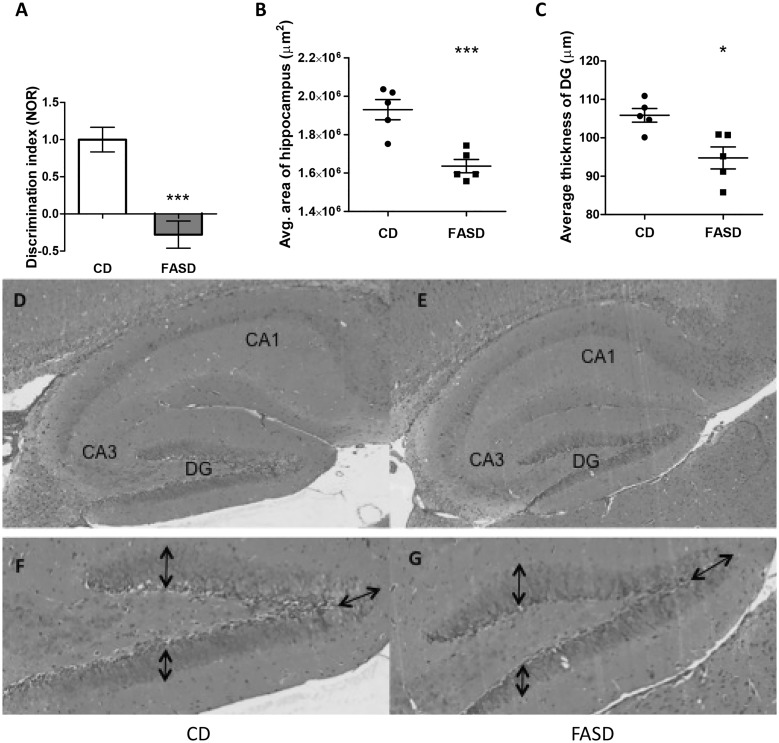
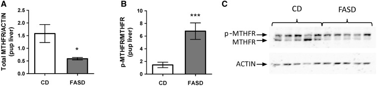
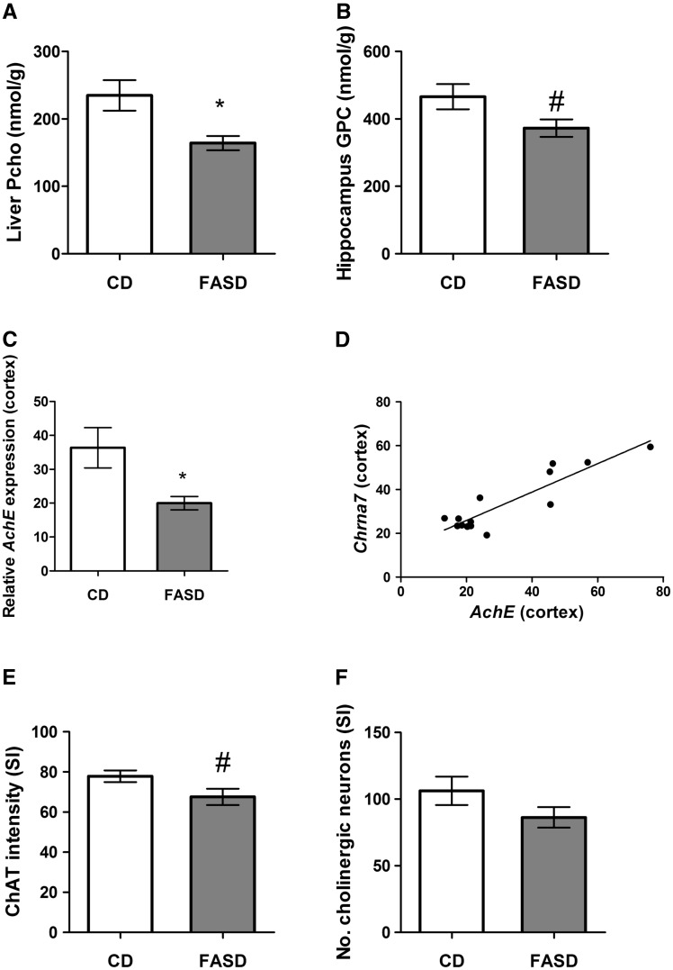
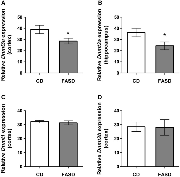
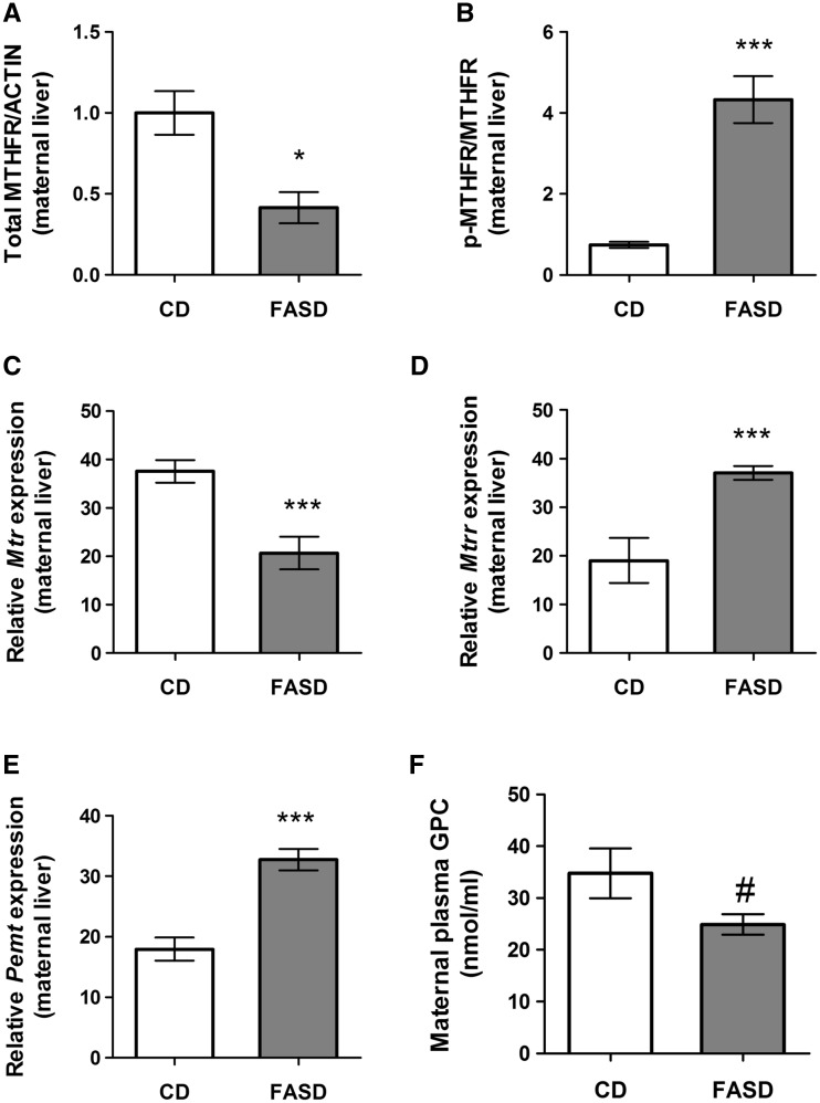
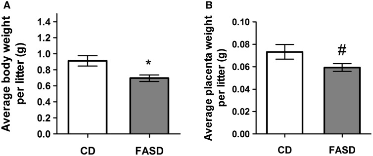
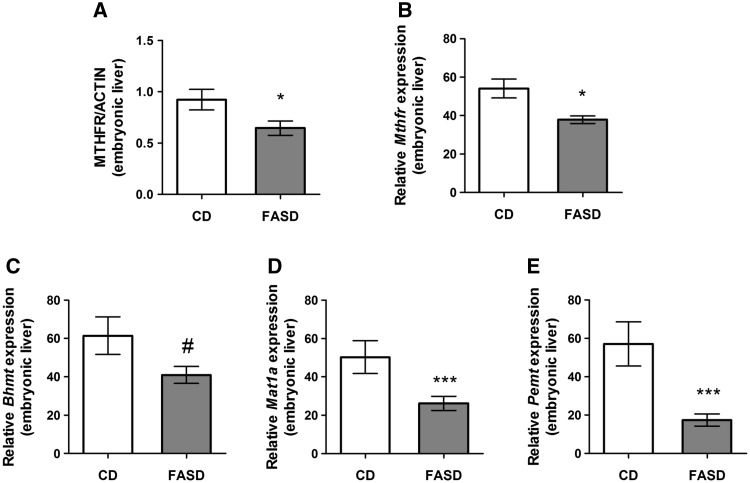
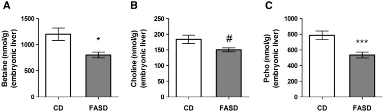
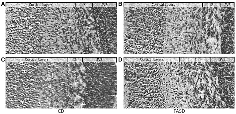
Methylenetetrahydrofolate reductase (MTHFR) generates methyltetrahydrofolate for methylation reactions. Severe MTHFR deficiency results in homocystinuria and neurologic impairment. Mild MTHFR deficiency (677C > T polymorphism) increases risk for complex traits, including neuropsychiatric disorders. Although low dietary folate impacts brain development, recent concerns have focused on high folate intake following food fortification and increased vitamin use. Our goal was to determine whether high dietary folate during pregnancy affects brain development in murine offspring. Female mice were placed on control diet (CD) or folic acid-supplemented diet (FASD) throughout mating, pregnancy and lactation. Three-week-old male pups were evaluated for motor and cognitive function. Tissues from E17.5 embryos, pups and dams were collected for choline/methyl metabolite measurements, immunoblotting or gene expression of relevant enzymes. Brains were examined for morphology of hippocampus and cortex. Pups of FASD mothers displayed short-term memory impairment, decreased hippocampal size and decreased thickness of the dentate gyrus. MTHFR protein levels were reduced in FASD pup livers, with lower concentrations of phosphocholine and glycerophosphocholine in liver and hippocampus, respectively. FASD pup brains showed evidence of altered acetylcholine availability and Dnmt3a mRNA was reduced in cortex and hippocampus. E17.5 embryos and placentas from FASD dams were smaller. MTHFR protein and mRNA were reduced in embryonic liver, with lower concentrations of choline, betaine and phosphocholine. Embryonic brain displayed altered development of cortical layers. In summary, high folate intake during pregnancy leads to pseudo-MTHFR deficiency, disturbed choline/methyl metabolism, embryonic growth delay and memory impairment in offspring. These findings highlight the unintended negative consequences of supplemental folic acid.


Figures

Figure 1
Figure 1
Pups born to mothers fed high folate diet had short-term memory impairment and reduced hippocampal area. (A) Three-week-old male mice were tested using the NOR. The discrimination index is the ratio of time spent with the novel object versus time with the familiar object; a negative discrimination index indicates short-term memory impairment. Data from two independent experiments are shown (n = 15–20/group). (B) The average area of the hippocampus and (C) the average thickness of the dentate gyrus (DG) were reduced in FASD pups (n = 5/group). Representative images of hippocampus and DG from CD pup (D and F, respectively) and FASD pup (E and G, respectively) are shown. Arrows in the DG indicate where the thickness measurements were performed. Values are means ± SEM; *P < 0.05, ***P < 0.005.
Figure 2
Figure 2
Pups born to mothers fed high folate diet had pseudo-MTHFR deficiency in liver. (A) Total immunoreactive MTHFR protein was reduced in livers of FASD pups. (B) The ratio of the phosphorylated isoform (p-MTHFR, less active) to the non-phosphorylated isoform (MTHFR) was increased in FASD pups. (C) Representative Western blot of MTHFR in offspring liver. Values are means ± SEM from 5 mice/group; *P<0.05, ***P<0.005.
Figure 3
Figure 3
Pups born to mothers fed high folate diet had disturbances in choline and acetylcholine metabolism. (A) The concentration of phosphocholine (PCho), the storage form of choline, was significantly decreased in liver (n = 6/group). (B) In hippocampus, there was a trend towards reduced glycerophosphocholine (GPC) in FASD pups (n = 7/group, #P = 0.06). (C) mRNA expression of acetylcholinesterase (AchE) was reduced in cortex of FASD pups (n = 7/group). (D) mRNA levels of the nicotinic receptor of acetylcholine, Chrna7 and AchE in cortex were significantly positively correlated (n = 7/group; r = 0.894, P < 0.05). ChAT staining to measure (E) staining intensity and (F) numbers of cholinergic neurons in the Substantia Innominata (SI). There was a trend towards reduced intensity of ChAT staining in the SI of FASD pups (n = 5/group, #P = 0.07) but no significant difference in number of cholinergic neurons. Values are means ± SEM; *P < 0.05.
Figure 4
Figure 4
Pups of FASD mothers had reduced expression of Dnmt3a mRNA in cortex and hippocampus. There was a significant decrease of Dnmt3a mRNA due to FASD in (A) cortex (n = 7/group) and (B) hippocampus of FASD pups (n = 5/group). (C) Dnmt1 and (D) Dnmt3b mRNA were not changed by diet in cortex. Values are means ± SEM; *P < 0.05.
Figure 5
Figure 5
High folic acid intake altered folate metabolism in liver of lactating mothers and led to changes in choline metabolism. (A) Total MTHFR protein was reduced in FASD mothers (n = 5/group). (B) The ratio of the phosphorylated MTHFR isoform (less active) to the non-phosphorylated isoform was increased in the FASD group (n = 5/group). mRNA expression of (C) methionine synthase (Mtr) was reduced, (D) methionine synthase reductase (Mtrr) was increased and (E) Pemt was increased due to FASD (n = 7/group). (F) There was a trend towards lower GPC due to diet in maternal plasma, (n = 7/group, #P = 0.06). Values are means ± SEM; *P < 0.05, ***P < 0.005.
Figure 6
Figure 6
Embryos and placentas from FASD mothers were smaller at E17.5. Weights of embryos and placentas were measured at sacrifice. (A) FASD led to lower mean body weights/litter of embryos. (B) There was a trend toward lower placental weights/litter (#P = 0.08). Values are means ± SEM of 6 l/group; *P < 0.05.
Figure 7
Figure 7
Maternal FASD altered expression of one-carbon enzymes in liver of E17.5 embryos. (A) Total MTHFR protein and (B) Mthfr mRNA was reduced in liver of E17.5 FASD embryos. (C) There was a trend towards reduced Bhmt mRNA in embryonic liver (#P = 0.08). (D) Mat1a and (E) Pemt mRNA were both significantly decreased due to diet in embryonic liver. Values are means ± SEM of 7–8 embryos/group; *P < 0.05, ***P < 0.005.
Figure 8
Figure 8
Choline metabolites decreased in E17.5 embryonic liver of FASD mothers. (A) Betaine concentration was significantly decreased. (B) There was a trend towards reduced choline (#P = 0.06) (C) Pcho concentration was significantly decreased. Values are means ± SEM of 6 embryos/group; *P < 0.05, ***P < 0.005.
Figure 9
Figure 9
Altered brain development in embryos of FASD mothers. Coronal sections of embryonic brain were stained with H & E. Representative images from embryos in the CD (A and C) and FASD (B and D) groups are depicted. Images show the cortical layers, the intermediate zone (IZ) and subventricular zone (SVZ). Cortical layers from FASD embryos appeared to be more organized and developed.

References

    1. Bottiglieri T. (1996) Folate, vitamin B12, and neuropsychiatric disorders. Nutr. Rev., 54, 382–390. - PubMed
    1. Frosst P., Blom H.J., Milos R., Goyette P., Sheppard C.A., Matthews R.G., Boers G.J., den Heijer M., Kluijtmans L.A., van den Heuvel L.P., et al. (1995) A candidate genetic risk factor for vascular disease: a common mutation in methylenetetrahydrofolate reductase. Nat. Genet., 10, 111–113. - PubMed
    1. Bailey L.B., Stover P.J., McNulty H., Fenech M.F., Gregory J.F., Mills J.L., Pfeiffer C.M., Fazili Z., Zhang M., Ueland P.M., et al. (2015) Biomarkers of nutrition for development—folate review. J. Nutr., 145, 1636S–1680S. - PMC - PubMed
    1. Zeisel S.H. (2006) Choline: critical role during fetal development and dietary requirements in adults. Annu. Rev. Nutr., 26, 229–250. - PMC - PubMed
    1. Whitehead V.M. (2006) Acquired and inherited disorders of cobalamin and folate in children. Br. J. Haematol., 134, 125–136. - PubMed

MeSH terms

Supplementary concepts