. 2017 Feb 17;292(7):2703-2713.
doi: 10.1074/jbc.M116.762849. Epub 2017 Jan 9.

Sensitive Versatile Fluorogenic Transmembrane Peptide Substrates for Rhomboid Intramembrane Proteases

Affiliations

Sensitive Versatile Fluorogenic Transmembrane Peptide Substrates for Rhomboid Intramembrane Proteases

Anežka Tichá et al. J Biol Chem. .

Abstract

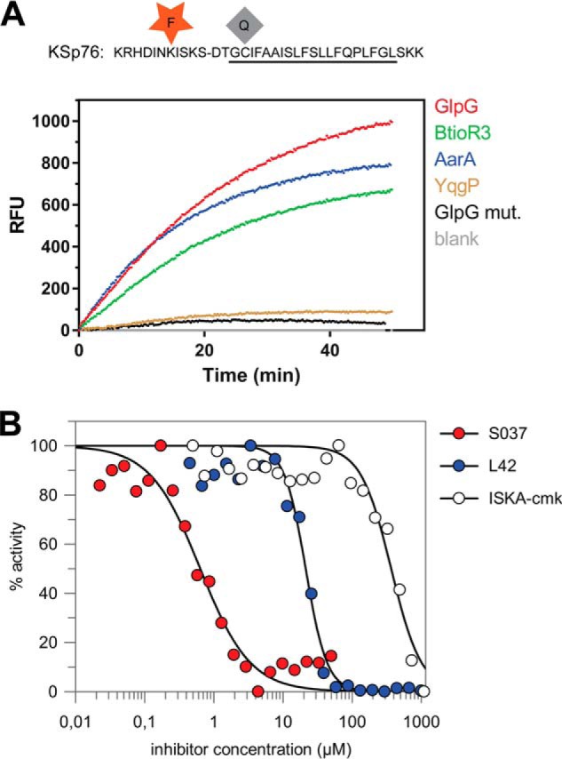
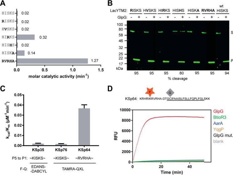
Rhomboid proteases are increasingly being explored as potential drug targets, but their potent and specific inhibitors are not available, and strategies for inhibitor development are hampered by the lack of widely usable and easily modifiable in vitro activity assays. Here we address this bottleneck and report on the development of new fluorogenic transmembrane peptide substrates, which are cleaved by several unrelated rhomboid proteases, can be used both in detergent micelles and in liposomes, and contain red-shifted fluorophores that are suitable for high-throughput screening of compound libraries. We show that nearly the entire transmembrane domain of the substrate is important for efficient cleavage, implying that it extensively interacts with the enzyme. Importantly, we demonstrate that in the detergent micelle system, commonly used for the enzymatic analyses of intramembrane proteolysis, the cleavage rate strongly depends on detergent concentration, because the reaction proceeds only in the micelles. Furthermore, we show that the catalytic efficiency and selectivity toward a rhomboid substrate can be dramatically improved by targeted modification of the sequence of its P5 to P1 region. The fluorogenic substrates that we describe and their sequence variants should find wide use in the detection of activity and development of inhibitors of rhomboid proteases.

Keywords: enzyme kinetics; enzyme mechanism; fluorescence resonance energy transfer (FRET); intramembrane proteolysis; membrane reconstitution; rhomboid protease; substrate specificity; transmembrane domain.


Conflict of interest statement

The authors declare that they have no conflicts of interest with the contents of this article

Figures

FIGURE 1.
FIGURE 1.
Identification of a widely accepted transmembrane substrate for rhomboid proteases. A, comparison of cleavage efficiency of model substrates LacYTM2, Gurken, TatA, and Spitz by bacterial rhomboid proteases GlpG (E. coli), AarA (P. stuartii), YqgP (B. subtilis), and BtioR3 (B. thetaiotaomicron) in vitro. Equal concentrations of purified recombinant substrates were exposed to purified recombinant rhomboid proteases. Cleavage products were separated by SDS-PAGE, stained, and quantified densitometrically to determine initial reaction rates, which were converted to molar catalytic activities to allow comparisons. Displayed values are representative of two independent experiments. B, cleavage of synthetic LacYTM2 transmembrane peptide KSp31 by GlpG. Purified synthetic peptide KSp31 was incubated with purified recombinant GlpG or its inactive mutant S201T in the presence of 0.05% (w/v) DDM, and the reaction mixtures were analyzed by MALDI mass spectrometry. The theoretical molecular masses of the expected cleavage products at the native cleavage site are denoted below the peptide sequence, and unambiguously match those experimentally determined and displayed in the mass spectra. The star-marked peak with molecular mass of 1893.3 is an unidentified minor contaminant in the preparation of KSp31. C, monitoring of cleavage of peptide substrate KSp31 by rhomboid protease GlpG using CE. The N-terminal cleavage product (P) of KSp31 was separated by free-flow CE in the background electrolyte composed of 100 mm H3PO4 and 69 mm Tris, pH 2.5, in bare fused silica capillary at separation voltage +25 kV. Samples for CE were prepared by mixing 20 μl of reaction mixture at selected reaction times (0–90 min) with 2 μl of 2.2 mm tyramine (T) as an internal standard. Samples were injected into the capillary by 20 mbar pressure for 10 s. Quantitative analysis was based on the ratio of corrected (migration time normalized) peak areas of peptides of interest and the internal standard. Analyses were performed in triplicate. P, cleaved N-terminal peptide; X, system peak. D, the importance of the transmembrane domain of the substrate for its recognition and cleavage by rhomboid. A series of synthetic peptides covering LacYTM2 with progressive truncations of its transmembrane domain from the C terminus was exposed to GlpG and initial rates of cleavage were quantified by capillary electrophoresis as denoted in panel C.
FIGURE 2.
FIGURE 2.
Fluorogenic transmembrane peptide substrate based on LacYTM2. A, fluorogenic variant of the LacYTM2 transmembrane helix-derived peptide (KSp31) with the P5 and P4′ positions replaced by Glu-EDANS and Lys-DABCYL, respectively, yielding fluorogenic substrate KSp35. B, solubility of KSp35 in 16 mm detergents DDM, DM, and nonyl glucoside (NG) and at 1 mm DDM. Note that the concentration of DDM micelles is about 100 μm at 16 mm DDM and about 10 μm at 1 mm DDM. The peptide was dissolved to the indicated concentration by dilution from a 10 mm stock solution in DMSO, and after a 2-h incubation at 37 °C the solution was centrifuged at 21,130 × g for 20 min. The absorbance of the supernatant at 455 nm indicated the concentration of the chromophore in solution. C, circular dichroism spectra of LacYTM2-derived transmembrane peptide KSp31 and its fluorogenic variant KSp35 in detergent micelles. Peptides were reconstituted into 0.5% (w/v) DDM to 135 μm (KSp31) and 82 μm (KSp35) concentrations. The spectra show similarly significant helical content for both peptides. D, identification of the cleavage site in KSp35 by GlpG. Purified 95 μm KSp35 was incubated with 26 μm GlpG for 20 h and analyzed by MALDI. The red peak of the mass of 2993.7 corresponds well to the expected size of the C-terminal cleavage product of 2990.690. The second peak lower by 130 Da is visible in both the blue and red traces is probably a deletion product of chemical synthesis lacking a C-terminal lysine. This variant has proven difficult to purify away, but it is cleaved by GlpG and probably does not influence the kinetics properties of the substrate significantly (see Fig. 1D). E, excitation and emission spectra of KSp35 and their change upon cleavage by rhomboid GlpG measured in detergent micelles. The spectra of 10 μm KSp35 substrate in reaction buffer (20 mm HEPES, pH 7.4, 150 mm NaCl, 0.05% (w/v) DDM, 10% (v/v) DMSO) were measured at 37 °C. Excitation wavelengths ranged from 235 to 435 nm with a 10-nm increment and the emission was measured at 493 nm. The emission wavelengths ranged from 365 to 595 nm with a 10-nm increment and excitation at 335 nm.
FIGURE 3.
FIGURE 3.
Kinetic characterization of fluorogenic transmembrane peptide substrate KSp35 in the detergent micelle system. A, dependence of the initial reaction rate on enzyme concentration. The fluorogenic substrate KSp35 (10 μm) was incubated with varying concentrations of GlpG in a reaction buffer composed of 20 mm HEPES, pH 7.4, 150 mm NaCl, 0.05% (w/v) DDM, and 10% (v/v) DMSO, and initial reaction rates were measured by following fluorescence at 493 nm. The displayed values are means from duplicate measurements with 2 × S.D. B, dependence of the initial reaction rate on substrate concentration. The rhomboid protease GlpG (0.4 μm) was incubated with varying concentrations of the fluorogenic substrate KSp35 in a reaction buffer composed of 20 mm HEPES, pH 7.4, 150 mm NaCl, 0.05% (w/v) DDM, 10% (v/v) DMSO, and the initial reaction rates were measured by following fluorescence at 493 nm. Representative values from one of three independent experiments are shown. C, dependence of the initial reaction rate on detergent concentration (solid circles, left and lower axes). The fluorogenic substrate KSp35 (10 μm) was incubated with 0.4 μm GlpG at varying concentrations of DDM in a reaction buffer composed of 20 mm HEPES, pH 7.4, 150 mm NaCl, 10% (v/v) DMSO, and initial reaction rates were measured by following fluorescence at 493 nm. Representative values from one of three independent experiments are shown. The open circles (right and upper axes) represent the same plot at the logarithmic scale. When this plot is fitted by second-order polynomial, the equation y = −0.1436x2 − 0.3906x + 2.8852 is obtained, the derivative of which, y′ = −0.2872x − 0.3906, is equal to the power of DDM concentration with which the reaction rate decreases. For high DDM concentrations the derivative tends to −1 (for x = 2, y′ = −0.965), whereas for lower DDM concentrations the absolute value of the power decreases (for x = 0, y′ = −0.3906). D, overall secondary structure of GlpG is not affected by high concentrations of DDM. CD spectra of GlpG at 0.05, 0.5, and 5% (w/v) (98 mm) DDM were recorded and show no variation in the secondary structure content of GlpG depending on DDM concentration. E, the pH dependence of GlpG activity on the LacYTM2-derived chimeric substrate MBP-LacYTM2-Trx. The substrate (2 μm) was incubated with 0.1 μm GlpG in a broad pH range buffer (38) composed of 40 mm H3PO4, 40 mm CH3COOH, and 40 mm H3BO3 adjusted to pH values between 2 and 12, and initial reaction rates were measured by SDS-PAGE and densitometry as described under “Experimental Procedures.” F, the pH dependence of cleavage of the fluorogenic LacYTM2-derived substrate KSp35 by GlpG. The substrate (10 μm) was incubated with 0.4 μm GlpG in a broad pH range buffer (38) composed of 40 mm H3PO4, 40 mm CH3COOH, and 40 mm H3BO3 adjusted to pH values between 2 and 12, and initial reaction rates were measured by recording fluorescence at 493 nm. G, selectivity of the fluorogenic substrate KSp35 for diverse bacterial rhomboid proteases. The purified recombinant rhomboid proteases GlpG, AarA, YqgP (all at 0.4 μm), and BtioR3 (at 0.04 μm) were incubated with 10 μm KSp35 in a reaction buffer composed of 20 mm HEPES, pH 7.4, 150 mm NaCl, 0.05% (w/v) DDM, and 10% (v/v) DMSO, and progress curves were measured by recording the increase in fluorescence at 493 nm.
FIGURE 4.
FIGURE 4.
The use of the transmembrane peptide substrate in liposomes. A, KSp35 was reconstituted into liposomes (LUVs) formed from E. coli polar lipid extract in the presence of GlpG or its inactive mutant S201A at pH 4.0. The resulting large unilamellar vesicles were analyzed by SDS-PAGE. B, the shape, lamellarity, and approximate size distribution of the KSp35+GlpG containing proteoliposomes formed at pH 4.0 were characterized by transmission electron microscopy. C, the integration of KSp35 into liposomes and its secondary structure content were analyzed by electronic CD. The substrate KSp35 (3 μm) was reconstituted with 2 mg/ml of E. coli polar lipid extract yielding an approximate peptide:lipid weight ratio of 1:500. D, activity of GlpG in liposomes detected by the KSp35 fluorogenic substrate. The substrate was co-reconstituted with wild type GlpG or its S201A/H254A mutant in a 30:1 molar ratio into LUVs made of E. coli polar lipid extract at pH 4.0, proteoliposomes were collected by ultracentrifugation and resuspended in 10 mm HEPES, 150 mm NaCl, pH 7.4, to start the cleavage reaction, which was then followed by measuring fluorescence at 493 nm. E, wild type GlpG or its H150A/H254A mutant were co-reconstituted with the substrate KSp35 in a 30:1 molar ratio into LUVs made of E. coli polar lipid extract at pH 4.0, proteoliposomes were collected by ultracentrifugation, resuspended in 50 mm sodium acetate, 150 mm NaCl, pH 4.0, and fluorescence was followed at 493 nm.
FIGURE 5.
FIGURE 5.
Red-shifted variant of the LacYTM2-based fluorogenic substrate. A, modification of Lys in the P5 position of KSp31 by the red-shifted TAMRA fluorophore and P4′ Cys by a dark quencher QXL610 yields highly fluorogenic substrate KSp76 that is efficiently cleaved by rhomboid proteases GlpG, AarA, YqgP, and BtioR3 at identical concentrations to those used in Fig. 3G. Excitation wavelength was 553 nm, and emission was followed at 583 nm. B, the red-shifted fluorogenic substrate KSp76 allows measurement of inhibition by compounds that absorb in the UV region, such as isocoumarin, and is thus suitable for high-throughput screening. The dose-response curves of the chloromethylketone ISKAcmk, β-lactam L42, and isocoumarin S037 were measured after a 60-min preincubation of enzyme with inhibitor. The curves were fitted in GraFit 7 to yield apparent IC50 values.
FIGURE 6.
FIGURE 6.
The effect of non-prime side substitutions on the catalytic parameters and selectivity of rhomboid substrates. A, preferred amino acids in the P5 to P1 positions of the LacYTM2 transmembrane substrate improve its cleavage by GlpG. The LacYTM2 embedded in the MBP-thioredoxin chimera (18) was point-mutated in the P5 to P1 positions according to the sequence preferences of E. coli GlpG (19). The recombinant substrates were expressed in E. coli ΔglpG, purified, and molar catalytic activity of GlpG in cleaving each of the substrates was determined using gel-based assay (see “Experimental Procedures” for details). The concentration of substrate was always 1.47 μm, concentration of DDM was 0.5%(w/v), the concentration of GlpG was 0.8 μm for wild type substrate (HISKS). and for the RISKS, HVSKS, and HISHS mutants the concentration was 0.08 μm for the HISKA mutant and 0.016 μm for the HIRKS and RVRHA variants (to ensure reliable measurement of the initial reaction rate). Representative values from one of three independent experiments are shown. B, the effects of the preferred amino acids in the P5 to P1 region of LacYTM2 on the steady-state level of cleavage by GlpG in biological membranes in vivo. Plasmids encoding individual mutant versions of the chimeric mutant LacYTM2 substrates described above were transformed into E. coli MC4100 expressing endogenous GlpG, and 2 h after induction of expression of the substrates, the cell lysates were analyzed by immunoblotting using antibody against His tag, located at the C terminus of the constructs. Detection by near-infrared laser scanning, exhibiting linearity over 6 orders of magnitude, enabled reliable quantitation. Integration of product and substrate band intensities yielded steady-state substrate conversion values that are listed below the image. A representative experiment is displayed. C, apparent kinetic parameters of fluorogenic rhomboid substrates derived from LacYTM2. Initial reaction rates at very low substrate concentrations were used to calculate catalytic efficiency values (kcat/Km) of substrates KSp35, KSp64, and Ksp76 cleaved by GlpG at 0.5% (w/v) DDM. The reaction buffer was 20 mm HEPES, pH 7.4, 150 mm NaCl, 10% (v/v) DMSO, enzyme concentration was 0.4 μm, and substrate concentration ranged from 0.5 to 20 μm. Note that a mere optimization of the P5 to P1 region of the substrate increases the catalytic efficiency (kcat/Km) of its cleavage by GlpG by 23-fold. D, influence of the optimization of the P5 to P1 region on the selectivity of a transmembrane substrate for rhomboids. KSp76 underwent cleavage by rhomboid proteases GlpG, AarA, YqgP, and BtioR3 at the same concentrations as described in the legends to Figs. 3G and 5A. Note that optimization of the P5 to P1 region of the substrate increases the selectivity for GlpG dramatically.

References

    1. Urban S., Lee J. R., and Freeman M. (2002) A family of rhomboid intramembrane proteases activates all Drosophila membrane-tethered EGF ligands. EMBO J. 21, 4277–4286 - PMC - PubMed
    1. Lee J. R., Urban S., Garvey C. F., and Freeman M. (2001) Regulated intracellular ligand transport and proteolysis control EGF signal activation in Drosophila. Cell 107, 161–171 - PubMed
    1. McQuibban G. A., Saurya S., and Freeman M. (2003) Mitochondrial membrane remodelling regulated by a conserved rhomboid protease. Nature 423, 537–541 - PubMed
    1. O'Donnell R. A., Hackett F., Howell S. A., Treeck M., Struck N., Krnajski Z., Withers-Martinez C., Gilberger T. W., and Blackman M. J. (2006) Intramembrane proteolysis mediates shedding of a key adhesin during erythrocyte invasion by the malaria parasite. J. Cell Biol. 174, 1023–1033 - PMC - PubMed
    1. Fleig L., Bergbold N., Sahasrabudhe P., Geiger B., Kaltak L., and Lemberg M. K. (2012) Ubiquitin-dependent intramembrane rhomboid protease promotes ERAD of membrane proteins. Mol. Cell 47, 558–569 - PubMed

Publication types

LinkOut - more resources