. 2017 Feb 24;292(8):3411-3419.
doi: 10.1074/jbc.M116.760074. Epub 2017 Jan 9.

Cooperative Substrate-Cofactor Interactions and Membrane Localization of the Bacterial Phospholipase A2 (PLA2) Enzyme, ExoU

Affiliations

Cooperative Substrate-Cofactor Interactions and Membrane Localization of the Bacterial Phospholipase A2 (PLA2) Enzyme, ExoU

Maxx H Tessmer et al. J Biol Chem. .

Abstract

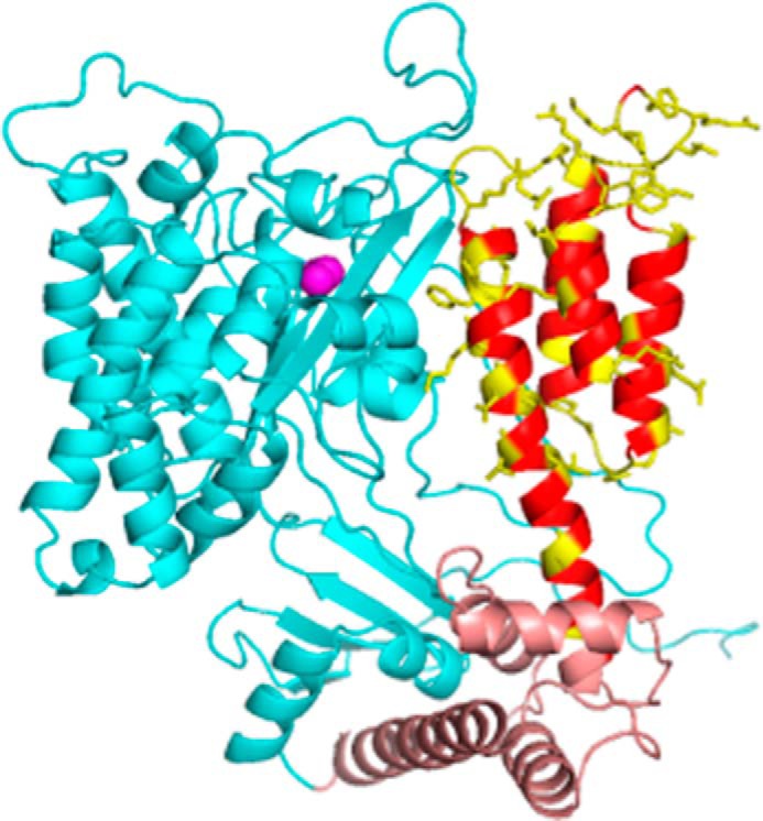
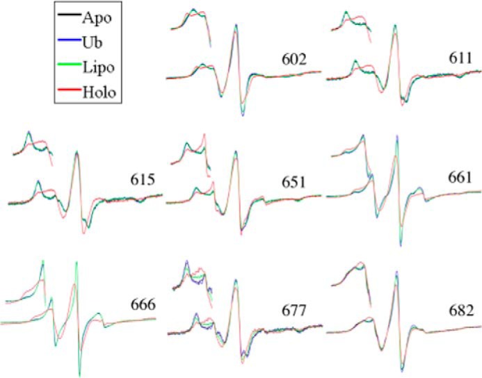
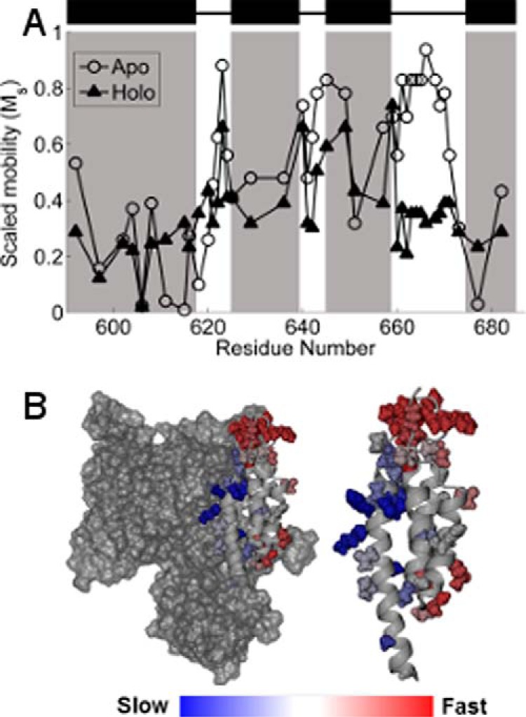
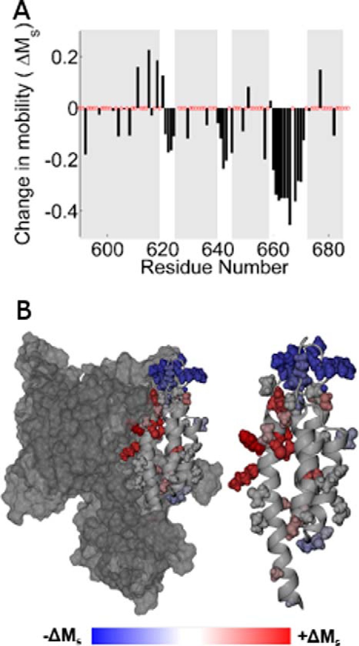
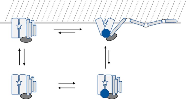
The ExoU type III secretion enzyme is a potent phospholipase A2 secreted by the Gram-negative opportunistic pathogen, Pseudomonas aeruginosa Activation of phospholipase activity is induced by protein-protein interactions with ubiquitin in the cytosol of a targeted eukaryotic cell, leading to destruction of host cell membranes. Previous work in our laboratory suggested that conformational changes within a C-terminal domain of the toxin might be involved in the activation mechanism. In this study, we use site-directed spin-labeling electron paramagnetic resonance spectroscopy to investigate conformational changes in a C-terminal four-helical bundle region of ExoU as it interacts with lipid substrates and ubiquitin, and to examine the localization of this domain with respect to the lipid bilayer. In the absence of ubiquitin or substrate liposomes, the overall structure of the C-terminal domain is in good agreement with crystallographic models derived from ExoU in complex with its chaperone, SpcU. Significant conformational changes are observed throughout the domain in the presence of ubiquitin and liposomes combined that are not observed with either liposomes or ubiquitin alone. In the presence of ubiquitin, two interhelical loops of the C-terminal four-helix bundle appear to penetrate the membrane bilayer, stabilizing ExoU-membrane association. Thus, ubiquitin and the substrate lipid bilayer act synergistically to induce a conformational rearrangement in the C-terminal domain of ExoU.

Keywords: Pseudomonas aeruginosa (P. aeruginosa); electron paramagnetic resonance (EPR); phospholipase; protein structure; ubiquitin.


Conflict of interest statement

The authors declare that they have no conflicts of interest with the contents of this article

Figures

FIGURE 1.
FIGURE 1.
Structural model of ExoU. Shown is a model based on PDB 4AKX (13) displaying the catalytic domain (cyan), bridging domain (brown), and C-terminal four-helix bundle (red/yellow). Native side chains at sites of spin label attachment are shown in yellow. The catalytic serine (Ser-142) is indicated by magenta spheres. Loops were modeled using Rosetta (see “Experimental Procedures”).
FIGURE 2.
FIGURE 2.
Continuous-wave EPR spectral overlays of several MTSL spin-labeled ExoU derivatives. Continuous-wave EPR spectra at representative sites along the four-helix bundle (site indicated by the residue number) are shown. Each overlay shows four conditions: ExoU alone (Apo); ExoU with a 12-fold excess diUb (Ub); ExoU in the presence of 100-fold molar excess liposomes (Lipo); and ExoU in the presence of both diUb and liposomes (Holo). Scan widths of the full spectra are 100 G. Insets show the low-field region for each spectrum.
FIGURE 3.
FIGURE 3.
A, scaled mobility of the R1 side chain. Open circles indicate the apo state, and filled triangles indicate the holo state. Higher values of Ms indicate greater mobility of the R1 side chain. Black rectangles and shaded regions indicate helices in the C-terminal domain as defined by the crystal structures (13, 14). B, Ms in the apo state mapped onto the crystal structure of ExoU. Color changes from blue to red indicate increasing motion of the R1 side chain. Left, the catalytic domain is shown in surface rendering. Right, the isolated C-terminal four-helix bundle.
FIGURE 4.
FIGURE 4.
A, histogram showing the change in scaled mobility, Ms(apo) − Ms(holo), as a function of the labeling site. Clusters of negative values indicating decreased mobility in the holo state correspond to the three interhelical loops. B, the change in Ms mapped onto the ExoU crystal structure. Sites that increased motion upon adoption of the holo state are shown in red, and sites that exhibit decreased motion are shown in blue.
FIGURE 5.
FIGURE 5.
Accessibilities and membrane localization as a function of labeling position for ExoU variants alone in solution (Apo, open circles), in the presence of liposomes (Lipo, filled circles), and in the presence of liposomes and diUb (Holo, red triangles). Shaded areas indicate helices in the C-terminal domain as defined by the crystal structures (13, 14). A, accessibility to NiEDDA. B, accessibility to oxygen. C, the EPR depth parameter. D, localization of the spin label side chain relative to the membrane bilayer. Negative values indicate exposure to the aqueous phase.
FIGURE 6.
FIGURE 6.
Schematic of ExoU membrane association and conformational changes. The ExoU four-helix bundle (cylinders) is depicted as dissociating and being displaced from the catalytic domain (rectangles) upon adoption of the holo state (upper right), accompanied by conformational changes in the bridging domain (oval) and catalytic domain that expose the catalytic site (star) to the membrane surface (located at the top of the figure). The binding of ubiquitin (octagon) and membrane association in the absence of ubiquitin (upper left) likely result in conformational changes relative to the apo state (lower left) that remain to be elucidated, and the location of the ubiquitin-binding site has not been fully identified.

References

    1. Galán J. E., Lara-Tejero M., Marlovits T. C., and Wagner S. (2014) Bacterial type III secretion systems: specialized nanomachines for protein delivery into target cells. Annu. Rev. Microbiol. 68, 415–438 - PMC - PubMed
    1. Yahr T. L., Goranson J., and Frank D. W. (1996) Exoenzyme S of Pseudomonas aeruginosa is secreted by a type III pathway. Mol. Microbiol. 22, 991–1003 - PubMed
    1. Yahr T. L., Mende-Mueller L. M., Friese M. B., and Frank D. W. (1997) Identification of type III secreted products of the Pseudomonas aeruginosa exoenzyme S regulon. J. Bacteriol. 179, 7165–7168 - PMC - PubMed
    1. Yahr T. L., Vallis A. J., Hancock M. K., Barbieri J. T., and Frank D. W. (1998) ExoY, an adenylate cyclase secreted by the Pseudomonas aeruginosa type III system. Proc. Natl. Acad. Sci. U.S.A. 95, 13899–13904 - PMC - PubMed
    1. Finck-Barbançon V., Goranson J., Zhu L., Sawa T., Wiener-Kronish J. P., Fleiszig S. M., Wu C., Mende-Mueller L., and Frank D. W. (1997) ExoU expression by Pseudomonas aeruginosa correlates with acute cytotoxicity and epithelial injury. Mol. Microbiol. 25, 547–557 - PubMed

MeSH terms

LinkOut - more resources