. 2017 Feb 24;292(8):3154-3163.
doi: 10.1074/jbc.M116.767699. Epub 2017 Jan 9.

Residues in the RecQ C-terminal Domain of the Human Werner Syndrome Helicase Are Involved in Unwinding G-quadruplex DNA

Affiliations

Residues in the RecQ C-terminal Domain of the Human Werner Syndrome Helicase Are Involved in Unwinding G-quadruplex DNA

Amit Ketkar et al. J Biol Chem. .

Abstract

The structural and biophysical properties typically associated with G-quadruplex (G4) structures render them a significant block for DNA replication, which must be overcome for cell division to occur. The Werner syndrome protein (WRN) is a RecQ family helicase that has been implicated in the efficient processing of G4 DNA structures. The aim of this study was to identify the residues of WRN involved in the binding and ATPase-driven unwinding of G4 DNA. Using a c-Myc G4 DNA model sequence and recombinant WRN, we have determined that the RecQ-C-terminal (RQC) domain of WRN imparts a 2-fold preference for binding to G4 DNA relative to non-G4 DNA substrates. NMR studies identified residues involved specifically in interactions with G4 DNA. Three of the amino acids in the WRN RQC domain that exhibited the largest G4-specific changes in NMR signal were then mutated alone or in combination. Mutating individual residues implicated in G4 binding had a modest effect on WRN binding to DNA, decreasing the preference for G4 substrates by ∼25%. Mutating two G4-interacting residues (T1024G and T1086G) abrogated preferential binding of WRN to G4 DNA. Very modest decreases in G4 DNA-stimulated ATPase activity were observed for the mutant enzymes. Most strikingly, G4 unwinding by WRN was inhibited ∼50% for all three point mutants and >90% for the WRN double mutant (T1024G/T1086G) relative to normal B-form dsDNA substrates. Our work has helped to identify residues in the WRN RQC domain that are involved specifically in the interaction with G4 DNA.

Keywords: DNA helicase; DNA repair; DNA replication; G-quadruplex; Werner syndrome helicase; enzyme mechanism.


Conflict of interest statement

The authors declare that they have no conflicts of interest with the contents of this article

Figures
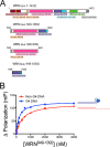
FIGURE 1.
FIGURE 1.
Schematic illustration of WRN constructs and preferential binding to G4 DNA. A, schematic illustration of WRN (amino acids (a.a.) 1–1432) domain orientation. Three truncated WRN constructs were prepared for the study as follows: 1) WRN(500–1092); 2) WRN(500–949), and 3) WRN(949–1092). B, change in fluorescence polarization for the FAM-labeled G4 (blue) and non-G4 (red) substrates is plotted as a function of WRN(949–1092) concentration. The results were fit to a quadratic equation to estimate the Kd, DNA. The results shown represent the mean ± S.D. (n = 3).
FIGURE 2.
FIGURE 2.
Identification of G4-specific amino acids in the WRN RQC domain. A, NMR spectra resulting from HSQC experiments titrating G4 DNA into a solution of 15N-labeled WRN RQC (amino acids 949–1092) domain are shown. Changes in peak intensity were monitored and quantified. B, peak intensity for each residue was monitored as a function of increasing amounts of DNA added and fit to a quadratic equation to estimate the apparent binding affinity (Kd) for each residue. C, Kd values for non-G4 (red bars) and G4 (blue bars) DNA substrates for Thr-1024 and Leu-1063 are shown. D, mean structure of the WRN RQC derived from NMR experiments (Protein Data Bank code 2AXL) was superimposed upon coordinates derived from the crystal structure of WRN RQC bound to dsDNA (Protein Data Bank code 3AAF). The 10 amino acids with the largest Kapp, non-G4 DNA/Kapp, G4 DNA (i.e. greatest affinity for G4 DNA) are shown (yellow). The α-helices, the α2-α3 loop, the strand-separating β-wing, and the unstructured C-terminal region of the WRN RQC domain are also labeled.
FIGURE 3.
FIGURE 3.
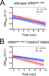
ATPase activity of WRN RQC mutant enzymes on G4 DNA is not greatly perturbed. The ATPase activity of wild-type (A) and T1024G/T1086G WRN(500–1092) (B) was measured using a coupled spectrophotometric assay (see “Experimental Procedures”). Results are shown for both G4 (blue squares) and non-G4 (red circles) DNA substrates.
FIGURE 4.
FIGURE 4.
Point mutations in the WRN RQC domain impair unwinding of c-MYC G4 DNA. A, schematic illustration of the helicase assay used to monitor unwinding of the c-MYC G4 DNA structure is shown. A control substrate containing a non-G4 sequence was used for comparison. B, helicase activity for wild-type (bright red) and T1024G/T1086G (dark red) WRN(500–1092) is shown for the non-G4 DNA substrate. C, helicase activity for wild-type (bright blue) and T1024G/T1086G (dark blue) WRN(500–1092) is shown for the c-MYC G4 DNA substrate. D, relative unwinding rate (mutant rate/wild-type rate × 100) on the non-G4 DNA substrate is plotted for each of the WRN RQC mutants. E, relative unwinding rate (mutant rate/wild-type rate × 100) on the c-MYC G4 DNA substrate is plotted for each of the WRN RQC mutants. The results shown represent the mean ± S.D. (n = 3). All p values were calculated using an unpaired Student's t test comparing wild-type and mutant WRN.
FIGURE 5.
FIGURE 5.
WRN RQC double mutant fails to unwind telomeric G4 DNA. A, schematic illustration of the FRET-based unwinding assay for the telomeric G4 DNA substrate (Tel22) is shown. B, change in Cy3 fluorescence over time was monitored for both wild-type (black circles) and T1024G/T1086G (red squares) WRN(500–1092). C, rate of Tel22 unwinding by wild-type (black) and T1024G/T1086G WRN(500–1092) was compared with that observed for the non-G4 DNA substrate to estimate the relative helicase activity of each enzyme against telomeric G4 DNA. The results shown represent the mean ± S.D. (n = 3). The reported p value was calculated using an unpaired Student's t test comparing wild-type and mutant WRN.

References

    1. Delagoutte E., and von Hippel P. H. (2002) Helicase mechanisms and the coupling of helicases within macromolecular machines. Part I: structures and properties of isolated helicases. Q. Rev. Biophys. 35, 431–478 - PubMed
    1. Eoff R. L., and Raney K. D. (2005) Helicase-catalysed translocation and strand separation. Biochem. Soc. Trans. 33, 1474–1478 - PubMed
    1. Wu C. G., and Spies M. (2013) Overview: what are helicases? Adv. Exp. Med. Biol. 767, 1–16 - PubMed
    1. Caruthers J. M., and McKay D. B. (2002) Helicase structure and mechanism. Curr. Opin. Struct. Biol. 12, 123–133 - PubMed
    1. Croteau D. L., Popuri V., Opresko P. L., and Bohr V. A. (2014) Human RecQ helicases in DNA repair, recombination, and replication. Annu. Rev. Biochem. 83, 519–552 - PMC - PubMed

Publication types

LinkOut - more resources