. 2017 Feb 23;85(3):e00961-16.
doi: 10.1128/IAI.00961-16. Print 2017 Mar.

Temporal Role for MyD88 in a Model of Brucella-Induced Arthritis and Musculoskeletal Inflammation

Affiliations

Temporal Role for MyD88 in a Model of Brucella-Induced Arthritis and Musculoskeletal Inflammation

Carolyn A Lacey et al. Infect Immun. .

Abstract

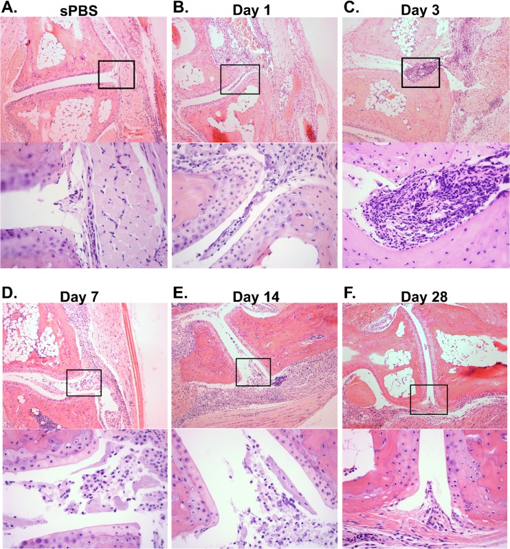
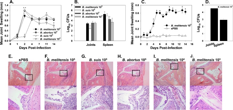
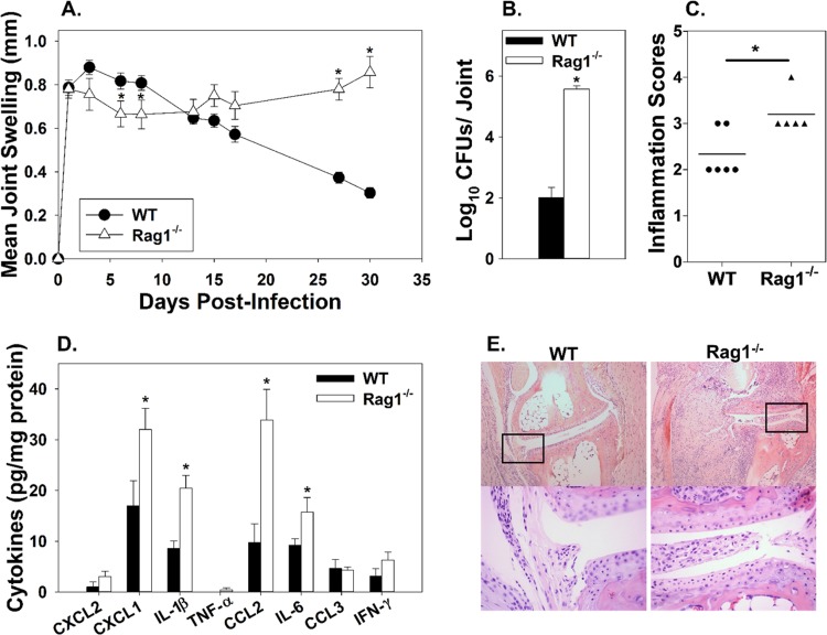
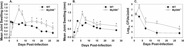
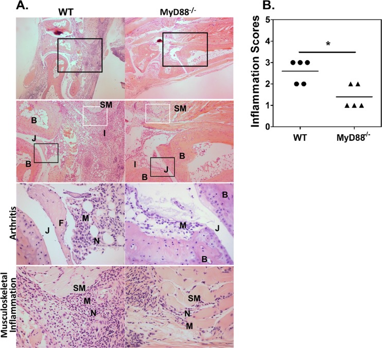
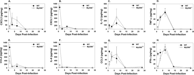
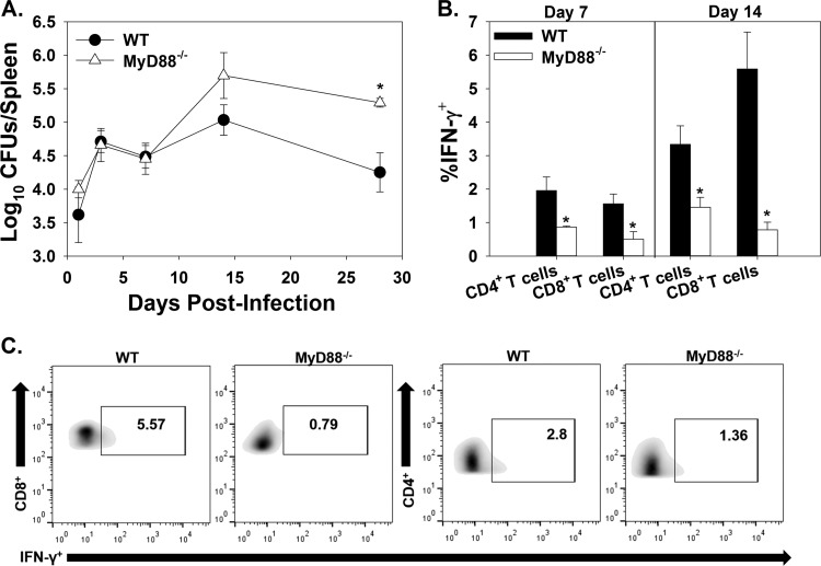
Brucella spp. are facultative intracellular Gram-negative bacteria that cause the zoonotic disease brucellosis, one of the most common global zoonoses. Osteomyelitis, arthritis, and musculoskeletal inflammation are common focal complications of brucellosis in humans; however, wild-type (WT) mice infected systemically with conventional doses of Brucella do not develop these complications. Here we report C57BL/6 WT mice infected via the footpad with 103 to 106 CFU of Brucella spp. display neutrophil and monocyte infiltration of the joint space and surrounding musculoskeletal tissue. Joint inflammation is detectable as early as 1 day postinfection and peaks 1 to 2 weeks later, after which WT mice are able to slowly resolve inflammation. B and T cells were dispensable for the onset of swelling but required for resolution of joint inflammation and infection. At early time points, MyD88-/- mice display decreased joint inflammation, swelling, and proinflammatory cytokine levels relative to WT mice. Subsequently, swelling of MyD88-/- joints surpassed WT joint swelling, and resolution of joint inflammation was prolonged. Joint bacterial loads in MyD88-/- mice were significantly greater than those in WT mice by day 3 postinfection and at all time points thereafter. In addition, MyD88-/- joint inflammatory cytokine levels on day 3 and beyond were similar to WT levels. Collectively these data demonstrate MyD88 signaling mediates early inflammatory responses in the joint but also contributes to subsequent clearance of Brucella and resolution of inflammation. This work also establishes a mouse model for studying Brucella-induced arthritis, musculoskeletal complications, and systemic responses, which will lead to a better understanding of focal complications of brucellosis.

Keywords: Brucella; MyD88; arthritis; brucellosis; musculoskeletal; osteomyelitis.


Figures

FIG 1
FIG 1
WT mice develop arthritis and musculoskeletal inflammation following footpad infection. C57BL/6 WT mice were either mock infected in each rear footpad with sterile PBS (sPBS) or infected with 1 × 10⁶ CFU of B. melitensis 16M. Mock-infected mice were euthanized at day 3 (A), and B. melitensis-infected mice were euthanized at days 1 (B), 3 (C), 7 (D), 14 (E), and 28 (F) postinfection. H&E staining was conducted on mouse joints. Representative images (100×) from mock-treated and infected mice are depicted. Amplified boxed regions (400×) showing inflammation within the joint space of B. melitensis-infected mice are displayed beneath the 100× image at each time point. Images are representative of joints from 4 to 5 mice per time point from one kinetic experiment. Similar pathology was observed at the day 3 and day 28 time points in 2 to 3 additional experiments.
FIG 2
FIG 2
WT mice infected via the footpad with B. melitensis, B. abortus, or B. suis develop arthritis and musculoskeletal inflammation. C57BL/6 WT mice were infected in each rear footpad with 1 × 10⁵ CFU of Brucella spp., 1 × 10⁴ CFU of B. melitensis 16M (A, B, F to I), or 1 × 10³ CFU of B. melitensis 16M or mock infected with sterile PBS (sPBS) (C and D) and evaluated for ankle swelling over time (A and C). Mice were euthanized on day 14 postinfection, and joint and spleen bacterial loads were enumerated (B and D). (A) *, P < 0.05 compared to WT mice infected with 1 × 10⁵ CFU of B. melitensis 16M. (C) *, P < 0.05 compared to mock-infected WT mice, and error bars depict standard errors of the mean. Representative plots of H&E-stained joint sections from mock-infected mice (E) or mice infected in the footpad with 1 × 10⁵ CFU of B. melitensis 16M (F) 1 × 10⁵ CFU of B. suis 1330 (G), 1 × 10⁵ CFU of B. abortus 2308 (H), 1 × 10⁴ CFU of B. melitensis 16M (I), or 1 × 10³ CFU of B. melitensis 16M (J) are shown. Images (100×) are depicted with amplified boxed regions (400×) displayed beneath each image. Data from mice infected with 1 × 10⁵ CFU of B. melitensis 16M is representative of 2 to 3 independent experiments, while data from other Brucella strains are representative of one experiment.
FIG 3
FIG 3
Adaptive immune responses are needed for inflammation resolution but not required for inflammation onset. C57BL/6 WT and Rag1−/− mice were infected in each rear footpad with 1 × 10⁶ CFU of B. melitensis 16M. Joint swelling was recorded over time (5 to 10 mice/group) (A). Mice were euthanized at day 30 postinfection, and ankle joint bacterial loads (n = 5 to 10/group) (B) and cytokine levels (n = 4 to 5 mice/group) (D) were enumerated. H&E staining was conducted on joints at day 30 postinfection (n = 5 to 6/group) and scored for inflammation severity as follows: 0 = none (no inflammation), 1 = minimal with inflammation involving <5% of tissue, 2 = moderate with focally extensive areas of inflammation (5% to 25% of tissue and involving 1 or more tissues), 3 = moderate to severe with focally extensive areas of inflammation (>25% to 50% of tissue and involving multiple tissues), and 4 = severe with large confluent areas of inflammation (>50% of tissue and involving multiple tissues) (C). Representative images are depicted (100×), and amplified boxed regions are displayed beneath each image (400×) (E). *, P < 0.05 compared to WT mice. Error bars depict standard errors of the mean. Data are representative of one experiment.
FIG 4
FIG 4
MyD88−/− mice have delayed joint swelling but increased joint Brucella burden. C57BL/6 WT and MyD88−/− mice were infected in each rear footpad with 1 × 10⁶ (A) or 1 × 10⁵ (B) CFU of B. melitensis 16M (4 to 5 mice/group), and swelling was recorded over time. At days 1, 3, 7, 14, and 28, mice infected with 1 × 10⁶ CFU of B. melitensis were euthanized, and bacterial loads were enumerated in the ankle joint (3 to 4 mice/group/time point) (C). *, P < 0.05 compared to WT mice. Error bars depict standard errors of the mean. CFU data in panel C are from one kinetic experiment. Swelling curves are representative of two independent experiments.
FIG 5
FIG 5
MyD88 mediates early arthritis and musculoskeletal inflammation. C57BL/6 WT and MyD88−/− mice were infected in each rear footpad with 1 × 10⁶ CFU of B. melitensis 16M. Mice were euthanized 3 days postinfection, and H&E staining was conducted on mouse joints. Representative images of joint sections from WT and MyD88−/− mice are depicted with amplified boxed regions displayed beneath each image (A). Magnifications are as follows: row 1, 40×; row 2, 100×; rows 3 and 4, 400×. The black boxes indicate areas of arthritis or inflammation within and around the joint, and the white boxes indicate areas of musculoskeletal inflammation. Tissue architecture is indicated as follows: B, bone; SM, skeletal muscle; J, joint space. Areas of pathology are indicated as follows: I, inflammation; M, macrophages; N, neutrophils. Day 3 WT and MyD88−/− ankle joint H&E staining was scored for total inflammation as follows: 0 = none (no inflammation), 1 = minimal with inflammation involving <5% of tissue, 2 = moderate with focally extensive areas of inflammation (5% to 25% of tissue and involving 1 or more tissues), 3 = moderate to severe with focally extensive areas of inflammation (>25% to 50% of tissue and involving multiple tissues), 4 = severe with large confluent areas of inflammation (>50% of tissue and involving multiple tissues) (B). Histology scores are from one of two independent experiments (5 mice/group) from which sections were obtained. *, P < 0.05 compared to sections from WT mice.
FIG 6
FIG 6
MyD88−/− joints have delayed cytokine production following footpad infection with B. melitensis. C57BL/6 WT and MyD88−/− mice (3 to 4 mice/group/time point) were infected in each rear footpad with 1 × 10⁶ CFU of B. melitensis 16M and sacrificed at days 1, 3, 7, 14, and 28. Rear joints were homogenized, and CXCL2 (A), CXCL1 (B), IL-1β (C), TNF-α (D), CCL2 (E), IL-6 (F), CCL3 (G), and IFN-γ (H) concentrations were quantified by Luminex and normalized to total protein via BCA. *, P < 0.05 compared to infected WT mice. Error bars depict standard errors of the mean. Data are representative of one kinetic experiment.
FIG 7
FIG 7
MyD88 aids in systemic Brucella clearance and T cell IFN-γ production. C57BL/6 WT and MyD88−/− mice were infected in each rear footpad with 1 × 10⁶ CFU of B. melitensis 16M. At days 1, 3, 7, 14, and 28, mice were euthanized, and bacterial loads were enumerated in the spleen (3 to 4 mice/group/time point) (A). At days 7 and 14, flow cytometry was performed to determine IFN-γ production by T cells from infected mouse spleens (B). (C) Representative plots showing IFN-γ production by gated CD4+ and CD8+ T cells from WT and MyD88−/− mice 14 days postinfection. *, P < 0.05 compared to WT mice. Error bars depict standard errors of the mean. Data are representative of one kinetic experiment.

Similar articles

Cited by

References

    1. Colmenero JD, Reguera JM, Fernandez-Nebro A, Cabrera-Franquelo F. 1991. Osteoarticular complications of brucellosis. Ann Rheum Dis 50:23–26. doi:10.1136/ard.50.1.23. - DOI - PMC - PubMed
    1. . 2012. Research priorities for zoonoses and marginalized infections. World Health Organ Tech Rep Ser 2012:ix–xi, 1–119, 2 p following p 119. - PubMed
    1. Zinsstag J, Roth F, Orkhon D, Chimed-Ochir G, Nansalmaa M, Kolar J, Vounatsou P. 2005. A model of animal-human brucellosis transmission in Mongolia. Prev Vet Med 69:77–95. doi:10.1016/j.prevetmed.2005.01.017. - DOI - PubMed
    1. Kaufmann AF, Fox MD, Boyce JM, Anderson DC, Potter ME, Martone WJ, Patton CM. 1980. Airborne spread of brucellosis. Ann N Y Acad Sci 353:105–114. doi:10.1111/j.1749-6632.1980.tb18912.x. - DOI - PubMed
    1. Mableson HE, Okello A, Picozzi K, Welburn SC. 2014. Neglected zoonotic diseases—the long and winding road to advocacy. PLoS Negl Trop Dis 8:e2800. doi:10.1371/journal.pntd.0002800. - DOI - PMC - PubMed

Publication types

MeSH terms

LinkOut - more resources