. 2017 Mar 18;79(3):492-501.
doi: 10.1292/jvms.16-0456. Epub 2017 Jan 6.

Injectable alginate-microencapsulated canine adipose tissue-derived mesenchymal stem cells for enhanced viable cell retention

Affiliations

Injectable alginate-microencapsulated canine adipose tissue-derived mesenchymal stem cells for enhanced viable cell retention

Eunji Koh et al. J Vet Med Sci. .

Abstract

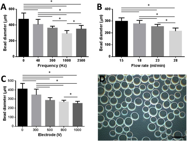
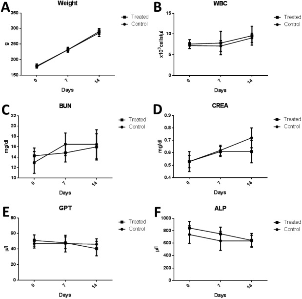
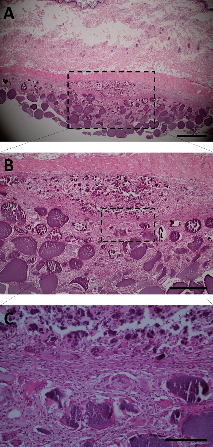
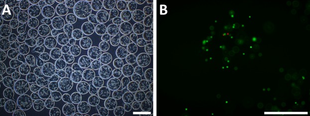
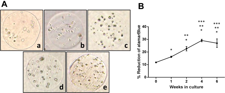
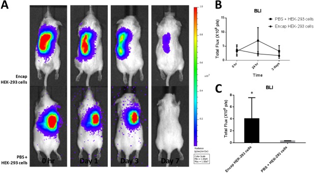
The purpose of this study was to establish an optimized protocol for the production of alginate-encapsulated canine adipose-derived mesenchymal stem cells (cASCs) and evaluate their suitability for clinical use, including viability, proliferation and in vivo cell retention. Alginate microbeads were formed by vibrational technology and the production of injectable microbeads was performed using various parameters with standard methodology. Microbead toxicity was tested in an animal model. Encapsulated cASCs were evaluated for viability and proliferation in vitro. HEK-293 cells, with or without microencapsulation, were injected into the subcutaneous tissue of mice and were tracked using in vivo bioluminescent imaging to evaluate the retention of transplanted cells. The optimized injectable microbeads were of uniform size and approximately 250 µm in diameter. There was no strong evidence of in vivo toxicity for the alginate beads. The cells remained viable after encapsulation, and there was evidence of in vitro proliferation within the microcapsules. In vivo bioluminescent imaging showed that alginate encapsulation improved the retention of transplanted cells and the encapsulated cells remained viable in vivo for 7 days. Encapsulation enhances the retention of viable cells in vivo and might represent a potential strategy to increase the therapeutic potency and efficacy of stem cells.


Figures

Fig. 1.
Fig. 1.
Standardization of optimal bead size for injection. (A) Microbead diameter (µm) as a function of increasing frequency (Hz). (B) Microbead diameter (µm) as a function of increasing flow rate (ml/min). (C) Microbead diameter (µm) as a function of increasing electrode (V). (D) Light microscope image of microbeads; bar=500 µm.
Fig. 2.
Fig. 2.
In vivo toxicology evaluation of alginate microbeads. Weight and blood chemistry evaluation of the PBS injected group and the alginate microbead injected group, which showed no significant difference between the groups at 0 days, 1 week and 2 weeks after injection. (A) Weight. (B) White blood cell count. (C) Blood urea nitrogen. (D) Creatinine. (E) Glutamic-pyruvic transaminase. (F) Alanine transaminase.
Fig. 3.
Fig. 3.
Photomicrograph of the H&E stained sections of the subcutaneous tissue injected with alginate microcapsules at week 2. Degradation and phagocytosis of the bead material are shown. (A) The alginate microcapsules were detectable 2 weeks after implantation; bar=500 µm. (B) Inset −higher magnification image of the boxed area; bar=250 µm. (C) Inset −higher magnification image of the boxed area; bar=125 µm.
Fig. 4.
Fig. 4.
Encapsulated canine ASCs. (A) Microscopic appearance of microencapsulated canine ASCs. Light microscopic appearance of encapsulated canine ASCs showing a bead diameter of approximately 250 µm; bar=500 µm. (B) Cell viability immediately after encapsulation. Live cell cytoplasm appeared green, while dead cells had a red-stained nucleus; bar=250 µm.
Fig. 5.
Fig. 5.
AlamarBlue percent reduction and associated photomicrographs, of the encapsulated canine ASCs at 0 hr (a), 1 week (b), 2 weeks (c), 4 weeks (d) and 6 weeks (e). (A) Light microscopic images of the canine ASCs showed an increase in cell proliferation within the beads with time. (B) The percent reduction of alamarBlue increased with time; however, there was no significant increase in reduction when comparing weeks 4 to 6.
Fig. 6.
Fig. 6.
In vivo optical bioluminescence imaging (BLI) showed greater retention and survival for encapsulated HEK-293 (Luc-2) cells compared to those of direct injection. (A) BLI image of representative animal from each group (n=5), showing increased cell retention in the animal administered encapsulated cells at day 7. (B) Quantification of BLI signal in regions of interest in the subcutaneous tissue at the total flux level of 108 p/s at 0 hr, 1 day and 3 days showed no significant difference between the groups. (C) Quantification of BLI signal in regions of interest in the subcutaneous tissue at the total flux level of 106 p/s showed significantly greater signal in the animals treated with encapsulated cells at day 7.

References

    1. Abbah S. A., Lu W. W., Chan D., Cheung K. M., Liu W. G., Zhao F., Li Z. Y., Leong J. C., Luk K. D.2006. In vitro evaluation of alginate encapsulated adipose-tissue stromal cells for use as injectable bone graft substitute. Biochem. Biophys. Res. Commun. 347: 185–191. doi: 10.1016/j.bbrc.2006.06.072 - DOI - PubMed
    1. Bhat A., Hoch A. I., Decaris M. L., Leach J. K.2013. Alginate hydrogels containing cell-interactive beads for bone formation. FASEB J. 27: 4844–4852. doi: 10.1096/fj.12-213611 - DOI - PubMed
    1. Bonnier F., Keating M. E., Wróbel T. P., Majzner K., Baranska M., Garcia-Munoz A., Blanco A., Byrne H. J.2015. Cell viability assessment using the Alamar blue assay: a comparison of 2D and 3D cell culture models. Toxicol. In Vitro 29: 124–131. doi: 10.1016/j.tiv.2014.09.014 - DOI - PubMed
    1. Capone S. H., Dufresne M., Rechel M., Fleury M. J., Salsac A. V., Paullier P., Daujat-Chavanieu M., Legallais C.2013. Impact of alginate composition: from bead mechanical properties to encapsulated HepG2/C3A cell activities for in vivo implantation. PLoS ONE 8: e62032. doi: 10.1371/journal.pone.0062032 - DOI - PMC - PubMed
    1. Gasperini L., Mano J. F., Reis R. L.2014. Natural polymers for the microencapsulation of cells. J. R. Soc. Interface 11: 20140817. doi: 10.1098/rsif.2014.0817 - DOI - PMC - PubMed

MeSH terms