. 2017 Apr;230(4):549-566.
doi: 10.1111/joa.12579. Epub 2017 Jan 10.

Evolutionary aspects of the development of teeth and baleen in the bowhead whale

Affiliations

Evolutionary aspects of the development of teeth and baleen in the bowhead whale

J G M Thewissen et al. J Anat. 2017 Apr.

Abstract

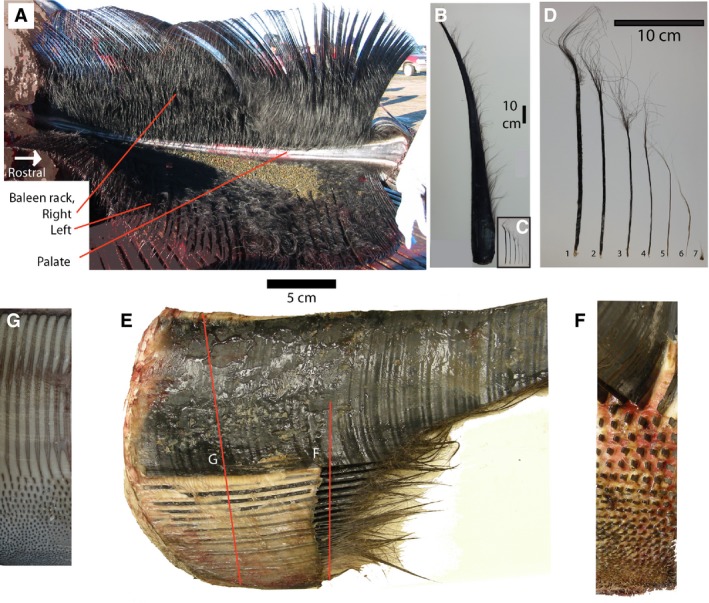
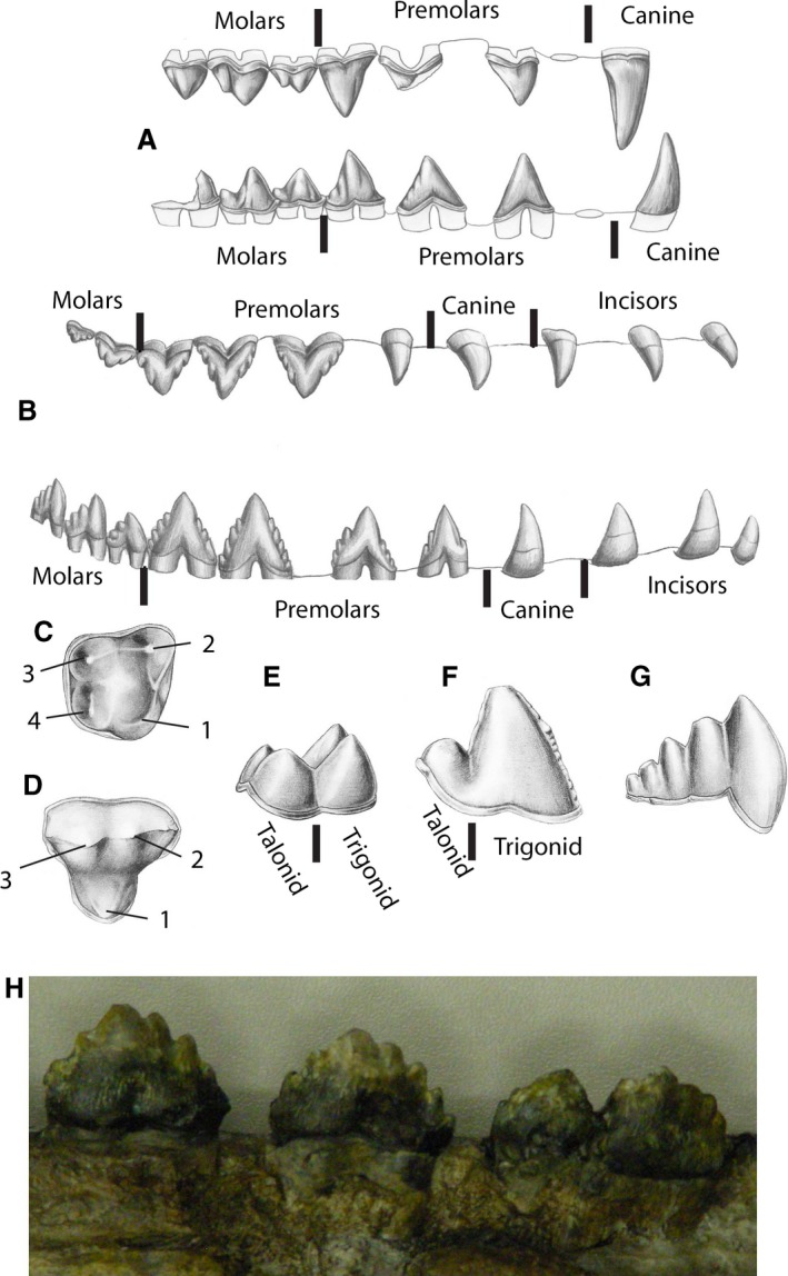
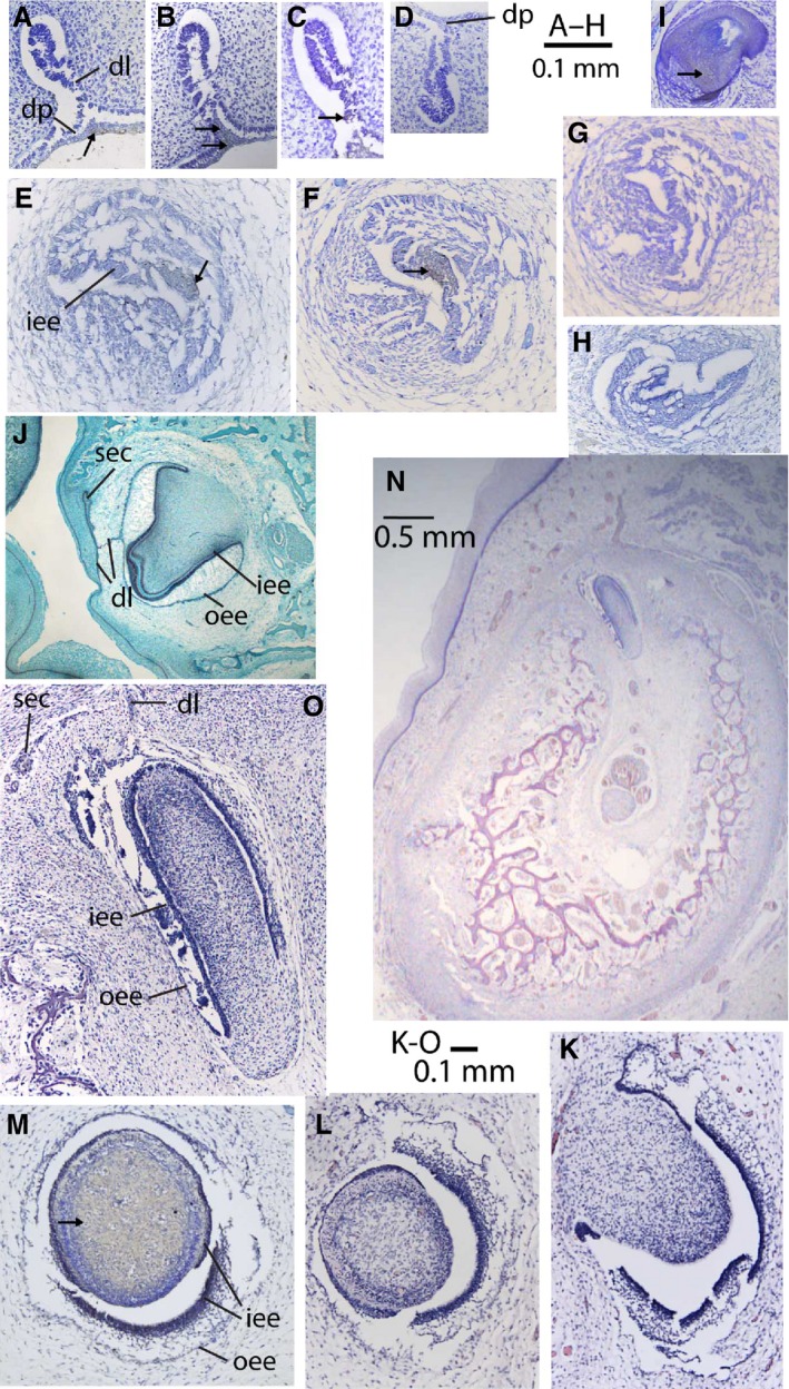
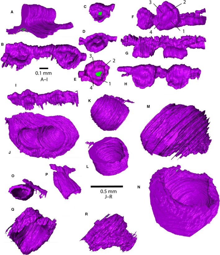
In utero, baleen whales initiate the development of several dozens of teeth in upper and lower jaws. These tooth germs reach the bell stage and are sometimes mineralized, but toward the end of prenatal life they are resorbed and no trace remains after birth. Around the time that the germs disappear, the keratinous baleen plates start to form in the upper jaw, and these form the food-collecting mechanism. Baleen whale ancestors had two generations of teeth and never developed baleen, and the prenatal teeth of modern fetuses are usually interpreted as an evolutionary leftover. We investigated the development of teeth and baleen in bowhead whale fetuses using histological and immunohistochemical evidence. We found that upper and lower dentition initially follow similar developmental pathways. As development proceeds, upper and lower tooth germs diverge developmentally. Lower tooth germs differ along the length of the jaw, reminiscent of a heterodont dentition of cetacean ancestors, and lingual processes of the dental lamina represent initiation of tooth bud formation of replacement teeth. Upper tooth germs remain homodont and there is no evidence of a secondary dentition. After these germs disappear, the oral epithelium thickens to form the baleen plates, and the protein FGF-4 displays a signaling pattern reminiscent of baleen plates. In laboratory mammals, FGF-4 is not involved in the formation of hair or palatal rugae, but it is involved in tooth development. This leads us to propose that the signaling cascade that forms teeth in most mammals has been exapted to be involved in baleen plate ontogeny in mysticetes.

Keywords: FGF; Cetacea; baleen; baleen whales; bowhead whale; embryology; keratin; mysticetes; ontogeny; tooth development.


Figures

Figure 1
Figure 1
(A) Palate of bowhead whale showing left and right sides of baleen rack, each consisting of approximately 320 plates. Note the narrow palate and baleen plates flaring laterally from it. The lingual side of the plates is frayed and makes a matted surface. (B) Single baleen plate, medial to right. (C) Minor plates from the posterior side of the palate, arranged from lateral to medial on the palate, to the same scale as (B). (D) Enlarged view of (C), showing flat, bilateral symmetrical plates (1, 2, 3); radially symmetrical plates (4, 5, 6) and baleen hair (7). (E) Cranial view of baleen plate (cut off) and the minor plates medial to it (bottom of image). The oral cavity is to the right in this image, and the full depth of gingiva (gums) is seen (white material). Red lines indicate sectional planes of (F) and (G). (F) Cut section through (E), occlusal view, with minor plates cut off to show pattern of implantation, medial to bottom. (G) Section through gingiva, showing cross‐sections of plate and minor plates as embedded in palate, medial to bottom.
Figure 2
Figure 2
Teeth of fossil cetaceans. (A) Right upper and lower dentition of Pakicetus, the oldest known whale, in labial (lateral) view. Incisors not shown, first premolar indicated as alveolus. (B) Right upper and lower dentition of Dorudon, a late Eocene whale, in labial view. (C, D) Right upper molars of Indohyus and Pakicetus, rostral to right, with homologous cusps identified by numbers. Lower molars of Indohyus (E), a middle Eocene protocetid whale (F), and late Eocene Zygorhiza (G), in lingual view. (H) Labial view of three cheek teeth (rostral to left) of toothed mysticete (aetiocetid), notice that the last tooth appears to consist of two teeth that are fused. Drawings from Thewissen (2014).
Figure 3
Figure 3
Histological sections through tooth germs and hair follicle of bowhead whale (A–I, K–O) and pig (only J). (A–D) Dental lamina stage (NSBDWM 1999B7F); (E–H) cap stage (NSBDWM 1999B6F); (K–O) bell stage (NSBDWM 2000B3F); staining indicated by arrows, absence of arrows indicates absence of staining. (A) Rostral part of upper dental lamina (slide 83, FGF‐8 stain). (B) Caudal part of upper dental lamina (slide 125, FGF‐8 stain). (C) Intermediate area of upper dental lamina (slide 103, SP‐6 stain for epiprofin). (D) Rostral part of lower dental lamina (slide 81, FGF‐8 stain). (E) Upper tooth cap (slide 111, SHH stain). (F) Upper tooth cap (slide 149, SHH stain). (G) Upper tooth cap (slide 195, SP‐6 stain). (H) Lower tooth cap (slide 111, SHH stain). (I) Hair follicle of NSBDWM 2003B3F, showing competency of antibody in bowheads (slide 29, SP‐6 stain). (J) Bell stage upper tooth germ of pig, showing complete dental lamina and germ for permanent tooth. (K, L) Bell stage upper tooth germ 4 (slide 49 and 77, no antibody stain). (M) Bell stage upper tooth germ 5 (slide 22, SHH stain). (N) Mandible with tooth germ of lower tooth 15 (slide c.30, no antibody stain), lingual to left. (O) Bell stage lower tooth germ, enlarged from (N). dl, dental lamina; dp, dental placode; iee, inner enamel epithelium; oee, outer enamel epithelium; sec, secondary tooth germ for permanent tooth.
Figure 4
Figure 4
Three‐dimensional reconstructions of tooth germs of bowhead whale, made using AMIRA, based on histological sections. The green areas indicate the presence of antibody stain. (A) Dental lamina stage in bowhead whale (NSBDWM 1999B7F); (B–I) cap stage (NSBDWM 1999B6F); (J–R) bell stage (NDBDWM 2000B3F). Scale bars are approximate because of foreshortening of images, and magnification of images of the same specimen are similar. (A) Rostral part of upper dental lamina, caudolingual view (slides 73–93). (B) Cap stage upper tooth germs 4 and 5, dorsolingual view, rostral to right (slides 64–81). (C–E) Cap stage upper tooth germs 9 (C, dorsolingual view, slides 104–114), 10 (D, dorsolabial view, slides 115–124) and 14 (E, dorsal view, slides 146–153); the green areas in (C) and (E) indicate antibody stain for SHH. (F–H) Cap stage upper tooth germs 23 and 24 in dorsorostral, lateral and dorsal views, respectively, showing topography of inner enamel epithelium; the numbers identify folds in the inner enamel epithelium as discussed in the text (slides 249–274). (I) Cap stage lower tooth germs 19 and 20 in ventrolingual view (slides 241–259). (J) Bell stage upper tooth germ 4 (slides P1:41–83) in dorsocaudal view. (K, L) Bell stage upper tooth germ 5 in lingual and dorsal view (slides P1:1–27). (M, N) Bell stage upper tooth germ 24 (slides P4:4–42) in lingual and dorsolabial view. (O, P) Bell stage lower tooth germ 15 (slides C:13–40) in ventral and lingual view. (Q, R) Bell stage lower tooth germ 40 (slides G:24–45) in labial and lingual view.
Figure 5
Figure 5
Palate and head of bowhead fetus (NSBDWM 2000B3F). (A) Palatal epithelium, as removed from skull, backlit showing transmitted light to show developing tooth germs (in the bell stage). (B) Head of fetus showing great extent of the lower lip. (C) Clear/stain preparation of head of the same fetus, showing the narrow dentary that does not match the outline of the lower lip. Cartilage is blue and bone is red in this preparation.
Figure 6
Figure 6
Histological sections showing baleen formation. (A) Cross‐section through half of the palate of fetus NSBDWM 2000B3F, showing bell‐shaped tooth germ and epithelium that will form baleen (slides P6:36). Medial to left, letters indicate approximate location of figures (B)–(G) in antibody‐stained sections of this figure. (B–D) Details of medial palatal epithelium of NSBDWM 2000B3F (B, slides P6:32, FGF‐4 stain; C, slides P6:33, FGF‐7 stain; D, slides P6:31, FGF‐8 stain). (E–G) Details of palatal epithelium near where baleen will form of NSBDWM 2000B3F (E, slides P6:32, FGF‐4 stain; F, slides P6:33, FGF‐7 stain; G, slides P6:31, FGF‐8). (H) Hair follicle of bowhead whale 2011B8 (slide 2, FGF‐7 stain). (I) Cross‐section through half of the palate of fetus NSBDWM 2009KK1F, same orientation as (A) (slides P12:26). (J, K) Detail of Zone 2 of the palatal epithelium of NSBDWM 2009KK1F (J, slides P12:24, cross‐section; K, slides P13:47, horizontal section; FGF‐4 stain). (L, M) Detail of Zone 3 of palatal epithelium of NSBDWM 2009KK1F with FGF‐4 stain (L, slides P12:24, cross‐section; M, slides P6:3, parasagittal section). (N) Detail of Zone 3 of palatal epithelium of NSBDWM 2009KK1F (slides P13:78) with FGF‐4 stain, horizontal section, labial to top. (O) Detail of Zone 2 of palatal epithelium of NSBDWM 2009KK1F (slides P12:29, cross‐section, FGF‐8 stain). Scale bars: 0.1 mm, unless otherwise indicated.
Figure 7
Figure 7
Palate and baleen in fetus. (A) Palate of fetus NSBDWM 2007B16F, rostral to left. (B) Rostral cross‐section through the posterior part of the unilateral palate of NSBDWM 2015B9F. (C) Ventral view of the posterior palate (same individual as B), showing baleen hairs and their root in the gingiva of the palate. Zones identify homologous areas in Figs 6 and 7.
Figure 8
Figure 8
Diagrams showing tooth and baleen development in bowhead whale. Upper and lower oral epithelium and structures derived from it are shown, mesenchyme is not shown. For baleen placode and postnatal stage, the lower jaw is not shown. Numbers refer to important features discussed in the text. (1) Upper dental lamina is higher than lower dental lamina. (2) Upper dental lamina extends further caudal than lower dental lamina. (3) FGF‐8 expression in caudal (molar) region of dental lamina. (4) Dental lamina lacks contact with oral epithelium. (5) More tooth caps occur in the upper than in the lower jaw. (6) Lower tooth caps are more variable in shape and size than upper tooth caps. (7–8) Inner enamel epithelium of tooth caps shows complex folds, whereas that of upper tooth bells (8) shows single fold that corresponds to single cusp of tooth. (9) Lower tooth bells are heterodont. (10) Secondary tooth buds develop in lower, but not upper jaw. (11) Dental lamina persists longer in lower than upper jaw. (12) FGF‐4 expression in upper jaw epithelium. The pattern of expression of fgf‐4 foreshadows the footprint of baleen plates.

Similar articles

Cited by

References

    1. Armfield BA (2010) The evolution and development of mammalian tooth class. PhD dissertation, Kent State University: Kent, Ohio, USA.
    1. Armfield BA, George JC, Vinyard CJ, et al. (2011) Allometric patterns of fetal head growth in mysticetes and odontocetes: comparison of Balaena mysticetus and Stenella attenuata . Mar Mam Sci 27, 819–827.
    1. Armfield BA, Zheng Z, Bajpai S, et al. (2013) Development and evolution of the unique cetacean dentition. PeerJ 1, e24. - PMC - PubMed
    1. Barnes LG, Kimura M, Furusawa H, et al. (1994) Classification and distribution of Oligocene Aetiocetidae (Mammalia, Cetacea, Mysticeti) from western North American and Japan. Island Arc 3, 392–431.
    1. Bhussry BR (1980) Development and growth of teeth In: Orban's Histology and Embryology, 9th edn (ed. Bhaskar SN.), pp. 24–45. St. Louis, Missouri: CV Mosby.

LinkOut - more resources