. 2017 Apr;38(4):2150-2164.
doi: 10.1002/hbm.23510. Epub 2017 Jan 10.

Convergent gustatory and viscerosensory processing in the human dorsal mid-insula

Affiliations

Convergent gustatory and viscerosensory processing in the human dorsal mid-insula

Jason A Avery et al. Hum Brain Mapp. 2017 Apr.

Abstract

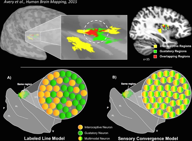
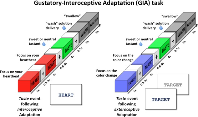
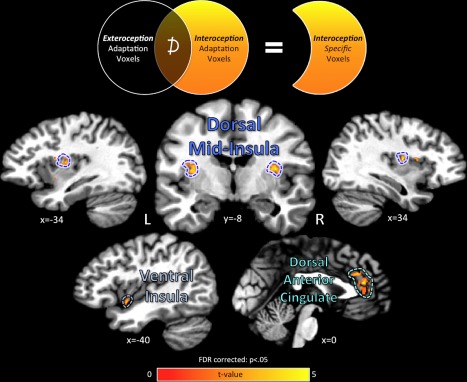
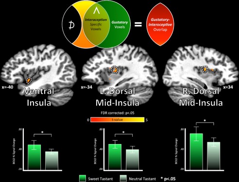
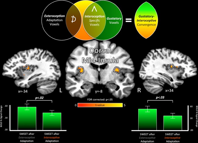
The homeostatic regulation of feeding behavior requires an organism to be able to integrate information from its internal environment, including peripheral visceral signals about the body's current energy needs, with information from its external environment, such as the palatability of energy-rich food stimuli. The insula, which serves as the brain's primary sensory cortex for representing both visceral signals from the body and taste signals from the mouth and tongue, is a likely candidate region in which this integration might occur. However, to date it has been unclear whether information from these two homeostatically critical faculties is merely co-represented in the human insula, or actually integrated there. Recent functional neuroimaging evidence of a common substrate for visceral interoception and taste perception within the human dorsal mid-insula suggests a model whereby a single population of neurons may integrate viscerosensory and gustatory signals. To test this model, we used fMRI-Adaptation to identify whether insula regions that exhibit repetition suppression following repeated interoception trials would then also exhibit adapted responses to subsequent gustatory stimuli. Multiple mid and anterior regions of the insula exhibited adaptation to interoceptive trials specifically, but only the dorsal mid-insula regions exhibited an adapted gustatory response following interoception. The discovery of this gustatory-interoceptive convergence within the neurons of the human insula supports the existence of a heretofore-undocumented neural pathway by which visceral signals from the periphery modulate the activity of brain regions involved in feeding behavior. Hum Brain Mapp 38:2150-2164, 2017. © 2017 Wiley Periodicals, Inc.

Keywords: fMRI; fMRI-adaptation; gustation; insular cortex; interoception.


Figures

Figure 1
Figure 1
Interoceptive and gustatory co‐activation of the dorsal mid‐insula. Top: A single group of subjects, performing very different tasks, displayed overlapping activation patterns in an identical region of the dorsal mid‐insula [Avery et al., 2015]. The image on the left depicts overlapping activations (red) on the cortical surface of the right dorsal mid‐insula when subjects performed a task requiring interoceptive attention to visceral sensations (yellow) or received sweet and neutral tastants during scanning (green). The image on the right depicts the same results, transformed into volumetric space for display purposes. Bottom: Two potential models for this gustatory‐interoceptive overlap. (A) Labeled Line Model—the voxels within that region of the insula contain groups of neurons separately responsible for either gustatory or interoceptive processing. (B) Sensory Convergence Model—the voxels within that region of the insula contain groups of multimodal neurons that support a shared representation of both gustation and interoception. [Color figure can be viewed at http://wileyonlinelibrary.com]
Figure 2
Figure 2
Gustatory‐interoceptive adaptation (GIA) task. The GIA task was composed of Interoception and Exteroception events presented in rapid succession (adaptation trains) in order to adapt the response of Taste events following those trains. During Interoception events, subjects focused on how intensely they could feel the sensation of their heart beating while the word “HEART” was presented on the screen. During Exteroception events, the word “TARGET” was presented in the middle of the screen and the font color would change to a lighter shade of gray every 500 ms. Subjects were instructed to focus their attention on the intensity of these color changes. Taste events involved the delivery of a sweet (0.4 mL of 0.6 M sucrose) or neutral (0.4 mL of distilled water) tasting liquid followed by a Wash period to wash the liquid from the subject's mouth. All Interoception, Exteroception, and Taste events were separated by variable duration interstimulus intervals (i.e. ‘jitters’) of 0.5 s to 3.0 s. These adaptation‐stimulus blocks were arranged in random order throughout each run of the GIA task. [Color figure can be viewed at http://wileyonlinelibrary.com]
Figure 3
Figure 3
Cortical regions exhibiting interoception‐specific adaptation. Regions of the cerebral cortex exhibiting a specific adaptation response to successive repetitions of Interoception events were located after subtracting out any cortical regions also displaying adaptation to Exteroception control events. Interoception‐specific regions were located in the bilateral dorsal mid‐insula as well as the left ventral insula. Outside of the insula, the bilateral dorsal anterior cingulate cortex also displayed specific adaptation for interoceptive attention. The unsmoothed fMRI data were plotted and analyzed on a standardized cortical surface model, and statistical contrasts were FDR corrected for multiple comparisons at P < 0.05. Data is plotted in volumetric space for viewing purposes. [Color figure can be viewed at http://wileyonlinelibrary.com]
Figure 4
Figure 4
Gustatory and interoceptive co‐activation. Prior to the GIA task, subjects performed a Gustatory Mapping (GM) task, which involved the presentation of sweet and neutral tastants during scanning [Avery et al. 2015; see Methods section for details]. Using imaging data acquired during the GM task, we next examined whether those interoceptive‐specific regions of the brain identified using the GIA task (see Fig. 3, Table 3) were also co‐activated by gustatory stimulation. Within each of those cortical surface regions, we compared the hemodynamic response to sweet vs. neutral tastants (t (14), two‐tailed paired t‐test). Neither region of the anterior cingulate cortex exhibited a significant response to the tastants, nor did their activity discriminate between them (P > 0.24; see Table 3). Only the ventral and mid‐insula regions exhibited co‐activation for gustatory and interoceptive processing. [Color figure can be viewed at http://wileyonlinelibrary.com]
Figure 5
Figure 5
Gustatory‐interoceptive convergence in the dorsal mid‐insula. The bilateral dorsal mid‐insula exhibited specific adaptation for interoception attention (Fig. 3; pictured here in volumetric cortical images) as well as significant activation to the taste of sucrose (Fig. 4). These regions also exhibited an adapted response to sweet tastants following Interoception adaptation trains compared to identical sweet tastants following Exteroception adaptation trains (SWEET after Exteroception—SWEET after interoception, t (14), two‐tailed paired t‐test; Table 4). This demonstrates that the dorsal mid‐insula contains a population of multimodal neurons that respond to both interoceptive and gustatory signals. Additionally, this gustatory adaptation by interoception was specific to the sweet tastant (0.6 M sucrose) and not the neutral tastant (distilled water) (Table 4; not pictured). The specificity of this interoceptive adaptation effect to energy‐rich gustatory stimuli suggests that these multimodal neurons may be involved in the homeostatic maintenance of energy intake. [Color figure can be viewed at http://wileyonlinelibrary.com]

References

    1. Argall BD, Saad ZS, Beauchamp MS (2006): Simplified intersubject averaging on the cortical surface using SUMA. Hum Brain Mapp 27:14–27. - PMC - PubMed
    1. Avery JA, Burrows K, Kerr KL, Bodurka J, Khalsa S, Paulus MP, Simmons WK (2017): How the brain wants what the body needs: The neural basis of positive alliesthesia. Neuropsychopharmacology. 27:14–27. - PMC - PubMed
    1. Avery JA, Drevets WC, Moseman SE, Bodurka J, Barcalow JC, Simmons WK (2014): Major depressive disorder is associated with abnormal interoceptive activity and functional connectivity in the insula. Biol Psychiatry 76:258–266. - PMC - PubMed
    1. Avery JA, Kerr KL, Ingeholm JE, Burrows K, Bodurka J, Simmons WK (2015): A common gustatory and interoceptive representation in the human mid‐insula. Hum Brain Mapp 36:2996–3006. - PMC - PubMed
    1. Berthoud HR, Neuhuber WL (2000): Functional and chemical anatomy of the afferent vagal system. Auton Neurosci 85:1–17. - PubMed

Publication types

MeSH terms