. 2017 Oct;35(10):2127-2134.
doi: 10.1002/jor.23517. Epub 2017 Jan 31.

Evidence that interfibrillar load transfer in tendon is supported by small diameter fibrils and not extrafibrillar tissue components

Affiliations

Evidence that interfibrillar load transfer in tendon is supported by small diameter fibrils and not extrafibrillar tissue components

Spencer E Szczesny et al. J Orthop Res. 2017 Oct.

Abstract

Collagen fibrils in tendon are believed to be discontinuous and transfer tensile loads through shear forces generated during interfibrillar sliding. However, the structures that transmit these interfibrillar forces are unknown. Various extrafibrillar tissue components (e.g., glycosaminoglycans, collagens XII and XIV) have been suggested to transmit interfibrillar loads by bridging collagen fibrils. Alternatively, collagen fibrils may interact directly through physical fusions and interfibrillar branching. The objective of this study was to test whether extrafibrillar proteins are necessary to transmit load between collagen fibrils or if interfibrillar load transfer is accomplished directly by the fibrils themselves. Trypsin digestions were used to remove a broad spectrum of extrafibrillar proteins and measure their contribution to the multiscale mechanics of rat tail tendon fascicles. Additionally, images obtained from serial block-face scanning electron microscopy were used to determine the three-dimensional fibrillar organization in tendon fascicles and identify any potential interfibrillar interactions. While trypsin successfully removed several extrafibrillar tissue components, there was no change in the macroscale fascicle mechanics or fibril:tissue strain ratio. Furthermore, the imaging data suggested that a network of smaller diameter fibrils (<150 nm) wind around and fuse with their neighboring larger diameter fibrils. These findings demonstrate that interfibrillar load transfer is not supported by extrafibrillar tissue components and support the hypothesis that collagen fibrils are capable of transmitting loads themselves. Conclusively determining how fibrils bear load within tendon is critical for identifying the mechanisms that impair tissue function with degeneration and for restoring tissue properties via cell-mediated regeneration or engineered tissue replacements. © 2017 Orthopaedic Research Society. Published by Wiley Periodicals, Inc. J Orthop Res 35:2127-2134, 2017.

Keywords: extrafibrillar proteins; interfibrillar load transfer; multiscale mechanics; tendon; trypsin digestion.


Figures

Figure 1
Figure 1
Immunostaining of extrafibrillar and collagenous proteins. Fluorescence intensity of decorin, tenascin C, and collagen XII were all reduced from fresh controls throughout the fascicle cross-section following trypsin digestion. Punctate staining was observed for elastin in fresh and control samples (characteristic of longitudinal fibers) but was more diffuse with greater background after trypsin digestion. No change in collagen type I or VI signal intensity was observed. Note that differences in fascicle cross-sectional area were due to sample selection and not effects of treatments. Scale bar, 100 μm.
Figure 2
Figure 2
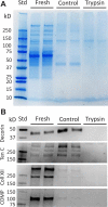
Analysis of protein extractions. (A) Coomassie staining of total protein extract demonstrates that a large amount of extrafibrillar protein is removed by buffer incubation alone, with further reductions observed after trypsin digestion. (B) Western blots show that, in particular, collagen XII and COMP are reduced in the buffer-incubated controls, while decorin and tenascin C are unaffected. All four proteins are absent in the trypsin digested samples. See Fig. S-1 in the Supplementary Material for full images of the Western blots. Std – molecular weight standards.
Figure 3
Figure 3
Desmosine content normalized by fascicle dry weight (DW) was not different between fresh (non-incubated) fascicles, buffer-incubated controls, and trypsin digested samples.
Figure 4
Figure 4
Effect of trypsin digestion on tendon fascicle multiscale mechanics. There was no significant difference in (A) macroscale mechanics or (B) fibril:tissue strain ratio between digested samples and buffer-incubated controls. (C) However, trypsin digestion did increase interfibrillar sliding (p<0.01).
Figure 5
Figure 5
Representative three-dimensional reconstruction of fibrils shows that the small diameter fibrils wind around and weave between their neighboring large diameter fibrils. See Movies S-1 and S-2 in the Supplementary Material for more detail.
Figure 6
Figure 6
Evidence of fusion between small and large diameter fibrils. (A) Initial two-dimensional image containing fibrils from Fig. 5 defining boundaries of each fibril (hatched areas). (B) Appearance of new small diameter fibril (single arrow). (C) Appearance of two new small diameter fibrils (double arrows). (D) Both the red and green unhatched small diameter fibrils fuse with the larger hatched fibrils. (E) The smaller fibrils remain fused as protrusions in the large diameter fibrils and begin to rotate clockwise in a right-hand fashion around the fibril periphery. (F) The smaller fibrils have completely incorporated into the larger fibrils. (G,H) Three-dimensional reconstructions of the fusions between the small and large diameter fibrils. The long arrows indicate the direction of increasing image number from panels A to F. Image numbers: (A) 1, (B) 10, (C) 14, (D) 25, (E) 50, (F) 82, with distance from initial image annotated in each panel. Scale bars, 200 nm. See the file ‘Segmentation_Images.zip’ in the Supplementary Material for the complete image stack used for fibril segmentation.

References

    1. Screen HRC, Lee DA, Bader DL, Shelton JC. An investigation into the effects of the hierarchical structure of tendon fascicles on micromechanical properties. [2012 May 9];Proc. Inst. Mech. Eng. [H] 2004 218(2):109–119. - PubMed
    1. Puxkandl R, Zizak I, Paris O, et al. Viscoelastic properties of collagen: synchrotron radiation investigations and structural model. [2012 May 9];Proc. R. Soc. B. 2002 357(1418):191–197. - PMC - PubMed
    1. Rigozzi S, Stemmer A, Müller R, Snedeker JG. Mechanical response of individual collagen fibrils in loaded tendon as measured by atomic force microscopy. [2012 May 9];J. Struct. Biol. 2011 176(1):9–15. - PubMed
    1. Szczesny SE, Elliott DM. Interfibrillar shear stress is the loading mechanism of collagen fibrils in tendon. Acta Biomater. 2014;10(6):2582–2590. - PMC - PubMed
    1. Szczesny SE, Caplan JL, Pedersen P, Elliott DM. Quantification of Interfibrillar Shear Stress in Aligned Soft Collagenous Tissues via Notch Tension Testing. Sci. Rep. 2015;5:14649. - PMC - PubMed

Publication types

LinkOut - more resources