. 2017 Jan 10;9(1):1.
doi: 10.1186/s13195-016-0227-5.

Tau passive immunization inhibits not only tau but also Aβ pathology

Affiliations

Tau passive immunization inhibits not only tau but also Aβ pathology

Chun-Ling Dai et al. Alzheimers Res Ther. .

Abstract

Background: Accumulation of hyperphosphorylated tau protein is a histopathological hallmark of Alzheimer's disease (AD) and related tauopathies. Currently, there is no effective treatment available for these progressive neurodegenerative diseases. In recent years, tau immunotherapy has shown great potential in animal models. We report the effect of immunization with tau antibodies 43D against tau 6-18 and 77E9 against tau 184-195 on tau and amyloid-β (Aβ) pathologies and cognition in triple-transgenic (3×Tg)-AD mice at mild to moderate stages of the disease.

Methods: We immunized 12-month-old female 3×Tg-AD mice with two to six or seven intravenous weekly doses of 15 μg of mouse monoclonal antibody 43D, 77E9, a combination of one-half dose each of 43D and 77E9, or as control of mouse immunoglobulin G (IgG). Age-matched wild-type mice treated with mouse IgG or a mixture of 43D and 77E9 were also used as controls. The effect of immunization with tau antibodies on tau and Aβ pathologies was assessed by Western blot and immunofluorescence analysis, and the effect on cognition was analyzed by using Morris water maze, one-trial novel object recognition, and novel object location tasks.

Results: We found that two doses of 43D and 77E9 reduced total tau but had no significant impact on hyperphosphorylation of tau. However, six doses of 43D reduced levels of both total tau and tau hyperphosphorylated at Ser262/356 and Ser396/404 sites in the hippocampus. Importantly, both 43D and 77E9 antibodies rescued spatial memory and short-term memory impairments in 3×Tg-AD mice. The beneficial effect of 43D and 77E9 antibodies on cognitive performance was sustained up to 3 months after the last dose. Six doses of immunization with 43D also decreased amyloid precursor protein (APP) level in CA1 and amyloid plaques in subiculum, and showed a trend toward reducing Aβ40 and Aβ42 in the forebrain. Immunization with 43D increased levels of complement components C1 and C9 and resulted in activation of microglia, especially surrounding Aβ plaques.

Conclusions: These findings suggest the potential of passive immunization targeting proximal N-terminal domain tau 6-18 as a disease-modifying approach to AD and related tauopathies.

Keywords: Alzheimer’s disease; Amyloid-β; Immunotherapy; Tau; Tauopathy.


Figures
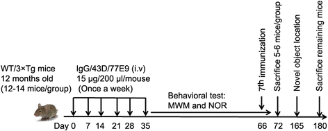
Fig. 1
Fig. 1
Study design. 3×Tg-AD mice were subjected to Morris water maze and one-trial novel object recognition tasks after immunization with six doses of mouse IgG (control), 43D, 77E9, and combination (50% each) of 43D and 77E9 antibodies. On day 66, the mice were administrated one more dose; five or six mice per group were killed on day 72; and the remaining animals were killed on day 180. The novel object location test was carried out from day 165 to day 167. WT mice immunized with mouse IgG or a combination of 43D and 77E9 were used as controls. AD Alzheimer’s disease, IgG Immunoglobulin G, MWM Morris water maze, NOR Novel object recognition, 3×Tg Triple-transgenic, WT Wild type
Fig. 2
Fig. 2
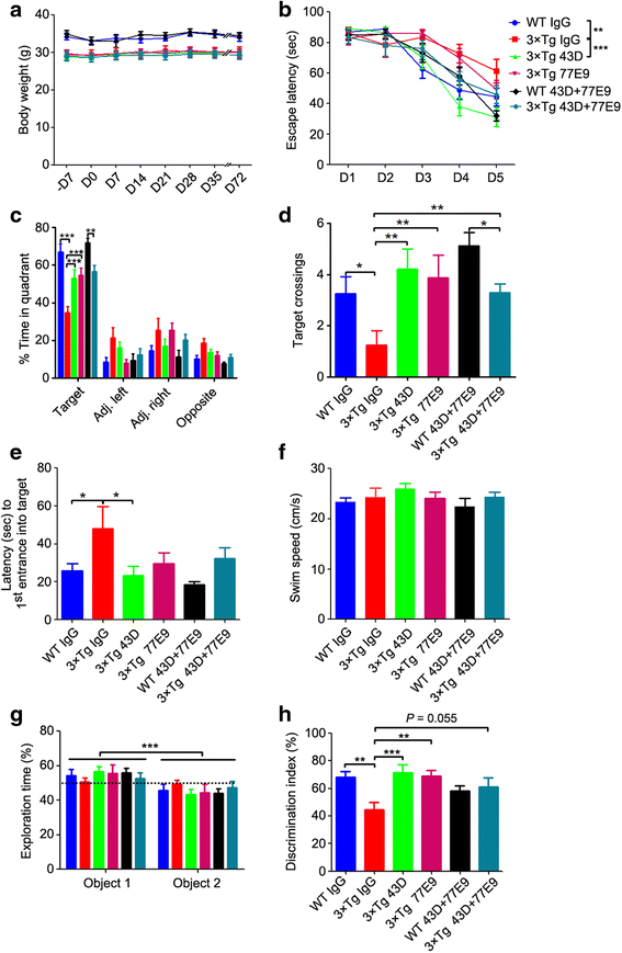
Immunization with tau antibodies 43D and 77E9 rescues cognitive impairment without any side effects in 3×Tg-AD mice. a The body weights of the mice were measured once per week. b–f The Morris water maze test was carried out after the sixth immunization (see Fig. 1). b The escape latency (in seconds) to reach the hidden platform during acquisition phase for 5 days (two-way ANOVA, WT-IgG vs. 3×Tg-IgG, F = 8.47, dfd = 90, p < 0.01; 3×Tg-IgG vs. 3×Tg-43D, F = 12.95, dfd = 75, p < 0.001; 3×Tg-IgG vs. 3×Tg-77E9, F = 0.23, dfd = 70, p = 0.63). c Percentage of time in the quadrant during the probe trial (two-way ANOVA, F = 241.59, dfd = 200, p < 0.0001; Bonferroni posttests for target quadrant, WT-IgG vs. 3×Tg-IgG, p < 0.01; 3×Tg-IgG vs. 3×Tg-43D, p < 0.001; 3×Tg-IgG vs. 3×Tg-77E9, p < 0.01). d Number of target crossings in the probe trial (one-way ANOVA, F = 3.666, p = 0.0067; Bonferroni multiple comparisons test, WT-IgG vs. 3×Tg-IgG, p < 0.05; 3×Tg-IgG vs. 3×Tg-43D, p < 0.01; 3×Tg-IgG vs. 3×Tg-77E9, p < 0.01). e Latency to first entrance into target zone (one-way ANOVA, F = 2.717, p = 0.0301; Bonferroni multiple comparisons test, WT-IgG vs. 3×Tg-IgG, p < 0.05; 3×Tg-IgG vs. 3×Tg-43D, p < 0.05). f The average swim speed during the probe trial (one-way ANOVA, F = 1.003, p = 0.4257). g and h, One-trial novel object recognition test (see Fig. 1). g The percentage of time spent exploring two identical objects during sample phase (two-way ANOVA, F = 18.93, dfd = 122, p < 0.0001). h Discrimination index (time spent exploring novel object/time spent exploring novel and familiar objects) × 100% in test phase (one-way ANOVA, F = 4.517, p = 0.0014; Bonferroni multiple comparisons test, WT-IgG vs. 3×Tg-IgG, p < 0.01; 3×Tg-IgG vs. 3×Tg-43D, p < 0.001; 3×Tg-IgG vs. 3×Tg-77E9, p < 0.01). n = 12 for WT mice treated with IgG or 43D + 77E9, 3×Tg-AD mice treated with 43D, 77E9, or 43D + 77E9; and n = 14 for 3×Tg-AD mice treated with IgG. Data are reported as mean ± SEM. *p < 0.05, **p < 0.01, ***p < 0.001 by ANOVA followed by a Bonferroni post hoc test. AD Alzheimer’s disease, ANOVA Analysis of variance, IgG Immunoglobulin G, 3×Tg Triple-transgenic, WT Wild type
Fig. 3
Fig. 3
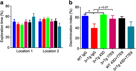
Immunization with tau antibodies 43D and 77E9 improves spatial memory beyond the period of immunization in 3×Tg-AD mice. Novel object location test was carried out 100 days after the last injection (see Fig. 1). a The percentage of time spent exploring two identical objects during sample phase (two-way ANOVA, F = 0.21, dfd = 56, p = 0.6484). b Discrimination index (time exploring object at novel location/time exploring object novel and familiar locations) × 100% in test phase (one-way ANOVA, F = 2.836, p = 0.0357; Bonferroni multiple comparisons test, WT-IgG vs. 3×Tg-IgG, p < 0.05; 3×Tg-IgG vs. 3×Tg-43D, p < 0.05). n = 6 and n = 5 for WT mice treated with IgG or 43D + 77E9, respectively; n = 6 for 3×Tg-AD mice treated with 43D, 77E9, or 43D + 77E9; and n = 8 for 3×Tg-AD mice treated with IgG. Data are reported as mean ± SEM. *p < 0.05 by one-way ANOVA followed by a Bonferroni post hoc test. ANOVA Analysis of variance, IgG Immunoglobulin G, 3×Tg Triple-transgenic, WT Wild type
Fig. 4
Fig. 4
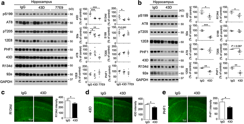
Immunization with 43D dose-dependently decreases tau pathology. Immunization with (a) two doses of 43D and 77E9 antibody or (b–e) six doses of 43D. Mice were killed 1 day after their last injection. a and b Representative Western blots of hippocampus developed with R134d and 92e against total tau, 43D against human transgenic tau, and several phosphorylation-dependent and site-specific tau antibodies. Densitometric quantification of blots after normalization with GAPDH is shown. Five of the 3×Tg-AD mice per group in (a) and three or five of the 3×Tg-AD mice for IgG or 43D treatment, respectively, are shown. Data are percentages of mouse IgG (100%)-treated animals reported as mean ± SEM. Immunostaining of CA1 from 3×Tg-AD mice immunized with six doses of 43D or control IgG with (c) R134d, (d) 43D, or (e) PHF1 antibodies. The quantification of R134d, 43D, and PHF1 immunostaining intensity in total CA1 region was based on two sections from each of three or five mice per group. Data are shown as mean ± SEM. *p < 0.05, **p < 0.01, ***p < 0.001 by ANOVA followed by a Bonferroni post hoc test for two doses of immunization or by unpaired two-tailed t test for six doses of immunization. AD Alzheimer’s disease, ANOVA Analysis of variance, GAPDH Glyceraldehyde 3-phosphate dehydrogenase, IgG Immunoglobulin G, 3×Tg Triple-transgenic
Fig. 5
Fig. 5
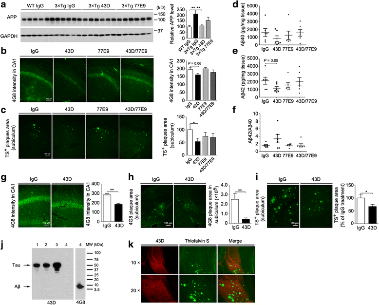
Immunization with 43D but not 77E9 decreases Aβ pathology in 3×Tg-AD mice. af Analysis of Aβ pathology 1 week after seventh dose (see Fig. 1). a Representative Western blots of forebrain developed with APP. Densitometric quantification of blots after normalization with GAPDH. Immunostaining of (b) CA1 with 4G8 and (c) subiculum with thioflavin S in 3×Tg-AD mice. Quantification of APP staining with 4G8 in CA1 and amyloid plaques in subiculum stained with thioflavin S from two sections per mouse of five or six mice per group. The levels of total (d) Aβ40 and (e) Aβ42 (unpaired t test, 3×Tg-IgG vs. 3×Tg-43D, p = 0.08) and (f) the ratio of Aβ42/Aβ40 in forebrain was quantified by ELISA. g–i Aβ pathology in 3×Tg-AD mice 1 day after the sixth injection. 4G8 staining in (g) CA1 and (h) subiculum and (i) thioflavin S staining in subiculum in 3×Tg-AD mice. 43D did not cross-react with Aβ (j) and Aβ plaques (k). j Thirty micrograms of 3×Tg-AD mouse brain homogenate (lanes 1–3) and 0.4 μg of purified Aβ (lane 4) were loaded. Nitrocellulose membrane was first developed with 43D antibody (left panel) and then developed with 4G8 antibody (right panel). k Double-stained 3×Tg-AD mouse brain sections with 43D antibody and thioflavin S. Top row: CA1 and subiculum area (original magnification × 10). Bottom row: Subiculum area (original magnification × 20). Quantification of APP staining in CA1 and amyloid plaques in subiculum stained with 4G8 and thioflavin S from two sections per mouse of three or five animals per group. Data are shown as mean ± SEM. *p < 0.05, **p < 0.01, ***p < 0.001 by ANOVA followed by a Bonferroni post hoc test for two doses of immunization or by unpaired two-tailed t test for six doses of immunization. AD Alzheimer’s disease, ANOVA Analysis of variance, APP Amyloid precursor protein, Amyloid-β, ELISA Enzyme-linked immunosorbent assay, GAPDH Glyceraldehyde 3-phosphate dehydrogenase, IgG Immunoglobulin G, MW Molecular weight, 3×Tg Triple-transgenic, TS Thioflavin S, WT Wild type
Fig. 6
Fig. 6
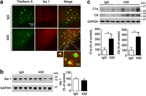
Immunization with 43D increases the activity of microglia and the complement system in 3×Tg-AD mice. The activation of microglia and the complement system was assessed 1 day after the sixth injection. a Thioflavin S staining and Iba1 immunofluorescence were performed in the subiculum area. White arrows highlight activated microglial cells aggregated around Aβ plaques. Representative Western blots of hippocampus developed (b) with Iba1 antibody and (c) with complement components C1q and C9 antibodies and densitometric quantification of blots after normalization with GAPDH. Three or five representative 3×Tg-AD mice in the IgG or 43D treatment group, respectively, are shown. Data are percentages of mouse IgG (100%)-treated animals, reported as mean ± SEM. *p < 0.05 by unpaired two-tailed Student’s t test. AD Alzheimer’s disease, Amyloid-β, GAPDH Glyceraldehyde 3-phosphate dehydrogenase, IgG Immunoglobulin G, 3×Tg Triple-transgenic
Fig. 7
Fig. 7
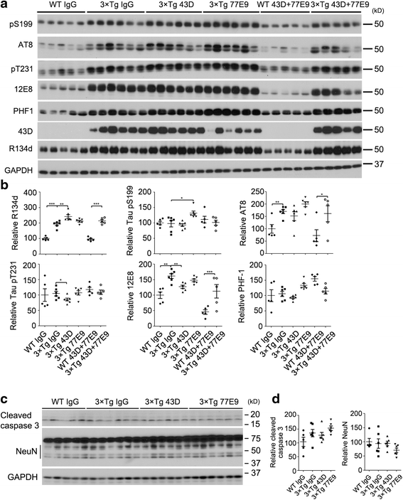
Effect of immunization with 43D and 77E9 antibodies on tau pathology lasts several weeks after the last treatment. a Representative Western blots of forebrain developed with R134d against total tau and with several phosphorylation-dependent and site-specific tau antibodies. b Densitometric quantification of phosphorylated tau after normalization with total tau (R134d). c Western blots of forebrain developed with antibodies to apoptotic marker cleaved caspase-3 and neuronal marker NeuN. d Quantification of cleaved caspase-3 and NeuN from (c). n = 5 for WT mice treated with IgG, 43D, and 77E9, and 3×Tg-AD mice treated with 43D + 77E9; n = 6 for 3×Tg-AD mice treated with IgG, 43D, and 77E9. Data are percentages of mouse IgG (100%)-treated animals, reported as mean ± SEM. *p < 0.05, **p < 0.01 by one-way ANOVA followed by Bonferroni post hoc test. AD Alzheimer’s disease, ANOVA Analysis of variance, Amyloid-β, GAPDH Glyceraldehyde 3-phosphate dehydrogenase, IgG Immunoglobulin G, pS199 Phospho-tau (Ser199), pT205 Phospho-tau (Thr205), 3×Tg Triple-transgenic, WT Wild type

References

    1. Alzheimer’s Association 2015 Alzheimer’s disease facts and figures. Alzheimers Dement. 2015;11:332–384. doi: 10.1016/j.jalz.2015.02.003. - DOI - PubMed
    1. Glenner GG, Wong CW. Alzheimer’s disease: initial report of the purification and characterization of a novel cerebrovascular amyloid protein. Biochem Biophys Res Commun. 1984;120:885–890. doi: 10.1016/S0006-291X(84)80190-4. - DOI - PubMed
    1. Grundke-Iqbal I, Iqbal K, Tung YC, Quinlan M, Wisniewski HM, Binder LI. Abnormal phosphorylation of the microtubule-associated protein tau (tau) in Alzheimer cytoskeletal pathology. Proc Natl Acad Sci U S A. 1986;83:4913–4917. doi: 10.1073/pnas.83.13.4913. - DOI - PMC - PubMed
    1. Hardy J, Selkoe DJ. The amyloid hypothesis of Alzheimer’s disease: progress and problems on the road to therapeutics. Science. 2002;297:353–356. doi: 10.1126/science.1072994. - DOI - PubMed
    1. Giacobini E, Gold G. Alzheimer disease therapy—moving from amyloid-β to tau. Nat Rev Neurol. 2013;9:677–686. doi: 10.1038/nrneurol.2013.223. - DOI - PubMed

MeSH terms