. 2017 Feb:50:22-35.
doi: 10.1016/j.dnarep.2016.12.003. Epub 2016 Dec 31.

PCNA dependent cellular activities tolerate dramatic perturbations in PCNA client interactions

Affiliations

PCNA dependent cellular activities tolerate dramatic perturbations in PCNA client interactions

Rosemary H C Wilson et al. DNA Repair (Amst). 2017 Feb.

Abstract

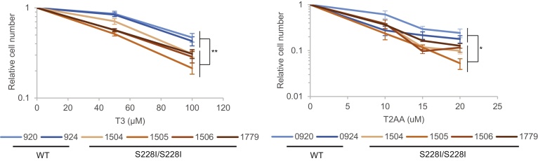
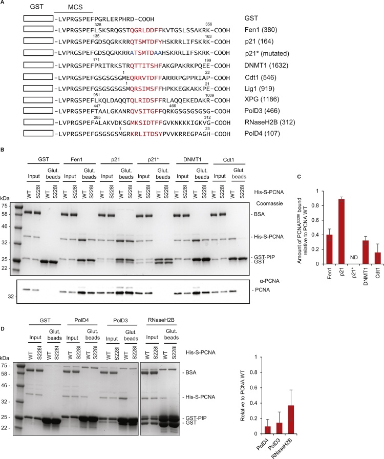
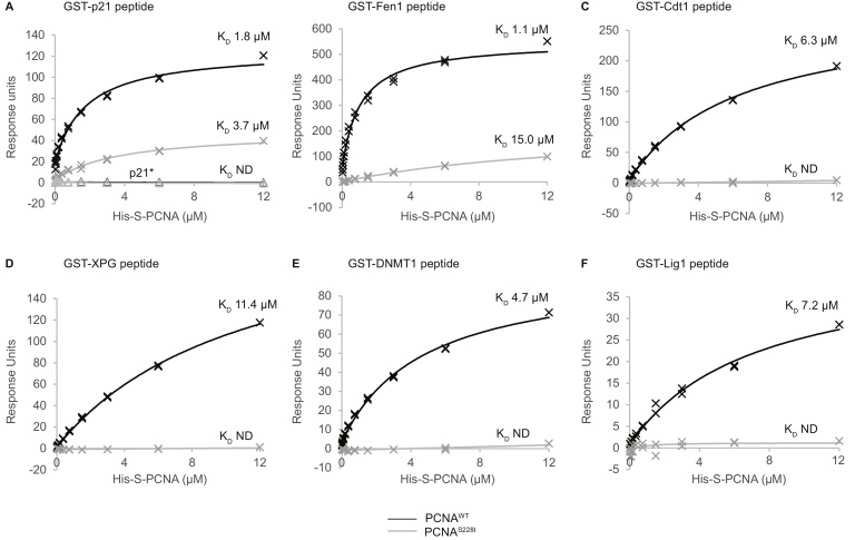
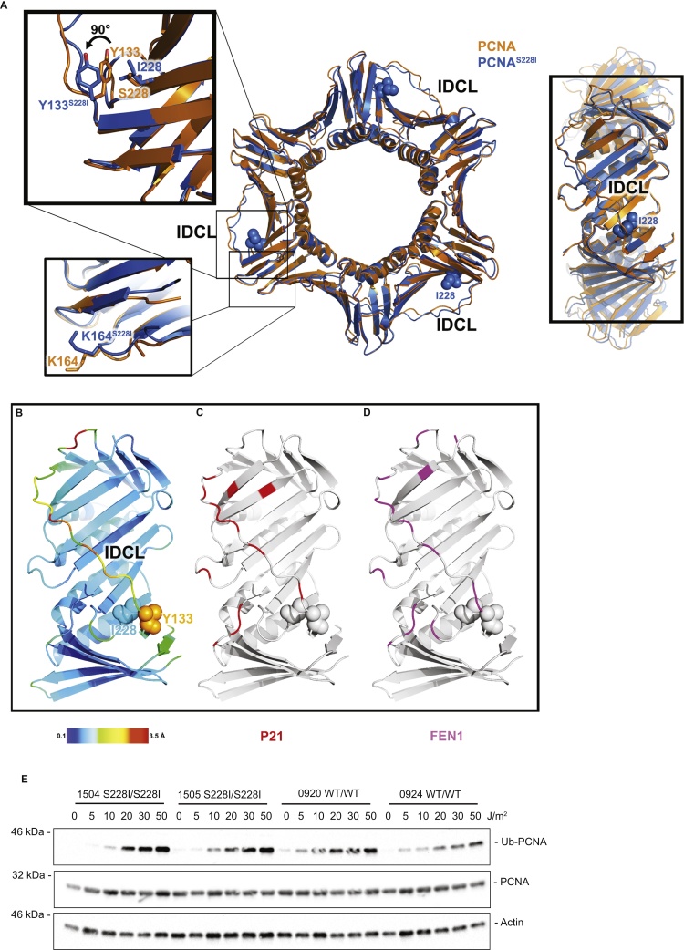
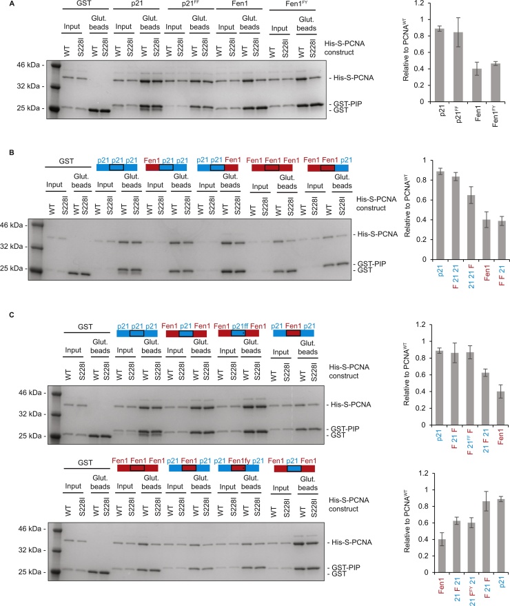
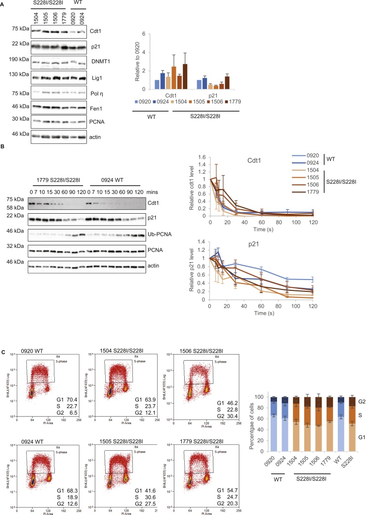
Proliferating cell nuclear antigen (PCNA) is an essential cofactor for DNA replication and repair, recruiting multiple proteins to their sites of action. We examined the effects of the PCNAS228I mutation that causes PCNA-associated DNA repair disorder (PARD). Cells from individuals affected by PARD are sensitive to the PCNA inhibitors T3 and T2AA, showing that the S228I mutation has consequences for undamaged cells. Analysis of the binding between PCNA and PCNA-interacting proteins (PIPs) shows that the S228I change dramatically impairs the majority of these interactions, including that of Cdt1, DNMT1, PolD3p66 and PolD4p12. In contrast p21 largely retains the ability to bind PCNAS228I. This property is conferred by the p21 PIP box sequence itself, which is both necessary and sufficient for PCNAS228I binding. Ubiquitination of PCNA is unaffected by the S228I change, which indirectly alters the structure of the inter-domain connecting loop. Despite the dramatic in vitro effects of the PARD mutation on PIP-degron binding, there are only minor alterations to the stability of p21 and Cdt1 in cells from affected individuals. Overall our data suggests that reduced affinity of PCNAS228I for specific clients causes subtle cellular defects in undamaged cells which likely contribute to the etiology of PARD.

Keywords: DNA repair; DNA replication; PCNA; PCNA-associated repair disorder (PARD).


Figures

Fig. 1
Fig. 1
PCNAS228I cells are sensitive to increased competition for IDCL binding. Graphs show relative cell number for PCNAS228I/S228I and PCNAWT/WT lymphoblasts treated with indicated concentrations of T3 (left) or T2AA (right) for three days. Data expressed as cell number relative to cells treated with vehicle. Data are average of at least three experiments, error bars are SEM, * p < 0.05, ** p < 0.005 using Student’s t-test.
Fig. 2
Fig. 2
The PCNAS228I mutation differentially affects PCNA binding to PIP box-containing proteins. A) Schematic showing GST-PIP peptide constructs generated during this study. GST represented by rectangle, PIP box sequences shown in red, mutated residues shown in blue. Numbers relate to amino acid position in full length protein. B) GST-PIP pull down of His-S-PCNAWT or PCNAS228I. Figure shows Coomassie stained gel of representative pull down (top) and anti-PCNA western blot of the same samples diluted 1:20 (bottom). Amount of ‘input’ loaded for Coomassie is equivalent to 1%, ‘Glut. beads’ (Glutathione sepharose 4B beads) is equivalent to 25%. Molecular weight markers are indicated. C) Quantification of binding. Histogram shows amount of PCNAS228I pulled down relative to PCNAWT for indicated PIP constructs. Fen1 (n = 8), p21 (n = 8), p21* (n = 1), DNMT1 (n = 3), Cdt1 (n = 6). Error bars are standard deviation. D) GST-PIP pull down of His-S-PCNAWT or PCNAS228I for weak binders with 100 μg GST-PIP construct used per condition. Coomassie as B, quantification as C, n = 3.
Fig. 3
Fig. 3
The affinity of PCNA-PIP box interactions is reduced by PCNAS228I variant. Affinity curves generated from SPR data for His-S-PCNAWT or His-S-PCNAS228I binding to CM5 chip coupled to indicated GST-PIP peptide. PCNAWT is shown in black, PCNAS228I in grey. GST-mutated p21 PIP peptide (GST-p21*) is also shown on the p21 graph (A) with triangle marker points; no binding of either PCNAWT or PCNAS228I to GST-p21* peptide lane was observed. KD calculated from Biacore T200 evaluation software indicated on relevant curves. ND = not determined.
Fig. 4
Fig. 4
PCNAS228I alters PCNA function via structural changes at the IDCL. A) Apo structure of PCNAS228I (5MOM) shown in blue overlayed on apo PCNAWT structure in orange. The mutated I228 residues are shown as spheres. Top left inset zoomed area shows the rotation of Tyr133, propagating along the IDCL. Bottom left inset zoom shows K164 and surrounding structural environment unchanged. Right panel shows the rotated view of the IDCL region highlighted in B, C, D below. B) Magnified view of the IDCL region of a single monomer of PCNAS228I colour coded according to the heat map of the rmsd between PCNAS228I (5MOM) and PCNAWT (1VYM). C) As B, but with residues found at the interface between PCNAWT and the p21 pip box (based on 4RJF) coloured in red. D) As C, but for the Fen1 PIP box (based on 4RJF), coloured in magenta. E) PARD affected and control lymphoblasts were treated with the indicated doses of UVC and harvested after 7 h. Western blot shows induction of mono-ubiquitinated PCNAWT and PCNAS228I.
Fig. 5
Fig. 5
The PIP-box motif is the major contributor which enables p21 binding to PCNAS228I. A) GST-PIP pull down of His-S-PCNAWT or His-S-PCNAS228I using wild type p21 and Fen1, and constructs with mutated PIP position 8 residues. Figure shows Coomassie stained gel of representative pull down. Amount of ‘input’ and ‘Glut. beads’ as Fig. 2. Molecular weight markers are indicated. Histogram (right) shows pulldown of PCNAS228I relative to PCNAWT for indicated PIP constructs. p21 and Fen1 (n = 8), p21FF (n = 4), Fen1FY (n = 2). Error bars are standard deviation. B) As A) except for single domain swaps. For histogram: all n = 3 except p21 and Fen1 n = 8. C) As A) except for double domains swaps. For histograms: all n = 3 except p21 and Fen1 n = 8.
Fig. 6
Fig. 6
PCNAS228I has subtle effects on cellular functions of PCNA. A) Example Western blots showing steady state levels of PCNA interacting proteins as indicated, also levels of PCNA and actin as a loading control. Histogram shows quantification of p21 and Cdt1 levels relative to actin, shown relative to 0920 levels. n = 8 (0920, 0924), n = 7 (1504, 1505), n = 6 (1506), n = 5 (1779). B) Example western blots showing degradation of Cdt1 and p21 at indicated times after 50 J/m2 254 nm UV exposure. Also levels of PCNA and actin as loading controls and appearance of ubiquitinated-PCNA at later time points. The graphs show quantification of p21 and Cdt1 levels relative to actin, expressed as relative to time zero. n = 3 (1504, 1505, 1506, 1779), n = 5 (0920, 0924). C) Representative dot plots from 3 independent FACS analysis for indicated cell lines plotting DNA content (PI, x-axis) against BrdU (y-axis) to determine percentage in G1, S and G2 cell cycle phases. Graph (right) shows average cell cycle proportion for each cell line (error bars are SEM) and each genotype (WT and S228I, error bars are STDEV).

Similar articles

Cited by

References

    1. Ducoux M., Urbach S., Baldacci G., Hubscher U., Koundrioukoff S., Christensen J., Hughes P. Mediation of proliferating cell nuclear antigen (PCNA)-dependent DNA replication through a conserved p21(Cip1)-like PCNA-binding motif present in the third subunit of human DNA polymerase delta. J. Biol. Chem. 2001;276:49258–49266. - PubMed
    1. Montecucco A., Rossi R., Levin D.S., Gary R., Park M.S., Motycka T.A., Ciarrocchi G., Villa A., Biamonti G., Tomkinson A.E. DNA ligase I is recruited to sites of DNA replication by an interaction with proliferating cell nuclear antigen: identification of a common targeting mechanism for the assembly of replication factories. EMBO J. 1998;17:3786–3795. - PMC - PubMed
    1. Levin D.S., McKenna A.E., Motycka T.A., Matsumoto Y., Tomkinson A.E. Interaction between PCNA and DNA ligase I is critical for joining of Okazaki fragments and long-patch base-excision repair. Curr. Biol. 2000;10:919–922. - PubMed
    1. Li X., Li J., Harrington J., Lieber M.R., Burgers P.M. Lagging strand DNA synthesis at the eukaryotic replication fork involves binding and stimulation of FEN-1 by proliferating cell nuclear antigen. J. Biol. Chem. 1995;270:22109–22112. - PubMed
    1. Chon H., Vassilev A., DePamphilis M.L., Zhao Y., Zhang J., Burgers P.M., Crouch R.J., Cerritelli S.M. Contributions of the two accessory subunits, RNASEH2B and RNASEH2C, to the activity and properties of the human RNase H2 complex. Nucleic Acids Res. 2009;37:96–110. - PMC - PubMed

MeSH terms

LinkOut - more resources