. 2017 Apr 1;312(4):E253-E263.
doi: 10.1152/ajpendo.00331.2016. Epub 2017 Jan 10.

Overexpression of PGC-1α increases peroxisomal activity and mitochondrial fatty acid oxidation in human primary myotubes

Affiliations

Overexpression of PGC-1α increases peroxisomal activity and mitochondrial fatty acid oxidation in human primary myotubes

Tai-Yu Huang et al. Am J Physiol Endocrinol Metab. .

Abstract

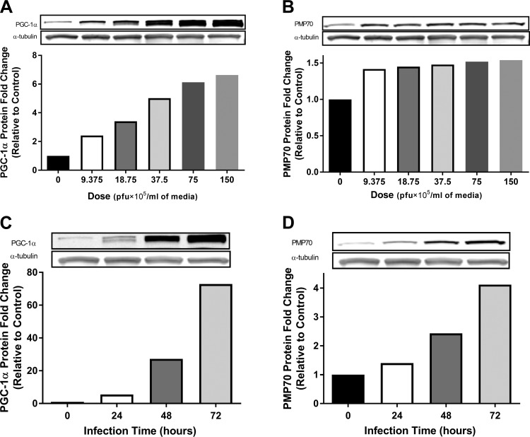
Peroxisomes are indispensable organelles for lipid metabolism in humans, and their biogenesis has been assumed to be under regulation by peroxisome proliferator-activated receptors (PPARs). However, recent studies in hepatocytes suggest that the mitochondrial proliferator PGC-1α (peroxisome proliferator-activated receptor gamma coactivator-1α) also acts as an upstream transcriptional regulator for enhancing peroxisomal abundance and associated activity. It is unknown whether the regulatory mechanism(s) for enhancing peroxisomal function is through the same node as mitochondrial biogenesis in human skeletal muscle (HSkM) and whether fatty acid oxidation (FAO) is affected. Primary myotubes from vastus lateralis biopsies from lean donors (BMI = 24.0 ± 0.6 kg/m2; n = 6) were exposed to adenovirus encoding human PGC-1α or GFP control. Peroxisomal biogenesis proteins (peroxins) and genes (PEXs) responsible for proliferation and functions were assessed by Western blotting and real-time qRT-PCR, respectively. [1-14C]palmitic acid and [1-14C]lignoceric acid (exclusive peroxisomal-specific substrate) were used to assess mitochondrial oxidation of peroxisomal-derived metabolites. After overexpression of PGC-1α, 1) peroxisomal membrane protein 70 kDa (PMP70), PEX19, and mitochondrial citrate synthetase protein content were significantly elevated (P < 0.05), 2) PGC-1α, PMP70, key PEXs, and peroxisomal β-oxidation mRNA expression levels were significantly upregulated (P < 0.05), and 3) a concomitant increase in lignoceric acid oxidation by both peroxisomal and mitochondrial activity was observed (P < 0.05). These novel findings demonstrate that, in addition to the proliferative effect on mitochondria, PGC-1α can induce peroxisomal activity and accompanying elevations in long-chain and very-long-chain fatty acid oxidation by a peroxisomal-mitochondrial functional cooperation, as observed in HSkM cells.

Keywords: human skeletal muscle cells; lignoceric acid oxidation; obesity; peroxisome proliferator-activated receptors; β-oxidation.


Figures

Fig. 1.
Fig. 1.
Overexpression of peroxisome proliferator-activated receptor-γ coactivator-1α (PGC-1α) increases peroxisomal membrane protein 70 kDa (PMP70) in a time-dependent fashion in human primary myotubes. Human skeletal muscle cells (HSkMC) from vastus lateralis muscle biopsies were exposed to varying doses [plaque-forming units (pfu)/ml media] or time (h) of adenovirus packed with human PGC-1α on days 4 and 5 post-differentiation and harvested and assayed on day 7 for PMP70 and PGC-1α proteins by Western blotting. Results are shown as Western blots and fold change vs. control (0 pfu/ml media) (pooled cells from lean donors, BMI = 23.3 ± 1.6; n = 3). A and C: dose and time-course responses of PGC-1α protein contents. B and D: dose and time-course responses of PMP70 protein. The data supported our hypothesis that PGC-1α drives the peroxisomal marker in human primary myotubes. Pfu: an indicator of active virus.
Fig. 2.
Fig. 2.
PGC-1α drives peroxisomal gene expression in human primary myotubes. HSkMCs from muscle biopsies were exposed to adenovirus packed with human PGC-1α (Ad-PGC-1α) or GFP (Ad-GFP) on days 4 and 5 post-differentiation and harvested on day 7 postdifferentiation and measured for peroxisomal biogenesis gene expression by qRT-PCR. A: PGC-1α mRNA expression (ΔΔCT value: −4.34). B: fluorescent images from fluorescent microscopy showed infection efficiency after 72-h infection with either Ad-PGC-1α (coexpressed GFP) or Ad-GFP in human primary myotubes (magnification ×20). C: peroxisomal biogenesis factor (PEX) mRNA expression. ABCD1, ATP-binding cassette subfamily D member 1 responsible for transporting very long-chain fatty acids (VLCFA) across the peroxisomal membrane (ΔΔCT value: 0.61). PMP70, peroxisomal membrane protein 70 kDa, a marker of peroxisomal biogenesis (ΔΔCT value: −1.97); PEX3, PEX11A, PEX11B, PEX13, and PEX19, peroxisomal biogenesis genes initiated to peroxisomal proliferation (ΔΔCT value: −3.20, −0.10, 0.30, −0.60, and −0.37); ACOX1, peroxisomal acyl-coenzyme A oxidase 1 (ΔΔCT value: −0.47); PBFE, peroxisomal bifunctional enzyme (ΔΔCT value: −1.73); PTHIO, peroxisomal 3-ketoacyl-CoA thiolase for the peroxisomal β-oxidation (ΔΔCT value: −2.61); CrAT, carnitine O-acetyltransferase for converting acetyl-CoA to acetyl-carnitine (ΔΔCT value: −3.45); CrOT, carnitine octanoyltransferase for converting acyl-CoA to acyl-carnitine (ΔΔCT value: 1.25); DNM1L, dynamin 1-like protein involved in fission of both peroxisomal and mitochondrial biogenesis (ΔΔCT value: −1.15). mRNA expression was determined using the comparative CT method (ΔΔCT) with an endogenous control (18S ribosomal RNA) for normalization and then compared with Ad-GFP. Results are shown as means ± SE (n = 6). *P < 0.05, **P < 0.005, and ***P < 0.001 vs. Ad-GFP groups by paired 2-tailed t-test. DAPI, 2-(4-amidinophenyl)-1H -indole-6-carboxamidine; GFP, green fluorescent protein. [(2R,3R,4S,5S,6R)-3-fluoro-4,5-dihydroxy-6-(hydroxymethyl)oxan-2-yl] dihydrogen phosphate.
Fig. 3.
Fig. 3.
PGC-1α induces both mitochondrial biogenesis marker and peroxisomal biogenesis proteins in human primary myotubes. Western blotting data of whole cell lysates from human primary myotubes incubated with differentiation media without any virus [nonvirus control (NVC; 0 pfu/ml media): 0 pfu/ml media] or infected with GFP or human PGC-1α adenovirus for 72 h. All values were normalized to α-tubulin and then compared with NVC. Results are shown as means ± SE (n = 6). *P < 0.05 vs. Ad-GFP groups by paired 2-tailed t-test. mtTFA, mitochondrial transcription factor A; CS, citrate synthetase; PMP70, peroxisomal membrane protein 70 kDa; PEX19, peroxisomal biogenesis protein initiated to peroxisomal proliferation.
Fig. 4.
Fig. 4.
PGC-1α overexpression increases both palmitic acid and lignoceric acid oxidation in human primary myotubes. Fatty acid oxidation (FAO) with radiolabeled substrates was performed by using primary myotubes homogenates. A: [1-14C]palmitate oxidation and acid-soluble metabolites (ASM). B: palmitate oxidation efficiency was expressed by an index of complete oxidation/acid-soluble metabolites (CO2/ASM); an increase in the index of CO2/ASM in the Ad-PGC-1α group indicates that oxidation efficiency is elevated post-PGC-1α overexpression. C: [1-14C]lignoceric acid (C24:0). VLCFA, which is necessarily oxidized first by peroxisomes, was used to assess mitochondrial oxidation, which utilized peroxisomal-derived fatty acid metabolites to boost mitochondrial/peroxisomal fatty acid disposal. D: lignoceric acid oxidation efficiency. E: indirect measure of total triglyceride (IMTG) content by measurement of triacylglycerol (TAG) content in cell lysates. F: lipid droplet metabolism and ether phospholipid synthesis gene expression by real-time qRT-PCR. mRNA expression was determined using the comparative CT method (ΔΔCT) with an endogenous control (18S ribosomal RNA) for normalization and then compared with the Ad-GFP group. Results are shown as means ± SE (n = 6). *P < 0.05, **P < 0.005, and ***P < 0.001 vs. Ad-GFP groups by paired, 2-tailed t-test. DGAT1, diacylglycerol acyltransferase 1 responsible for TAG synthesis (ΔΔCT value: −2.48); ATGL, adipose triglyceride lipase (ΔΔCT value: −1.54); PLIN5, perilipin 5 involved in lipid droplet mobilization (ΔΔCT value: −4.04); GNPAT/DHAPAT, glyceronephosphate acyltransferase-dihydroxyacetone phosphate acyltransferase involved in ether phospholipid synthesis (ΔΔCT value: −0.46) vs. Ad-GFP groups by paired 2-tailed t-test.
Fig. 5.
Fig. 5.
A schematic model of PGC-1α-induced peroxisomal activity and mitochondrial biogenesis and subsequent functional cooperation for enhanced FAO in human skeletal muscle. PGC-1α overexpression enhanced nuclear transcription of peroxisomal proliferator genes and subsequent peroxisomal activity and mitochondrial biogenesis. Elevated peroxisomal function/activity leads to elevated peroxisomal β-oxidation. Increased export of peroxisomal acyl- and acetyl-carnitine products is reconverted by mitochondrial CPT II and undergoes β-oxidation, thereby increasing lipid disposal under conditions of mitochondrial metabolites overload (low energy demand). Further mechanistic investigations are needed to define potential downstream targets of PGC-1α or involved transcriptional control networks such as specific PPAR isoforms and/or other unidentified transcriptional cofactors (TF?) involved in both peroxisomal activity, and mitochondrial biogenesis in HSkM represents fruitful areas for future scientific exploration. LCFA, long-chain fatty acid; VLCFA, very-long-chain fatty acid; CrOT, carnitine octanoyltransferase; CrAT, carnitine O-acetyltransferase.

References

    1. Adams JM II, Pratipanawatr T, Berria R, Wang E, DeFronzo RA, Sullards MC, Mandarino LJ. Ceramide content is increased in skeletal muscle from obese insulin-resistant humans. Diabetes 53: 25–31, 2004. doi: 10.2337/diabetes.53.1.25. - DOI - PubMed
    1. Amati F, Dubé JJ, Alvarez-Carnero E, Edreira MM, Chomentowski P, Coen PM, Switzer GE, Bickel PE, Stefanovic-Racic M, Toledo FGS, Goodpaster BH. Skeletal muscle triglycerides, diacylglycerols, and ceramides in insulin resistance: another paradox in endurance-trained athletes? Diabetes 60: 2588–2597, 2011. doi: 10.2337/db10-1221. - DOI - PMC - PubMed
    1. Anderson EJ, Lustig ME, Boyle KE, Woodlief TL, Kane DA, Lin CT, Price JW III, Kang L, Rabinovitch PS, Szeto HH, Houmard JA, Cortright RN, Wasserman DH, Neufer PD. Mitochondrial H2O2 emission and cellular redox state link excess fat intake to insulin resistance in both rodents and humans. J Clin Invest 119: 573–581, 2009. doi: 10.1172/JCI37048. - DOI - PMC - PubMed
    1. Austin S, St-Pierre J. PGC1α and mitochondrial metabolism—emerging concepts and relevance in ageing and neurodegenerative disorders. J Cell Sci 125: 4963–4971, 2012. doi: 10.1242/jcs.113662. - DOI - PubMed
    1. Bagattin A, Hugendubler L, Mueller E. Transcriptional coactivator PGC-1alpha promotes peroxisomal remodeling and biogenesis. Proc Natl Acad Sci USA 107: 20376–20381, 2010. doi: 10.1073/pnas.1009176107. - DOI - PMC - PubMed

Publication types

MeSH terms

Substances

LinkOut - more resources