. 2017 Mar 9;129(10):1357-1367.
doi: 10.1182/blood-2016-09-741298. Epub 2017 Jan 10.

Platelets and neutrophil extracellular traps collaborate to promote intravascular coagulation during sepsis in mice

Affiliations

Platelets and neutrophil extracellular traps collaborate to promote intravascular coagulation during sepsis in mice

Braedon McDonald et al. Blood. .

Erratum in

Abstract

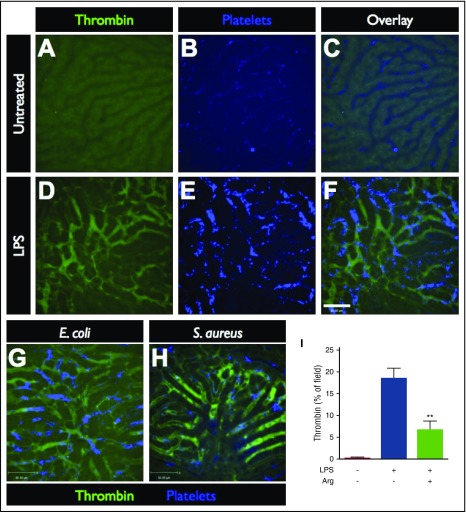
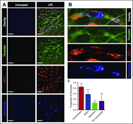
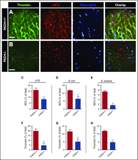
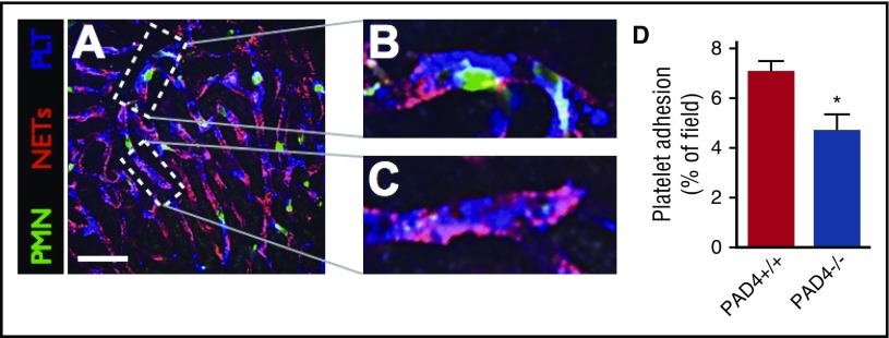
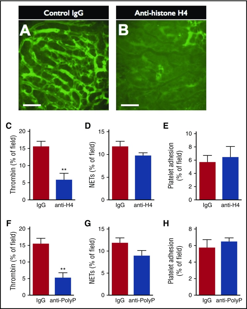
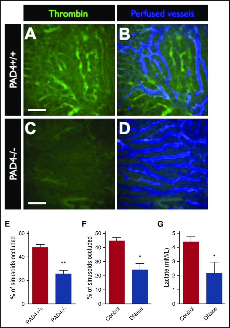
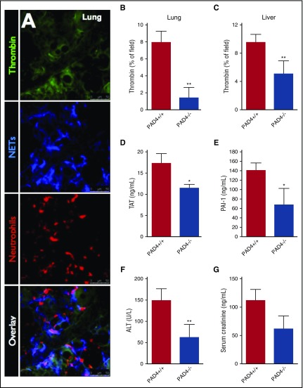
Neutrophil extracellular traps (NETs; webs of DNA coated in antimicrobial proteins) are released into the vasculature during sepsis where they contribute to host defense, but also cause tissue damage and organ dysfunction. Various components of NETs have also been implicated as activators of coagulation. Using multicolor confocal intravital microscopy in mouse models of sepsis, we observed profound platelet aggregation, thrombin activation, and fibrin clot formation within (and downstream of) NETs in vivo. NETs were critical for the development of sepsis-induced intravascular coagulation regardless of the inciting bacterial stimulus (gram-negative, gram-positive, or bacterial products). Removal of NETs via DNase infusion, or in peptidylarginine deiminase-4-deficient mice (which have impaired NET production), resulted in significantly lower quantities of intravascular thrombin activity, reduced platelet aggregation, and improved microvascular perfusion. NET-induced intravascular coagulation was dependent on a collaborative interaction between histone H4 in NETs, platelets, and the release of inorganic polyphosphate. Real-time perfusion imaging revealed markedly improved microvascular perfusion in response to the blockade of NET-induced coagulation, which correlated with reduced markers of systemic intravascular coagulation and end-organ damage in septic mice. Together, these data demonstrate, for the first time in an in vivo model of infection, a dynamic NET-platelet-thrombin axis that promotes intravascular coagulation and microvascular dysfunction in sepsis.


Figures

Figure 1.
Figure 1.
Imaging intravascular coagulation in sepsis. Representative SD-IVM images of the liver microcirculation in untreated (A-C), endotoxemic (D-F), E coli–infected (G), and S aureus–infected (H) mice. Thrombin probe fluorescence is shown in green (note the level of background autofluorescence of the liver parenchyma shown in panel A), and platelets are shown in blue (AF647 anti-CD49b). Bar represents 50 μm. (I) Quantitative analysis of thrombin probe fluorescence within the liver microcirculation of untreated control mice, LPS-treated mice, or LPS-treated mice that received a direct thrombin inhibitor (argatroban). Data are shown as mean ± SEM. **P < .01; N = 3-6 mice per group.
Figure 2.
Figure 2.
Spatial relationship between thrombin activity and NETs in the microvasculature. (A) Representative SD-IVM images of the liver microcirculation of untreated and LPS-treated mice. Thrombin probe fluorescence is in green, NETs are shown in red (AF555 anti-H2Ax), and neutrophils are shown in blue (AF647 anti-Gr1). Bars represent 50 μm. (B) Magnified inset showing a liver sinusoid containing an adherent neutrophil (blue), surrounding NETs (red), and the associated thrombin activity (green). (C) The regions of the highest thrombin probe signal within each liver sinusoid (per field of view) were identified in LPS-treated mice and categorized according to the spatial relationship with the nearest NET relative to the direction of blood flow: downstream, within NET, upstream, or not associated (>100 μm from the nearest NET). Data are represented as mean ± SEM. *P < .05; N = 4 mice per group.
Figure 3.
Figure 3.
Reducing NET production in PAD4-deficient mice reduces intravascular coagulation. Representative SD-IVM images of the liver microcirculation of wild-type (A, PAD4+/+) and PAD4−/− (B) mice 4 hours after LPS administration. Thrombin probe fluorescence is in green, NETs are shown in red (AF555 anti-H2Ax), and neutrophils are shown in blue (AF647 anti-Gr1). Bars represent 50 μm. Quantitative analysis of NETs (C-E) and thrombin probe fluorescence (F-H) within the liver microvasculature of wild-type (PAD4+/+) and PAD4−/− mice after administration of LPS, E coli infection, or S aureus infection. Data are represented as mean ± SEM. **P < .01; N = 3-6 mice per group.
Figure 4.
Figure 4.
Minor contribution of NET-platelet interaction to microvascular platelet adhesion. (A) Representative SD-IVM image of the liver microvasculature of an endotoxemic wild-type (PAD4+/+) mouse showing neutrophils (green, AF488 anti-Gr1), NETs (red, AF555 anti-H2Ax), and platelets (blue, AF647 anti-CD49b). Bar represents 50 μm. (B) Magnified inset showing platelets (blue) aggregating on the surface of an adherent neutrophil (green). (C) Magnified inset showing platelets (blue) adhering to a NET (red). (D) Quantitative analysis of platelet adhesion within the liver sinusoids of wild-type (PAD4+/+) and PAD4−/− mice. Data are represented as mean ± SEM. *P < .05; N = 4-6 mice per group.
Figure 5.
Figure 5.
Neutralization of histone H4 or PolyP reduces intravascular thrombin activity, but not platelet adhesion or NET production. Representative SD-IVM images of thrombin probe activity (green) within the liver microcirculation of endotoxemic wild-type mice treated with (A) control IgG versus (B) anti-histone H4 IgG. Bars represent 50 μm. (C-D) Quantitative analysis of (C) thrombin probe fluorescence, (D) NETs, and (E) platelet adhesion within the liver sinusoids of endotoxemic wild-type mice treated with control IgG or anti-histone H4 IgG. (F-H) Quantitative analysis of (F) thrombin probe fluorescence, (G) NETs, and (H) platelet adhesion within the liver sinusoids of endotoxemic wild-type mice treated with control IgG or antipolyphosphate IgG. Data are represented as mean ± SEM. **P < .01; N = 5 mice per group.
Figure 6.
Figure 6.
Blocking NETs-induced intravascular coagulation restores microvascular perfusion. Representative SD-IVM images of the liver microcirculation of endotoxemic wild-type (PAD4+/+) (A-B) and PAD4−/− (C-D) mice. Thrombin probe activity is shown in green. Mice were then injected with AF647-albumin (blue) as a contrast material to identify perfused versus occluded vessels. Bars represent 50 μm. The proportion of occluded vessels was quantified per field of view in (E) endotoxemic wild-type (PAD4+/+) versus PAD4−/− mice and (F) endotoxemic mice treated with IV DNase versus vehicle control. (G) Serum lactate levels were quantified in blood samples from septic mice (24 hours after i.p. infection with E coli) treated with IV DNase versus vehicle control. Data are represented as mean ± SEM. **P < .01, *P < .05; N = 3-6 mice per group.
Figure 7.
Figure 7.
Inhibition of NETs attenuates intravascular coagulation and end-organ damage in sepsis. (A) Representative resonance-scanning confocal intravital microscopy images of the lung microvasculature of endotoxemic mice showing intravascular thrombin activity (green), NETs (blue, AF647 anti-NE), and neutrophils (red, PE-anti-Gr1). Microvascular thrombin probe fluorescence was quantified in the lung microvasculature in response to intratracheal LPS (B), and liver microvasculature in response to i.p. E coli (C) in PAD+/+ and PAD4−/− mice. (D) Plasma TAT levels were quantified in PAD4+/+ and PAD4−/− mice 6 hours after IV S aureus infection. Plasma levels of (E) PAI-1, (F) ALT, and (G) creatinine were measured in PAD4+/+ and PAD4−/− mice with E coli peritonitis (24 hours postinfection). Data are represented as mean ± SEM. **P < .01, *P < .05; N = 5-10 mice per group.

Comment in

References

    1. Dombrovskiy VY, Martin AA, Sunderram J, Paz HL. Rapid increase in hospitalization and mortality rates for severe sepsis in the United States: a trend analysis from 1993 to 2003. Crit Care Med. 2007;35(5):1244-1250. - PubMed
    1. Rittirsch D, Flierl MA, Ward PA. Harmful molecular mechanisms in sepsis. Nat Rev Immunol. 2008;8(10):776-787. - PMC - PubMed
    1. Singer M, Deutschman CS, Seymour CW, et al. . The Third International Consensus Definitions for Sepsis and Septic Shock (Sepsis-3). JAMA. 2016;315(8):801-810. - PMC - PubMed
    1. Dempfle C-E. Coagulopathy of sepsis. Thromb Haemost. 2004;91(2):213-224. - PubMed
    1. Clark SR, Ma AC, Tavener SA, et al. . Platelet TLR4 activates neutrophil extracellular traps to ensnare bacteria in septic blood. Nat Med. 2007;13(4):463-469. - PubMed

Publication types