. 2017 Sep;66(9):1697-1708.
doi: 10.1136/gutjnl-2016-312473. Epub 2017 Jan 10.

Selective targeting of lysyl oxidase-like 2 (LOXL2) suppresses hepatic fibrosis progression and accelerates its reversal

Affiliations

Selective targeting of lysyl oxidase-like 2 (LOXL2) suppresses hepatic fibrosis progression and accelerates its reversal

Naoki Ikenaga et al. Gut. 2017 Sep.

Abstract

Background/aims: We studied the role of lysyl oxidase-like 2 (LOXL2) in collagen crosslinking and hepatic progenitor cell (HPC) differentiation, and the therapeutic efficacy of a LOXL2-blocking monoclonal antibody on liver fibrosis progression/reversal in mice.

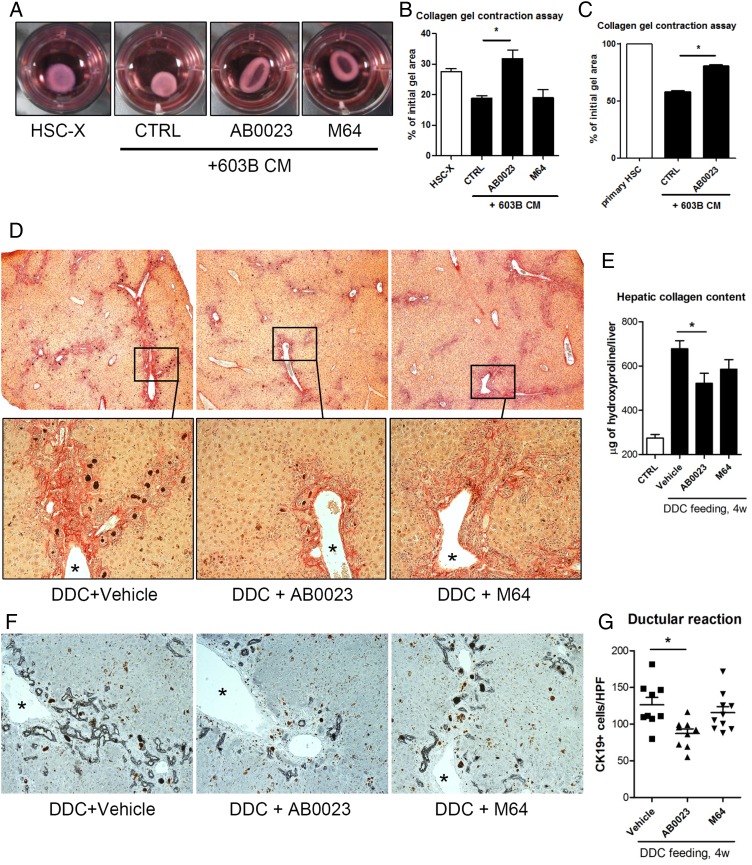
Methods: Anti-LOXL2 antibody, control antilysyl oxidase antibody or placebo was administered during thioacetamide (TAA)-induced fibrosis progression or during recovery. Therapeutic efficacy in biliary fibrosis was tested in BALB/c.Mdr2-/- and 3,5-diethoxycarbonyl-1,4-dihydrocollidine (DDC)-fed mice. Collagen crosslinking, fibrosis progression and reversal were assessed histologically and biochemically. HPC differentiation was studied in primary EpCAM(+) liver cells in vitro.

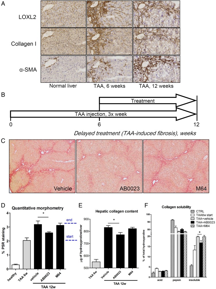
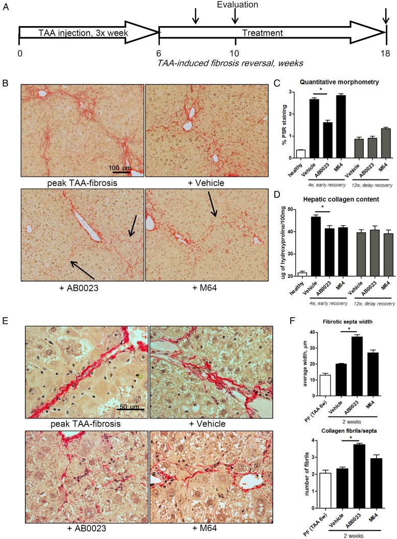
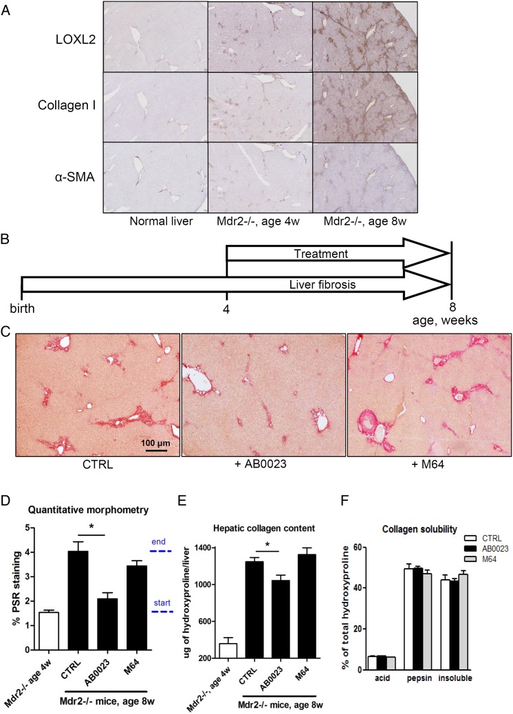
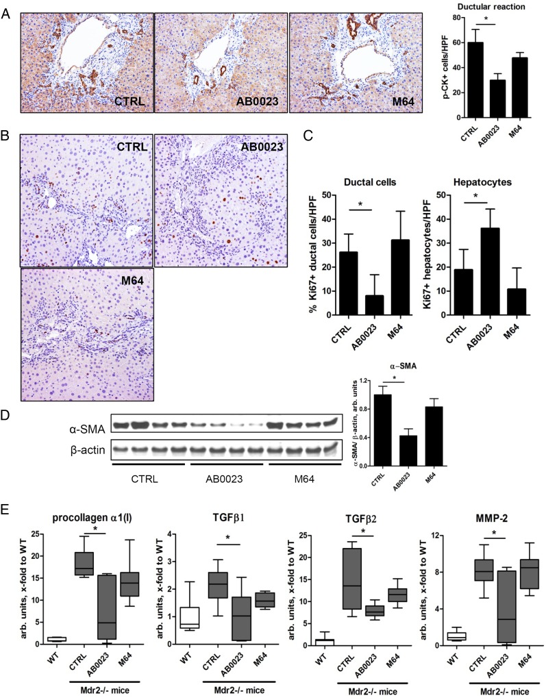
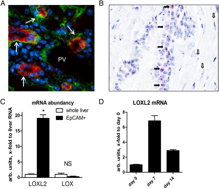
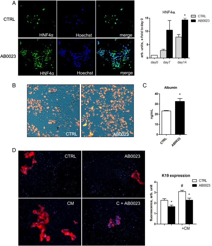
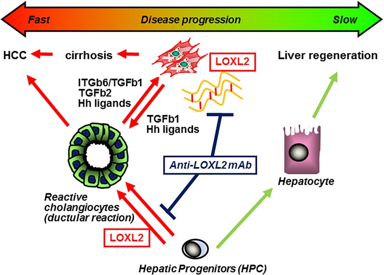
Results: LOXL2 was virtually absent from healthy but strongly induced in fibrotic liver, with predominant localisation within fibrotic septa. Delayed anti-LOXL2 treatment of active TAA fibrosis significantly reduced collagen crosslinking and histological signs of bridging fibrosis, with a 53% reduction in morphometric collagen deposition. In established TAA fibrosis, LOXL2 inhibition promoted fibrosis reversal, with enhanced splitting and thinning of fibrotic septa, and a 45% decrease in collagen area at 4 weeks of recovery. In the Mdr2-/- and DDC-induced models of biliary fibrosis, anti-LOXL2 antibody similarly achieved significant antifibrotic efficacy and suppressed the ductular reaction, while hepatocyte replication increased. Blocking LOXL2 had a profound direct effect on primary EpCAM(+) HPC behaviour in vitro, promoting their differentiation towards hepatocytes, while inhibiting ductal cell lineage commitment.

Conclusions: LOXL2 mediates collagen crosslinking and fibrotic matrix stabilisation during liver fibrosis, and independently promotes fibrogenic HPC differentiation. By blocking these two convergent profibrotic pathways, therapeutic LOXL2 inhibition attenuates both parenchymal and biliary fibrosis and promotes fibrosis reversal.

Keywords: CIRRHOSIS; HEPATIC FIBROSIS; HEPATITIS; PRIMARY BILIARY CIRRHOSIS; PRIMARY SCLEROSING CHOLANGITIS.


Conflict of interest statement

Competing interests: AM-V and VS are employees of Gilead Sciences which develops anti-LOXL2 antibody and holds related patents. YVP received research funding from Gilead Sciences.

Figures

Figure 1
Figure 1
Anti-LOXL2 antibody attenuates the progression of advanced pan-lobular thioacetamide (TAA)-induced fibrosis. (A) Immunohistochemistry for lysyl oxidase-like 2 (LOXL2), collagen I and α-smooth muscle actin (α-SMA) (200×). (B) Liver fibrosis was induced in C57Bl/6J mice with repeated TAA injections, and treatment with anti-LOXL2 monoclonal antibody (mAb) (AB0023, 30 mg/kg), vehicle or anti-LOX mAb (M64, 30 mg/kg) performed two times a week from week 7 to 12. (C) AB0023 inhibits bridging fibrosis compared with vehicle and M64 treatment (Sirius Red staining, 50΄). (D) Quantitative morphometry of collagen area (Sirius Red, n=5–8 for fibrotic groups, n=3 in healthy controls). Dotted bar indicates start and end of treatment. (E) Hepatic collagen content as determined biochemically via hydroxyproline (n=13–15). (F) Fibrotic matrix stability assessed ex vivo as described in M&M. AB0023 reduced the accumulation of highly crosslinked (insoluble) hepatic collagen at advanced liver fibrosis stages (n=4). Data are expressed as means±SEM. *p<0.05 (analysis of variance). CTRL, control.
Figure 2
Figure 2
Lysyl oxidase-like 2 inhibition accelerates the reversal of thioacetamide (TAA)-induced hepatic fibrosis. (A) C57BL/6 mice with pre-established liver fibrosis (TAA for 6 weeks) were allowed to recover for up to 12 weeks, while treated with AB0023 (30 mg/kg), M64 (30 mg/kg) or vehicle intraperitoneally two times per week. (B) Collagen staining and morphometry (C, n=10) show that AB0023 promotes early resolution of bridging fibrosis with widening and disruption of fibrotic septa (arrows) (4 weeks of recovery, Sirius Red, 200×). (D) Hepatic collagen content (via hydroxyproline, 4 weeks of recovery, n=15–16). (E and F) Septal morphology demonstrates widening of septa and splitting of collagen bundles into thinner fibrils (E, 630×) in AB0023-treated mice. (F) Septal thickness and number of fibrils assessed in 10 randomly selected fibrotic septa (Sirius Red, 630×, 2 weeks recovery, n=4). *p<0.05 vs vehicle controls (analysis of variance).
Figure 3
Figure 3
Targeting lysyl oxidase-like 2 (LOXL2) with a therapeutic antibody suppresses the progression of biliary fibrosis in BALB/c.Mdr2−/− mice. (A) Immunohistochemistry for LOXL2, collagen I and α-smooth muscle actin (α-SMA) in BALB/c.Mdr2−/− livers (40΄). (B) BALBc.Mdr2−/− mice were administered AB0023 or M64 (30 mg/kg) intraperitoneally two times per week from 4 to 8 weeks of age. (n=9–10) (C) Diminished fibrosis in AB0023-treated mice (representative Sirius Red staining, 50΄). (D) Quantitative collagen morphometry (n=4–6). Dotted bar indicates start and end of treatment. (E) Hepatic hydroxyproline content (n=8–15). (F) Fibrotic matrix stability as assessed ex vivo via serial collagen extraction (n=4 for each bar). Data are expressed as mean±SEM. *p<0.05 (analysis of variance). CTRL, control.
Figure 4
Figure 4
Profibrogenic ductular reaction is attenuated upon lysyl oxidase-like 2 (LOXL2) inhibition in BALBc.Mdr2−/− mice. (A) Representative pan-cytokeratin (p-CK) immunohistochemistry and p-CK+ cell numbers (>7 random portal fields, n=4, 200×). (B) Cell proliferation (Ki-67 immunohistochemistry). Note the increased proliferation in ductular structures in untreated controls, whereas labelled hepatocytes predominate in AB0023-treated Mdr2−/− mice. (C) Quantification of Ki-67+ hepatocytes and ductal cells (>7 HPF, n=4, 200΄). (D) α-smooth muscle actin (α-SMA) expression in liver lysates (representative western blot and densitometry, with β-actin as a loading control, n=8–9). (E) Hepatic messenger RNA expression of procollagen α1(I), transforming growth factor β (TGFβ)1, TGFβ2 and matrix metalloproteinase 2 (MMP2) (n=9–15). *p<0.05 (analysis of variance). CTRL, control; HPF, high-power field; WT, wild type.
Figure 5
Figure 5
Lysyl oxidase-like 2 (LOXL2) is expressed by hepatic progenitor cells (HPCs) in fibrotic livers. (A) Double immunofluorescence for LOXL2 (green) and EpCAM (red) in Mdr2−/− mice reveals prominent LOXL2 expression adjacent to EpCAM(+) cells (arrows). PV, portal vein. (B) In situ hybridisation reveals LOXL2 messenger RNA (mRNA)-specific signal (magenta) within HPC/ductal cells forming pseudoduct (closed arrows). Note spindle-shaped myofibroblast-like cells in periductular scar, also positive for LOXL2 transcripts (open arrows). Representative images from four Mdr2−/− mice shown (original magnification, ×40). (C) LOXL2 and LOX mRNA expression in freshly isolated EpCAM(+) cells compared with whole liver RNA from Mdr2−/− mice, and during prolonged culture in vitro (D). Data are means±SEM. *p<0.05 (Student's t-test).
Figure 6
Figure 6
Function-blocking experiments reveal that lysyl oxidase-like 2 (LOXL2) regulates lineage commitment of EpCAM+ hepatic progenitor cell (HPC) in vitro. EpCAM(+) progenitor cells were grown in differentiation medium for up to 14 days in the presence of AB0023 (30 μg/mL, added 48 hours after plating) or isotype control IgG (CTRL, 30 μg/mL). (A–C) In vitro LOXL2 neutralisation promotes HPC differentiation into functional hepatocytes. (A) Accumulation of HNF4α-expressing cells in EpCAM(+)-derived colonies (left, green HNF4α, blue nuclei) and HNF4α messenger RNA levels (right) in cell lysates. (B) Low-density lipoprotein (LDL) uptake analysed using Dil-AcLDL (red fluorescence overlayed on phase-contrast image). (C) Albumin levels in the EpCAM(+) cell supernatant after 12 days of culture with AB0023 or control IgG. (D) Proliferation of K19(+) ductular-like cell clusters (red) from EpCAM(+) progenitors is promoted by hepatic stellate cell-conditioned medium (CM), and inhibited by anti-LOXL2 AB0023 antibody. Quantification of K19 expression in EpCAM(+) HPC-derived colonies (ImageJ, n=3). Data are means±SEM. *p<0.05 (Student's t-test).
Figure 7
Figure 7
Collagen gel contraction by hepatic stellate cells is inhibited by lysyl oxidase-like 2 (LOXL2) blocking antibody. (A–C) Collagen gel contraction assay using the activated rat hepatic stellate cell (HSC)-X cell line (A, representative gel images after 4 days) and primary murine HSC in the presence of AB0023, isotype control IgG or M64 (all 10 μg/mL). The per cent reduction in collagen gel surface area was analysed at days 4 in HSC-X cells (B) or 15 in primary HSC (C) and quantified using ImageJ software in triplicates. *p<0.05 (Student's t-test); CM, conditioned medium. (D–F) Anti-LOXL2 antibody achieves antifibrotic efficacy in a 3,5-diethoxycarbonyl-1,4-dihydrocollidine (DDC)-feeding model. (D) Low-magnification representative images of collagen staining (50΄, upper row) show decrease in periportal and bridging fibrosis in AB0023-treated mice. Lower panel shows high magnification of selected portal areas (200×, asterisk marks portal vein). (E) Hepatic collagen deposition (via hydroxyproline content, n=9–10 in fibrotic groups, n=5 in healthy controls). (F) The ductular reaction is attenuated in mice with LOXL2 inhibition (K19 immunohistochemistry, left) with (G) quantification of K19+ cells (>7 random portal fields/liver, n=9–10, 200×). Data expressed as means±SEM. *p<0.05 (analysis of variance). CTRL, control.
Figure 8
Figure 8
Scheme of the proposed role of lysyl oxidase-like 2 (LOXL2) in promoting hepatic fibrogenesis. (1) LOXL2-mediated crosslinking stabilises collagen into a more rigid and degradation-resistant ECM. The stiff crosslinked matrix microenvironment further promotes hepatic stellate cell activation via mechanosensing. (2) Progenitor cell responses are controlled by autocrine/paracrine LOXL2, which promotes progenitor commitment towards a (profibrogenic) cholangiocyte lineage, while suppressing differentiation into (proregeneration) hepatocytes. (3) Blocking of LOXL2 activity in the extracellular space with AB0023 monoclonal antibody (mAb) (a) attenuates collagen crosslinking and (b) redirects hepatic progenitor cell (HPC) differentiation from profibrogenic cholangiocytes (ductular reaction) to hepatocytes, promoting fibrosis resolution and liver regeneration. HCC, hepatocellular carcinoma; TGF, transforming growth factor.

Comment in

References

    1. Friedman SL. Mechanisms of hepatic fibrogenesis. Gastroenterology 2008;134:1655–69. 10.1053/j.gastro.2008.03.003 - DOI - PMC - PubMed
    1. Schuppan D, Afdhal NH. Liver cirrhosis. Lancet 2008;371:838–51. 10.1016/S0140-6736(08)60383-9 - DOI - PMC - PubMed
    1. Popov Y, Schuppan D. Targeting liver fibrosis: strategies for development and validation of antifibrotic therapies. Hepatology 2009;50:1294–306. 10.1002/hep.23123 - DOI - PubMed
    1. Iredale JP. Models of liver fibrosis: exploring the dynamic nature of inflammation and repair in a solid organ. J Clin Invest 2007;117:539–48. 10.1172/JCI30542 - DOI - PMC - PubMed
    1. Popov Y, Sverdlov DY, Sharma AK, et al. . Tissue transglutaminase does not affect fibrotic matrix stability or regression of liver fibrosis in mice. Gastroenterology 2011;140:1642–52. 10.1053/j.gastro.2011.01.040 - DOI - PMC - PubMed

Publication types

MeSH terms