A Truncated Granulocyte Colony-stimulating Factor Receptor (G-CSFR) Inhibits Apoptosis Induced by Neutrophil Elastase G185R Mutant: IMPLICATION FOR UNDERSTANDING CSF3R GENE MUTATIONS IN SEVERE CONGENITAL NEUTROPENIA
- PMID: 28073911
- PMCID: PMC5336180
- DOI: 10.1074/jbc.M116.755157
A Truncated Granulocyte Colony-stimulating Factor Receptor (G-CSFR) Inhibits Apoptosis Induced by Neutrophil Elastase G185R Mutant: IMPLICATION FOR UNDERSTANDING CSF3R GENE MUTATIONS IN SEVERE CONGENITAL NEUTROPENIA
Abstract
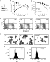
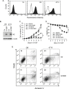
Mutations in ELANE encoding neutrophil elastase (NE) have been identified in the majority of patients with severe congenital neutropenia (SCN). The NE mutants have been shown to activate unfolded protein response and induce premature apoptosis in myeloid cells. Patients with SCN are predisposed to acute myeloid leukemia (AML), and progression from SCN to AML is accompanied by mutations in CSF3R encoding the granulocyte colony-stimulating factor receptor (G-CSFR) in ∼80% of patients. The mutations result in the expression of C-terminally truncated G-CSFRs that promote strong cell proliferation and survival. It is unknown why the CSF3R mutations, which are rare in de novo AML, are so prevalent in SCN/AML. We show here that a G-CSFR mutant, d715, derived from an SCN patient inhibited G-CSF-induced expression of NE in a dominant negative manner. Furthermore, G-CSFR d715 suppressed unfolded protein response and apoptosis induced by an SCN-derived NE mutant, which was associated with sustained activation of AKT and STAT5, and augmented expression of BCL-XL. Thus, the truncated G-CSFRs associated with SCN/AML may protect myeloid precursor cells from apoptosis induced by the NE mutants. We propose that acquisition of CSF3R mutations may represent a mechanism by which myeloid precursor cells carrying the ELANE mutations evade the proapoptotic activity of the NE mutants in SCN patients.
Keywords: apoptosis; cell differentiation; cell surface receptor; leukemia; neutrophil.
© 2017 by The American Society for Biochemistry and Molecular Biology, Inc.
Conflict of interest statement
The authors declare that they have no conflicts of interest with the contents of this article
Figures


Similar articles
-
Effect of the unfolded protein response and oxidative stress on mutagenesis in CSF3R: a model for evolution of severe congenital neutropenia to myelodysplastic syndrome/acute myeloid leukemia.Mutagenesis. 2020 Dec 1;35(5):381-389. doi: 10.1093/mutage/geaa027. Mutagenesis. 2020. PMID: 33511998 Free PMC article.
-
Inducible expression of a disease-associated ELANE mutation impairs granulocytic differentiation, without eliciting an unfolded protein response.J Biol Chem. 2020 May 22;295(21):7492-7500. doi: 10.1074/jbc.RA120.012366. Epub 2020 Apr 16. J Biol Chem. 2020. PMID: 32299910 Free PMC article.
-
G-CSF resistance of ELANE-mutant neutropenia depends on SERF1-containing truncated-neutrophil elastase aggregates.J Clin Invest. 2024 Nov 19;135(2):e177342. doi: 10.1172/JCI177342. J Clin Invest. 2024. PMID: 39560992 Free PMC article.
-
[Gene Mutation and Acute Leukemia Transformation of Severe Congenital Neutropenia- Review].Zhongguo Shi Yan Xue Ye Xue Za Zhi. 2017 Oct;25(5):1580-1584. doi: 10.7534/j.issn.1009-2137.2017.05.053. Zhongguo Shi Yan Xue Ye Xue Za Zhi. 2017. PMID: 29070147 Review. Chinese.
-
ELANE mutations in cyclic and severe congenital neutropenia: genetics and pathophysiology.Hematol Oncol Clin North Am. 2013 Feb;27(1):19-41, vii. doi: 10.1016/j.hoc.2012.10.004. Epub 2012 Nov 7. Hematol Oncol Clin North Am. 2013. PMID: 23351986 Free PMC article. Review.
Cited by
-
JAK/STAT: Why choose a classical or an alternative pathway when you can have both?J Cell Mol Med. 2022 Apr;26(7):1865-1875. doi: 10.1111/jcmm.17168. Epub 2022 Mar 3. J Cell Mol Med. 2022. PMID: 35238133 Free PMC article. Review.
-
Mutation, drift and selection in single-driver hematologic malignancy: Example of secondary myelodysplastic syndrome following treatment of inherited neutropenia.PLoS Comput Biol. 2019 Jan 7;15(1):e1006664. doi: 10.1371/journal.pcbi.1006664. eCollection 2019 Jan. PLoS Comput Biol. 2019. PMID: 30615612 Free PMC article.
-
Effect of the unfolded protein response and oxidative stress on mutagenesis in CSF3R: a model for evolution of severe congenital neutropenia to myelodysplastic syndrome/acute myeloid leukemia.Mutagenesis. 2020 Dec 1;35(5):381-389. doi: 10.1093/mutage/geaa027. Mutagenesis. 2020. PMID: 33511998 Free PMC article.
-
Upregulation of nuclear protein Hemgn by transcriptional repressor Gfi1 through repressing PU.1 contributes to the anti-apoptotic activity of Gfi1.J Biol Chem. 2024 Nov;300(11):107860. doi: 10.1016/j.jbc.2024.107860. Epub 2024 Oct 5. J Biol Chem. 2024. PMID: 39374784 Free PMC article.
-
Inducible expression of a disease-associated ELANE mutation impairs granulocytic differentiation, without eliciting an unfolded protein response.J Biol Chem. 2020 May 22;295(21):7492-7500. doi: 10.1074/jbc.RA120.012366. Epub 2020 Apr 16. J Biol Chem. 2020. PMID: 32299910 Free PMC article.
References
-
- Bellanné-Chantelot C., Clauin S., Leblanc T., Cassinat B., Rodrigues-Lima F., Beaufils S., Vaury C., Barkaoui M., Fenneteau O., Maier-Redelsperger M., Chomienne C., and Donadieu J. (2004) Mutations in the ELA2 gene correlate with more severe expression of neutropenia: a study of 81 patients from the French Neutropenia Register. Blood 103, 4119–4125 - PubMed
-
- Aprikyan A. A., Kutyavin T., Stein S., Aprikian P., Rodger E., Liles W. C., Boxer L. A., and Dale D. C. (2003) Cellular and molecular abnormalities in severe congenital neutropenia. Exp. Hematol. 31, 372–381 - PubMed
-
- Massullo P., Druhan L. J., Bunnell B. A., Hunter M. G., Robinson J. M., Marsh C. B., and Avalos B. R. (2005) Aberrant subcellular targeting of the G185R neutrophil elastase mutant associated with severe congenital neutropenia induces premature apoptosis of differentiating promyelocytes. Blood 105, 3397–3404 - PMC - PubMed
MeSH terms
Substances
Supplementary concepts
Grants and funding
LinkOut - more resources
Full Text Sources
Other Literature Sources
Research Materials
Miscellaneous

