. 2017 Mar;15(3):269-280.
doi: 10.1158/1541-7786.MCR-16-0227-T. Epub 2017 Jan 10.

DNA Polymerase Beta Germline Variant Confers Cellular Response to Cisplatin Therapy

Affiliations

DNA Polymerase Beta Germline Variant Confers Cellular Response to Cisplatin Therapy

Antonia A Nemec et al. Mol Cancer Res. 2017 Mar.

Abstract

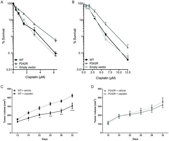
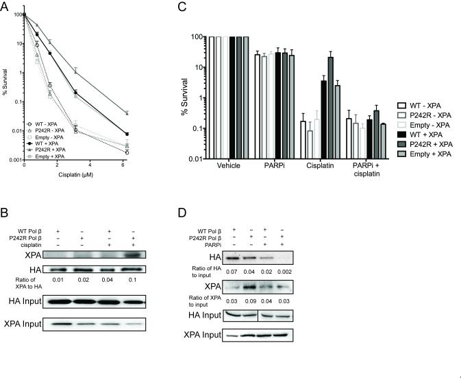
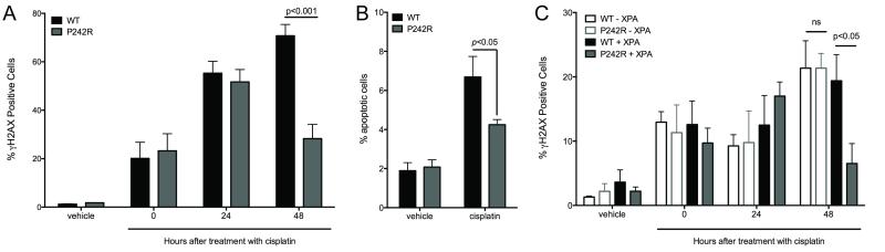
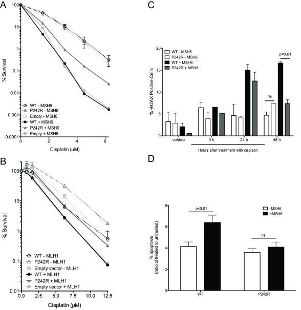
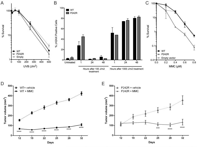
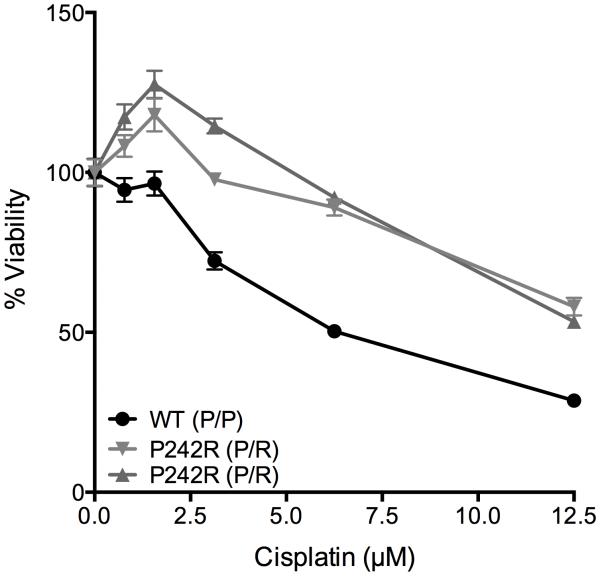
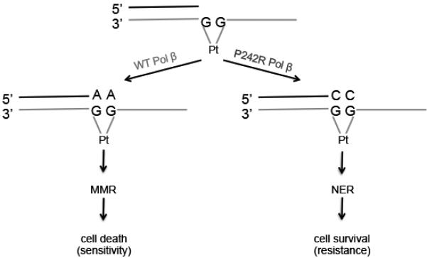
Resistance to cancer chemotherapies leads to deadly consequences, yet current research focuses only on the roles of somatically acquired mutations in this resistance. The mutational status of the germline is also likely to play a role in the way cells respond to chemotherapy. The carrier status for the POLB rs3136797 germline mutation encoding P242R DNA polymerase beta (Pol β) is associated with poor prognosis for lung cancer, specifically in response to treatment with cisplatin. Here, it is revealed that the P242R mutation is sufficient to promote resistance to cisplatin in human cells and in mouse xenografts. Mechanistically, P242R Pol β acts as a translesion polymerase and prefers to insert the correct nucleotide opposite cisplatin intrastrand cross-links, leading to the activation of the nucleotide excision repair (NER) pathway, removal of crosslinks, and resistance to cisplatin. In contrast, wild-type (WT) Pol β preferentially inserts the incorrect nucleotide initiating mismatch repair and cell death. Importantly, in a mouse xenograft model, tumors derived from lung cancer cells expressing WT Pol β displayed a slower rate of growth when treated with cisplatin, whereas tumors expressing P242R Pol β had no response to cisplatin. Pol β is critical for mediating crosstalk in response to cisplatin. The current data strongly suggest that the status of Pol β influences cellular responses to crosslinking agents and that Pol β is a promising biomarker to predict responses to specific chemotherapies. Finally, these results highlight that the genetic status of the germline is a critical factor in the response to cancer treatment.Implications: Pol β has prognostic biomarker potential in the treatment of cancer with cisplatin and perhaps other intrastrand crosslinking agents. Mol Cancer Res; 15(3); 269-80. ©2017 AACR.


Figures

Figure 1
Figure 1. Cells expressing P242R are resistant to cisplatin in vitro and in vivo
MCF7 (A) or A549 (B) cells expressing WT or P242R Pol β or containing empty vector were treated with varying concentrations of cisplatin (0-12.5 μM). Clonogenic survival assays were performed, and data are presented as mean ± SEM of the percent survival (n = 3-6). C-D. Mouse xenografts of A549 tumors expressing either WT (circles) or P242R (triangles) Pol β were treated by intraperitoneal injection once per week with either saline (dashed lines, open symbols) or 4 mg/kg cisplatin (solid line, closed symbols). Tumor growth measurements are presented as mean ± SEM (n=5). *, **, ***, or **** represent p<0.05, 0.01, 0.001, or 0.0001, respectively, comparing cisplatin-treated tumors to saline at each time point.
Figure 2
Figure 2. NER is required for P242R-mediated cisplatin resistance
A. XP20S cells deficient in XPA (−XPA; dashed lines, open symbols) or complemented with XPA (+XPA; solid lines, filled symbols) expressing WT, P242R, or empty vector were treated with various concentrations of cisplatin (0-6.25 μM). Clonogenic survival assays were performed and data are presented as mean ± SEM of the percent survival (n = 3). B. Whole cell extracts from A549 cells expressing either HA-tagged WT or P242R Pol β were immunoprecipated with anti-HA antibody as described in Experimental Procedures. Samples were separated by SDS-PAGE and the membrane was probed with antibodies against XPA or HA. The ratio of XPA to HA was quantified and shown under the blot. Co-immunoprecipitations were performed 3 times. C. XP20S deficient in XPA (−XPA; open bars) or complemented with XPA (+XPA; solid bars) expressing WT, P242R, or empty vector were treated with 1.6 μM cisplatin in the presence or absence of 10 μM PARPi (olaparib; AZD2281). Clonogenic survival assays were performed and data are presented as mean ± SEM of the percent survival (n = 3). D. A whole cell extract binding assay was performed with biotinylated Pt-GG DNA and lysates from A549 cells expressing either HA-tagged WT or P242R Pol β as described in Experimental Procedures. Westerns were performed and membranes were probed with anti-HA or anti-XPA antibodies. The ratios of HA or XPA to 10% HA input were quantified and are shown under the blots (n=3). XPA input is also shown.
Figure 3
Figure 3. Cisplatin-induced damage is repaired more efficiently in P242R-expressing cells
A-B. A549 cells expressing WT or P242R were treated with 6.25 μM cisplatin for 24 h and allowed to recover for 0-96 h. Double-strand breaks (A) or apoptotic cells (B) were measured by staining with γH2AX antibody or Annexin V, respectively using flow cytometry. C. XP20S cells deficient in XPA (−XPA; open bars) or complemented with XPA (+XPA; filled bars) expressing WT or P242R were treated with 1.6 μM cisplatin and allowed to recover for 0-48 h. Double-strand breaks were measured by staining with γH2AX antibody using flow cytometry. All data are presented as mean ± SEM (n = 3-4). Two-way ANOVA with Bonferroni’s post hoc test was performed to determine significance.
Figure 4
Figure 4. Mismatch repair is required for cisplatin-induced apoptosis in WT cells
A. DLD1 cells deficient in MSH6 (−MSH6; dashed lines, open symbols) or complemented with MSH6 (+MSH6; solid lines, filled symbols) expressing WT or P242R Pol β or empty vector, were treated with various concentrations of cisplatin (0-6.25 μM). B. HCT116 cells deficient in MLH1 (−MLH1; dashed lines, open symbols) or complemented with MLH1 (+MLH1; solid lines, filled symbols) were treated with various concentrations of cisplatin (0-12.5 μM). A-B. Clonogenic survival assays were performed. C-D. DLD1 cells deficient in MSH6 (−MSH6; open bars) or complemented with MSH6 (+MSH6; filled bars) were treated with 6.25 μM cisplatin and allowed to recover for 0-48 h (C) or 0 and 96 h (D). C. Double-strand breaks were measured by staining with γH2AX antibody using flow cytometry. D. Apoptotic cells were measured by staining with Annexin V antibody. Data are presented as the ratio of apoptotic cells in cisplatin-treated cells vs untreated cells for each cell line. All data are presented as mean ± SEM (n = 3). Two-way ANOVA with Bonferroni’s post hoc test (C) or two-tailed t-tests (D) were performed to determine significance.
Figure 5
Figure 5. P242R-mediated resistance to cisplatin is lesion-specific
A-B. A549 cells expressing WT or P242R Pol β or empty vector were treated with varying doses of UVB and allowed to recover for up to 48 h. A. Clonogenic survival assays were performed, and data are presented as mean ± SEM of the percent survival (n = 3). B. Double-strand breaks were measured by staining with γH2AX antibody using flow cytometry. C. Cells were treated various concentrations of mitomycin C (MMC) and clonogenic survival assays were performed. Data are presented as mean ± SEM of the percent survival (n = 3). D-E. Mouse xenografts of A549 tumors expressing either WT (circles) or P242R (triangles) Pol β were treated by intraperitoneal injection once per week with either saline (dashed lines, open symbols) or 5 mg/kg MMC (solid line, closed symbols). Tumor growth measurements are presented as mean ± SEM (n=5). *** or **** represents p< 0.001 or 0.0001, respectively, comparing MMC-treated tumors to saline at each time point.
Figure 6
Figure 6. Heterozygosity for P242R confers resistance to cisplatin in population-derived cells
Human lymphoblastoid cells derived from individuals with the genotype P/P (WT) or P/R (heterozygous for P242R) at residue 242 were plated at a density of 20,000 cells/well in a 96 well plate. Cells were treated with various concentrations (0-12.5 μM) of cisplatin for 24 h and cell viability was measured using CellTiter-Glo®. Data are presented as mean ± SEM of the percent viability (n = 3).
Figure 7
Figure 7. Model for P242R-medicated cisplatin resistance
After DNA is damaged by cisplatin, and intrastrand crosslink is formed. If WT Pol β bypasses the lesion, it preferentially inserts the incorrect A opposite the crosslink. This mismatch is recognized by the mismatch repair pathway (MMR), but cannot be repaired leading to cell death. In contrast, if the intrastrand crosslink is bypassed by the P242R Pol β variant, the correct nucleotide, C, is inserted across the lesion. This is now recognized by the nucleotide excision repair pathway (NER), which properly excises and repairs the DNA, resulting in cell survival.

References

    1. Weiss RB, Christian MC. New cisplatin analogues in development. A review. Drugs. 1993;46(3):360–77. - PubMed
    1. Wang D, Lippard SJ. Cellular processing of platinum anticancer drugs. Nat Rev Drug Discov. 2005;4(4):307–20. - PubMed
    1. Eastman A. The formation, isolation and characterization of DNA adducts produced by anticancer platinum complexes. Pharmacol Ther. 1987;34(2):155–66. - PubMed
    1. Hoffmann JS, Pillaire MJ, Maga G, Podust V, Hubscher U, Villani G. DNA polymerase beta bypasses in vitro a single d(GpG)-cisplatin adduct placed on codon 13 of the HRAS gene. Proc Natl Acad Sci U S A. 1995;92(12):5356–60. - PMC - PubMed
    1. Kartalou M, Essigmann JM. Mechanisms of resistance to cisplatin. Mutat Res. 2001;478(1-2):23–43. - PubMed

LinkOut - more resources