Review
. 2016:2016:6170243.
doi: 10.1155/2016/6170243. Epub 2016 Dec 15.

A Comprehensive Review of Portosystemic Collaterals in Cirrhosis: Historical Aspects, Anatomy, and Classifications

Affiliations
Review

A Comprehensive Review of Portosystemic Collaterals in Cirrhosis: Historical Aspects, Anatomy, and Classifications

Cyriac Abby Philips et al. Int J Hepatol. 2016.

Abstract

Portosystemic collateral formation in cirrhosis plays an important part in events that define the natural history in affected patients. A detailed understanding of collateral anatomy and hemodynamics in cirrhotics is essential to envisage diagnosis, management, and outcomes of portal hypertension. In this review, we provide detailed insights into the historical, anatomical, and hemodynamic aspects to portal hypertension and collateral pathways in cirrhosis with emphasis on the various classification systems.


Conflict of interest statement

The authors declare that they have no competing interests.

Figures

Figure 1
Figure 1
(a) Coronal-oblique MIP image demonstrating multiple collaterals in the esophagus (arrowheads) as well as paraesophageal region (arrow). Asterisk denotes the gastroesophageal junction; (b) coronal-oblique maximum-intensity-projection (MIP) CECT image showing a dilated left gastric vein (arrow) which is serving as an afferent for multiple paraesophageal collaterals (arrowheads); (c) axial MIP image showing multiple gastric fundal collaterals (arrows) being drained by a tortuous gastrorenal shunt (arrowheads) into the left renal vein (not shown). Asterisk denotes the gastric lumen. This corresponds to IGV-1 in Sarin classification of gastric varices; (d) coronal MIP image showing multiple esophageal collaterals (arrowheads) continuing along the cardia to form collaterals in the lesser curvature of stomach. This corresponds to GOV-1 in Sarin classification of gastric varices.
Figure 2
Figure 2
(a) Coronal-oblique MIP image showing a tortuous gastro-lieno-renal shunt (asterisks) draining multiple gastroesophageal collaterals (arrowheads) into the superior aspect of the left renal vein (arrow); (b) coronal MIP image demonstrating a mesentericorenal and a mesentericocaval shunt in the same patient (black and white arrowheads, resp.). Asterisk: right renal vein, black arrow: IVC, and white arrow: superior mesenteric vein (double portosystemic shunts); (c) coronal-oblique MIP image demonstrating a dilated and tortuous mesentericocaval shunt (arrowheads) communicating between the superior mesenteric vein (white arrow) and the inferior vena cava (black arrow); (d) axial CECT image showing multiple duodenal and paraduodenal collaterals (asterisks). Arrowhead denotes the duodenum.
Figure 3
Figure 3
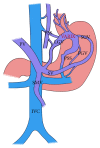
The basic portosystemic venous anatomy of gastric varices. PV: portal vein, SV: splenic vein, SMV: superior mesenteric vein, IVC: inferior vena cava, PSS: portosystemic shunt, SGV: short gastric vein, LGV: left gastric vein, and PGV: posterior gastric vein. Modified from [34].
Figure 4
Figure 4
The Kiyosue classification of gastric varices. (a) Classification based on drainage pathway. Type A consists of a portosystemic shunt as the only drainage; Type B portosystemic shunts along with additional small portosystemic collaterals; in Type C, there is presence of multiple large portosystemic shunts; and Type D consists of multiple small portosystemic collaterals as the drainage pathways without proper shunt formation. (b) Classification based on the inflow pathway: Type 1 consists of single afferent vein for the varices; Type 2 has multiple afferent vessels contributing to the variceal formation; Type 3 is similar to Type 2 but with additional small collateral/shunts directly communicating with outflow tract. Modified and redrawn from [17].
Figure 5
Figure 5
Classification of ectopic varices. SC: systemic circulation; PC: portal circulation; P-P: portoportal collaterals, P-S: portosystemic collaterals; PvB: portal venous branch; P-O: portal outflow; S-O: systemic outflow. Modified and redrawn from [35].
Figure 6
Figure 6
(a) Axial MIP image demonstrating multiple jejunal collaterals (arrows); (b) axial-oblique MIP CECT image showing multiple pericolonic collaterals (arrowheads) arising from the superior mesenteric vein (arrow); (c) axial MIP CECT image showing multiple rectal collaterals (arrow); (d) axial CECT image showing multiple paracholedochal collaterals (asterisks) encircling the common bile duct (arrow) in a patient with EHPVO.
Figure 7
Figure 7
(a) Axial CECT image showing multiple pericholecystic collaterals (arrows); (b) axial-oblique MIP image showing a right infradiaphragmatic type of shunt (arrowhead) arising from the left portal vein branch (asterisk) and draining into the intercostal vein; (c) coronal-oblique MIP image demonstrating a prominent recanalized paraumbilical vein (arrowheads) arising from the left branch of portal vein (black arrow) and draining into the right internal iliac vein (white arrow); (d) caput medusa, multiple periumbilical abdominal wall varices (asterisks).

References

    1. Cárdenas A., Ginès P. Portal hypertension. Current Opinion in Gastroenterology. 2009;25(3):195–201. doi: 10.1097/MOG.0b013e328329e154. - DOI - PubMed
    1. Cichoz-Lach H., Celiński K., Słomka M., Kasztelan-Szczerbińska B. Pathophysiology of portal hypertension. Journal of Physiology and Pharmacology. 2008;59(2):231–238. - PubMed
    1. Moreau R. VEGF-induced angiogenesis drives collateral circulation in portal hypertension. Journal of Hepatology. 2005;43(1):6–8. doi: 10.1016/j.jhep.2005.04.002. - DOI - PubMed
    1. Gilbert A., Villaret M. Contribution al'etude du syndrome d'hypertension portale; Cytologie des liquides d’ascite dans less cirrhoses. Comptes Rendus Societe de Biologie. 1906;60:820–823.
    1. Doehner G. A., Ruzicka F. F., Jr., Rousselot L. M., Hoffman G. The portal venous system: on its pathological roentgen anatomy. Radiology. 1956;66(2):206–217. doi: 10.1148/66.2.206. - DOI - PubMed

LinkOut - more resources