. 2016:2016:7534571.
doi: 10.1155/2016/7534571. Epub 2016 Dec 15.

Delayed Tension Pneumocephalus following Gunshot Wound to the Head: A Case Report and Review of the Literature

Affiliations

Delayed Tension Pneumocephalus following Gunshot Wound to the Head: A Case Report and Review of the Literature

Arthur Wang et al. Case Rep Surg. 2016.

Abstract

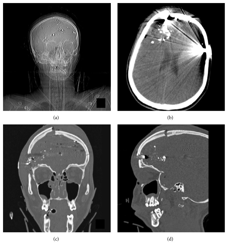
Tension pneumocephalus is a rare complication of head trauma and neurosurgical procedures, amongst other causes. It is defined by the combination of intracranial air, increased intracranial pressure, and mass effect. Although it often presents soon after surgery, it can also rarely present in a delayed fashion. We present a case of delayed tension pneumocephalus, occurring approximately 16 weeks after bifrontal craniectomy for a self-inflicted gunshot wound. Following a month of rhinorrhea, postnasal drip, and cough, the patient presented with a sensation of expansion in the area of the right forehead. As tension pneumocephalus is an emergency that can be fatal, this patient was treated expediently and avoided severe neurological deficits. The case recounted here is important as a demonstrative example that tension pneumocephalus does not always follow a defined course immediately after trauma or neurosurgery but rather can develop insidiously without obvious signs.


Conflict of interest statement

The authors declare that there is no conflict of interests regarding the publication of this paper.

Figures

Figure 1
Figure 1
Preoperative computed tomography (CT) of the head and neck. (a) Scout image demonstrating multiple metallic fragments intracranially and in the bifrontal soft tissue and bone. (b) Axial noncontrast CT demonstrating an extensive right frontal skull fracture with intracranial acute blood products including intraparenchymal, subarachnoid, subdural, and epidural components. (c, d) Coronal and sagittal reconstructions demonstrating the entry and exit sites of the bullet and the comminuted displaced free floating right frontal bone fragment.
Figure 2
Figure 2
Readmission CT head and maxillofacial-sinus scan. (a) Scout image demonstrating a large pocket of hypodensity in the right frontal region consistent with pneumocephalus. (b) Axial CT demonstrating right frontal tension pneumocephalus with significant mass effect including effacement of the right lateral ventricle, midline shift, and subfalcine herniation. (c, d) Coronal reconstructed images demonstrating air communicating through the skin in the right orbital-frontal region and a small defect with shard of bone in the superior aspect of the right frontal sinus.
Figure 3
Figure 3
Postcranioplasty CT head. (a) Scout image showing reduction in size of pneumocephalus. (b) Axial noncontrast head CT demonstrates marked resolution of pneumocephalus and decreased midline shift and mass effect on the right lateral ventricle.

References

    1. Solomiichuk V., Lebed V., Drizhdov K. Posttraumatic delayed subdural tension pneumocephalus. Surgical Neurology International. 2013;4, article 37 doi: 10.4103/2152-7806.109537. - DOI - PMC - PubMed
    1. Rao V., Fredriksli O., Gulati S. Post-traumatic epidural tension pneumocephalus: a case report. Journal of Medical Case Reports. 2015;9, article 151 doi: 10.1186/s13256-015-0633-5. - DOI - PMC - PubMed
    1. Dabdoub C. B., Salas G., Silveira E., et al. Review of the management of pneumocephalus. Surgical Neurology International. 2015;6, article 155 - PMC - PubMed
    1. Leong K. M., Vijayananthan A., Sia S. F., Waran V. Pneumocephalus: an uncommon finding in trauma. Medical Journal of Malaysia. 2008;63(3):256–258. - PubMed
    1. Kon T., Hondo H., Kohno M., Kasahara K. Severe tension pneumocephalus caused by opening of the frontal sinus by head injury 7 years after initial craniotomy—case report. Neurologia Medico-Chirurgica. 2003;43(5):242–245. doi: 10.2176/nmc.43.242. - DOI - PubMed

LinkOut - more resources