. 2017 Jan 11:8:13944.
doi: 10.1038/ncomms13944.

The mito-DAMP cardiolipin blocks IL-10 production causing persistent inflammation during bacterial pneumonia

Affiliations

The mito-DAMP cardiolipin blocks IL-10 production causing persistent inflammation during bacterial pneumonia

Krishnendu Chakraborty et al. Nat Commun. .

Abstract

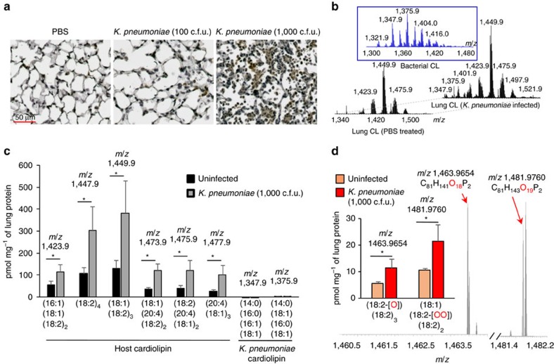
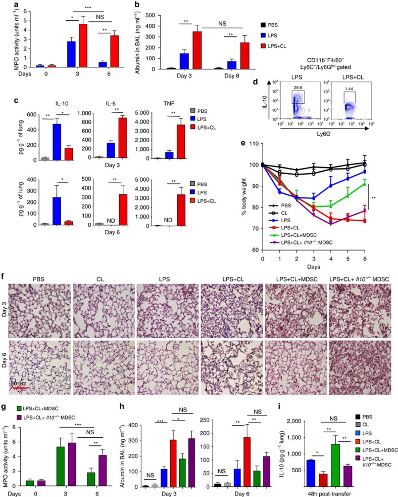
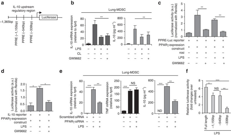
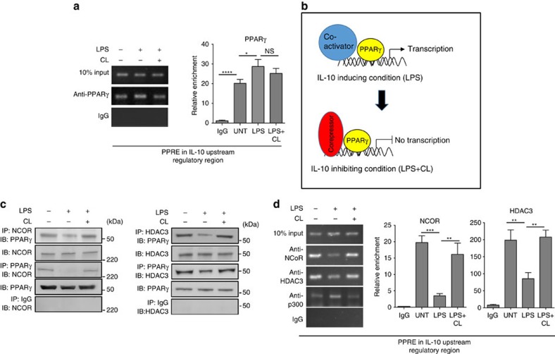
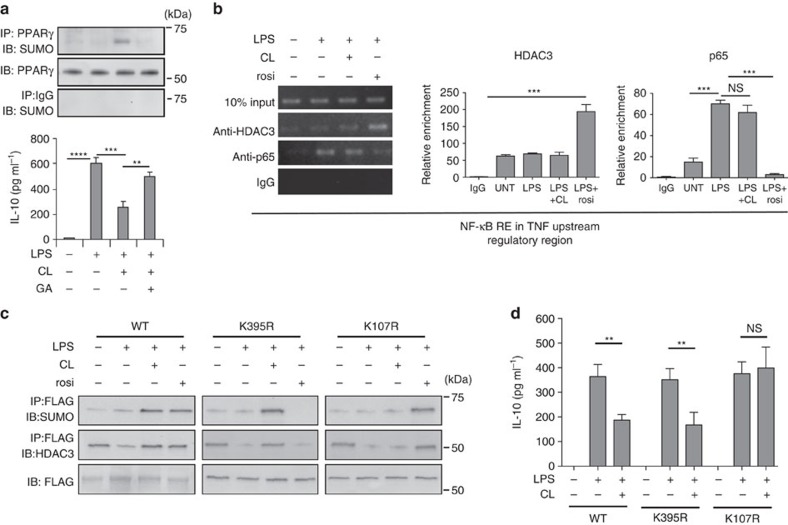
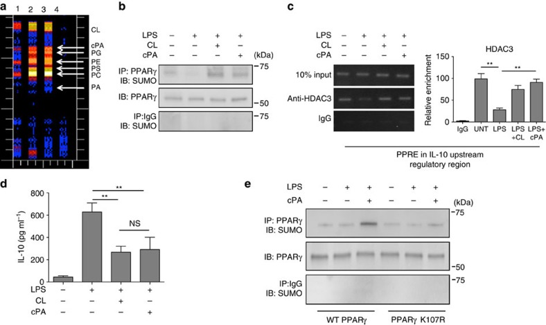
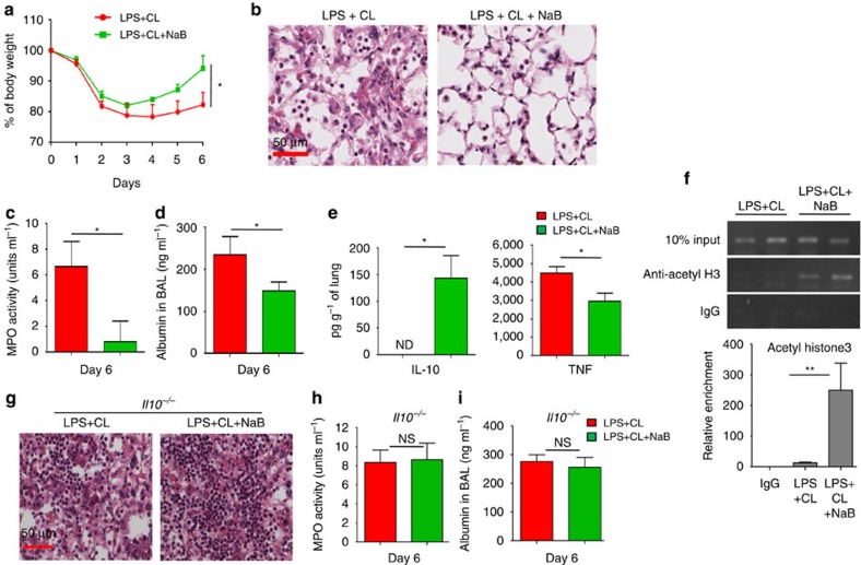
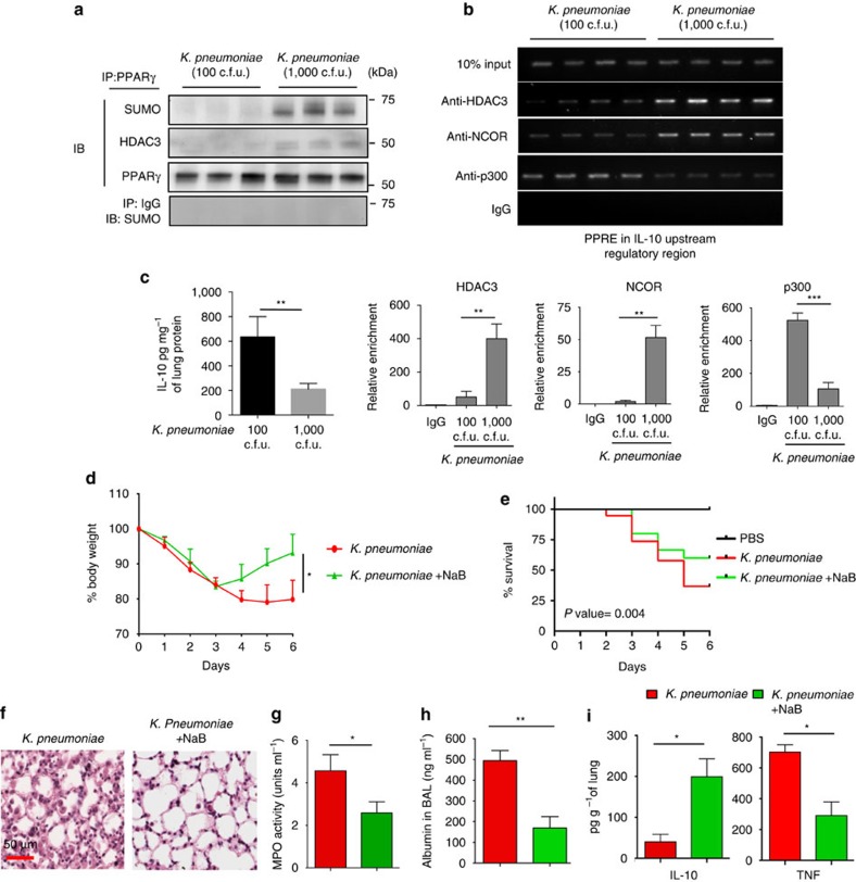
Bacterial pneumonia is a significant healthcare burden worldwide. Failure to resolve inflammation after infection precipitates lung injury and an increase in morbidity and mortality. Gram-negative bacteria are common in pneumonia and increased levels of the mito-damage-associated molecular pattern (DAMP) cardiolipin can be detected in the lungs. Here we show that mice infected with Klebsiella pneumoniae develop lung injury with accumulation of cardiolipin. Cardiolipin inhibits resolution of inflammation by suppressing production of anti-inflammatory IL-10 by lung CD11b+Ly6GintLy6CloF4/80+ cells. Cardiolipin induces PPARγ SUMOylation, which causes recruitment of a repressive NCOR/HDAC3 complex to the IL-10 promoter, but not the TNF promoter, thereby tipping the balance towards inflammation rather than resolution. Inhibition of HDAC activity by sodium butyrate enhances recruitment of acetylated histone 3 to the IL-10 promoter and increases the concentration of IL-10 in the lungs. These findings identify a mechanism of persistent inflammation during pneumonia and indicate the potential of HDAC inhibition as a therapy.


Figures

Figure 1
Figure 1. Increased level of cardiolipin in lungs of mice infected with K. pneumoniae.
(a) C57BL/6J mice were either left uninfected or infected intratracheally (i.t.) with K.pneumoniae (100 or 1,000 c.f.u.). Day 3 post infection, lung tissue sections of mice were stained for oxidized phospholipids. (b) LC/MS spectra of host cardiolipin obtained from lungs of PBS-treated mice (left) and infected mice (right). Middle panel depicts LC/MS spectrum of bacterial cardiolipin. (c) Quantitation of major host and bacterial cardiolipin molecular species from spectral data shown in b. (d) LC–MS spectra of mono- (m/z 1,463.9654) and di- (m/z 1,481.9760) oxygenated cardiolipin molecular species and their quantitative assessments in the lung. Data shown are mean±s.d. and all data are representative of two independent experiments with n=3 mice per group. Significance was calculated using Student's unpaired two-tailed t test for comparisons involving two groups or one-way ANOVA with Tukey's post hoc test for multiple pairwise comparisons. *P<0.05. CL, cardiolipin.
Figure 2
Figure 2. Induction of non-resolving lung inflammation with cardiolipin and low dose of LPS.
(a,b) C57BL/6J mice were treated intratracheally (i.t.) with LPS (20 μg per mouse), either alone or in combination with cardiolipin (100 μg per mouse). Cardiolipin was administered 1 h post LPS instillation. PBS-treated mice were used as controls. On days 3 and 6 post-treatment, the mice were analysed for (a) myeloperoxidase activity (MPO) activity in BAL fluid, (b) BAL albumin levels, a measure of microvascular permeability and (c) levels of cytokines in lung homogenates measured using magnetic bead-based assay. (d) Mice were treated with LPS±cardiolipin for three consecutive days. Day 6 post-treatment, IL-10 expression in CD11b+Ly6Cloy6GintF4/80+ lung MDSCs was assayed by flow cytometry. Gating is shown in Supplementary Fig. 1. (eh) Mice were treated with LPS either alone or in combination with cardiolipin. Twenty-four hour post-treatment, 1 × 10⁶ bone marrow-derived MDSCs from wild type or Il10−/− mice were adoptively transferred i.t. into mice that had received both LPS and cardiolipin and the following parameters were studied day 3 and 6 post LPS±cardiolipin administration: (e) Change in body weight monitored over six days (presented as % change from initial body weight). (f) lung pathology by H&E staining of lung tissue sections, (g) MPO activity in BAL fluid and (h) BAL albumin level. (i) Level of IL-10 in lung homogenates measured 48 h post adoptive transfer. The experiment was initiated with the following numbers of mice: 3 for PBS, cardiolipin and LPS groups, 8 for LPS+cardiolipin, 7 for LPS+cardiolipin+MDSC (WT) and 8 for LPS+cardiolipin+MDSC (Il10−/−) groups. At the conclusion of the experiment on day 6, 3 animals in PBS, cardiolipin, LPS and LPS+cardiolipin groups, 5 mice in the LPS+cardiolipin+MDSC (WT) group and 3 mice in the LPS cardiolipin+MDSC (Il10−/−) were available for analysis. Statistical significance was calculated using one-way ANOVA or two-way ANOVA with Bonferroni's post hoc test was used for multiple pairwise comparisons. Data shown are mean±s.d. and data are representative of two independent experiments. *P≤0.05, **P≤0.01, ***P≤0.001; NS, not significant.
Figure 3
Figure 3. PPARγ is critical for IL-10 regulation in lung MDSCs.
(a) Schematic of IL-10 upstream regulatory region showing multiple PPAR/RXR response elements (PPAR-RE). (b) Lung MDSCs were treated ex vivo with LPS (1 μg ml−1) either alone or in combination with cardiolpin (10 μg ml−1)±GW9662 (10 μM) for 6 h, cardiolipin being added 1 h post LPS treatment. Levels of IL-10 mRNA and secreted IL-10 in the culture supernatant were measured by qPCR (left panel) and ELISA (right panel) respectively. Addition of cardiolipin alone did not promote IL-10 gene expression (data not shown). (c) RAW 264.7 cells were transfected with both PPRE-Luciferase reporter construct and PPARγ expression vector. Twenty-four hour post-transfection, cells were stimulated with rosiglitazone (rosi) or LPS± GW9662. Two hour post-stimulation, reporter activity was measured. (d) RAW 264.7 cells were transfected with both IL-10 reporter and PPARγ expression constructs. Twenty-four hour post-transfection, cells were stimulated as indicated and reporter activity was measured. (e) Il10 and Tnf mRNA expression in lung MDSCs isolated from LPS-treated mice and transfected (1 × 10⁶ cells per condition) with scrambled or pparg-targeted siRNA (10 nM) was studied by qPCR (left and middle panel). IL-10 protein in culture supernatant was measured by ELISA (right panel). (f) Reporter assay of peritoneal macrophages transfected with plasmid constructs containing WT or deletion mutants of the IL-10 promoter (WT and mutants with deletions of indicated PPAR/RXR sites) linked to the luciferase gene. Statistical significance was calculated using one-way ANOVA with Bonferroni's post hoc test for multiple pairwise comparisons. Data shown are mean±s.d. and all data are representative of two independent experiments. *P≤0.05, **P≤0.01 and ***P≤0.001; NS, not significant.
Figure 4
Figure 4. Cardiolipin promotes association of corepressor complex with PPARγ.
Lung MDSCs were treated ex vivo with LPS (1 μg ml−1)±cardiolipin (10 μg ml−1) for 2 h and binding of PPARγ to the IL-10 promoter and association with corepressor proteins were studied. (a) ChIP assay was performed using PPARγ-specific antibody. Immunoprecipitated chromatin was used as template to amplify the region containing the PPRE at −339 bp in the IL-10 URR (left panel). qPCR quantification of three independent ChIP experiments is shown (right panel). (b) Schematic of hypothesis of IL-10 regulation in lung MDSCs by LPS±cardiolipin. (c) NCOR- or HDAC3-associated protein complexes were immunoprecipitated using their respective antibodies and probed with PPARγ-specific antibody or vice versa. (d) ChIP assay using NCOR-, HDAC3- or p300-specific antibody (left panel). qPCR quantitation of three independent ChIP experiments is shown (right panel). Statistical significance was calculated using one-way ANOVA with Bonferroni's post hoc test for multiple pairwise comparisons. Data shown are mean±s.d. and all data are representative of two independent experiments. *P≤0.05, **P≤0.01, ***P≤0.001 and ****P≤0.0001; NS, not significant.
Figure 5
Figure 5. Cardiolipin-induced PPARγ-SUMOylation is required to recruit corepressors to the IL-10 URR.
(a) Lung MDSCs were treated individually or in combination with LPS, cardiolipin or ginkgolic acid (GA, 50 μM) for 6 h. SUMOylation of immunoprecipitated PPARγ was studied by western blot analysis (upper panel) and IL-10 in the culture supernatant was measured by ELISA (lower panel). (b) Lung MDSCs were treated with LPS, cardiolipin or rosiglitazone (rosi) either alone or with the indicated combinations. ChIP assay was performed with antibodies against either HDAC3 or the p65 subunit of NF-κB (RelA). The region containing the NF-κB response element at −214 bp in the TNF promoter was amplified with specific primers. qPCR quantitation represents analysis of two independent ChIP experiments (middle and right panels) (c,d) RAW macrophages were transfected with plasmid expression constructs containing either FLAG-tagged WT PPARγ gene (left panel) or a variant containing either K395R (middle panel) or K107R (right panel) mutation in PPARγ. Transfected cells were treated individually or in combination with LPS, cardiolipin or rosi. FLAG-tagged PPARγ was immunoprecipitated with anti-FLAG antibody. SUMOylation and HDAC3 association with PPARγ was studied by western blot analysis. (d) Secreted IL-10 in the culture supernatant was measured by ELISA. Statistical significance was calculated using one-way ANOVA with Bonferroni's post hoc test for multiple pairwise comparisons. Data shown are mean±s.d. and all data are representative of two independent experiments. **P≤0.01 and ***P≤0.001; NS, not significant.
Figure 6
Figure 6. The cardiolipin metabolite cPA induces SUMOylation of PPARγ and recruitment of corepressor complex.
(a) Lung MDSCs were incubated with 3H-labelled cardiolipin for 10, 30 and 60 min and cellular lipids were extracted using the Bligh and Dyer method. Lipids were then resolved by thin layer chromatography to detect individual phospholipids. The 3H metabolites of cardiolipin were quantified using a plate reader. Lanes 1, 2 and 3 depict incubation of 3H-labelled cardiolipin for 10, 30 and 60 min, respectively, lane 4—blank. (bd) Lung MDSCs were treated with either LPS (1 μg ml−1) alone or in combination with cardiolipin (10 μg ml−1) or cPA (50 μM) and analysed. (b) PPARγ was immunoprecipitated and SUMOylation was studied by western blot methods. (c) HDAC3 recruitment to the IL-10 URR was investigated by ChIP assay (left panel). qPCR quantitation represents analysis of three independent ChIP experiments (right panel). (d) IL-10 in cell-free culture supernatant was measured by ELISA. (e) RAW 264.7 cells were transfected with either wild type or mutated (K107R) PPARγ (FLAG-tagged) expression construct. Twenty-four hour post-transfection, cells were stimulated with LPS±cPA as indicated. Two hours post-stimulation, PPARγ was immunoprecipitated and SUMOylation was examined by western blot methods. Statistical significance was calculated using one-way ANOVA with Bonferroni's post hoc test for multiple pairwise comparisons. Data shown in all experiments are mean±s.d. and data are representative of two independent experiments. **P≤0.01; NS, not significant.
Figure 7
Figure 7. Inhibition of HDAC3 by a short-chain fatty acid promotes resolution of lung inflammation.
C57BL/6J mice were treated i.t with both LPS (20 μg per mouse) and cardiolipin (100 μg per mouse). Day 1 post-treatment, mice were randomly divided into two groups. One group was treated i.p. with an HDAC3 inhibitor, sodium butyrate (NaB) (200 mg kg−1 body weight) for five consecutive days while the other group received only vehicle (PBS). Day 6 post-treatment, the following parameters of inflammation were studied: (a) Change in body weight, (b) lung pathology by H&E staining of lung tissue sections, (c) MPO activity in BAL fluid, (d) BAL albumin level and (e) Secreted cytokines (IL-10 and TNF) assayed in lung homogenates using magnetic bead-based assay. (f) Lung MDSCs were isolated from the two groups of mice and ChIP assay was performed to study recruitment of acetyl-histone to the IL-10 URR (upper panel). qPCR quantitation represents analysis of two independent ChIP experiments (lower panel). (gi) Il10−/− mice were treated i.t. with both LPS and cardiolipin as described above. Day 1 post-treatment, mice were randomly divided into two groups. One group was treated (i.p.) with an HDAC3 inhibitor, sodium butyrate (NaB), while the other group received only vehicle (PBS). Day 6 post-treatment, the following parameters of inflammation were studied: (g) lung pathology by H&E staining of lung tissue sections, (h) MPO activity in BAL fluid, (i) BAL albumin level by ELISA. Statistical significance was calculated using Student's unpaired two-tailed t test for comparisons involving two groups or one-way ANOVA with Bonferroni's post hoc test for multiple pairwise comparisons. The experiment was initiated with 8 mice in LPS+cardiolipin group and 6 mice in LPS+cardiolipin+NaB group with 3 and 4 mice respectively in each group available for analysis at the conclusion of the experiment. Data shown are mean±s.d. and are representative of two independent experiments. *P≤0.05, **P≤0.01; NS, not significant.
Figure 8
Figure 8. Infection with high dose of K. pneumoniae recruits corepressor complex to the IL-10 URR and amelioration of pathogenicity by short-chain fatty acid.
(ac) C57BL/6J mice were infected i.t. with either low dose (100 c.f.u.) or high dose (1,000 c.f.u.) of K. pneumoniae (n=3–4 mice per group at the conclusion of experiment). Four days post-infection, lung MDSCs were purified. (a) Coimmunoprecipitation and (b) ChIP experiments were performed using NCOR-, HDAC3- or p300-specific antibodies. Data shown are representative of two independent experiments. qPCR quantitation represents analysis of two independent ChIP experiments (lower panel). (c) IL-10 protein level in the lung on day 6 was measured by magnetic bead-based cytokine assay. Data shown are mean±s.d. and representative of two independent experiments. (di) C57BL/6J mice were infected with 1,000 c.f.u. of K. pneumonia. Day 1 post-infection, mice were randomly divided into two groups. One group was treated i.p. with NaB (200 mg kg−1 body weight) for five consecutive days while the other group received only vehicle (PBS). Day 6 post-treatment, the following parameters of inflammation were studied: (d) Change in body weight. (e) Percentage of survival represented by Kaplan–Meier survival curves of PBS-treated mice or mice infected with Kleb±NaB treatment. Data shown are mean±s.d. from three pooled experiments with n=12 mice in PBS group, 15 in bacteria only group and 20 in bacteria+NaB group. The P value was calculated using log-rank (Mantel-Cox) test. (f) Lung pathology by H&E staining of lung tissue sections. (g) MPO activity in BAL fluid, (h) BAL albumin level by ELISA, and (i) secreted cytokines assayed in lung homogenates using magnetic bead-based assay. Experiments in c,d and fi were initiated with n=8 and 6 mice in Klebsiella and Klebsiella+NaB groups, respectively. At the conclusion of the experiment, 3 mice in bacteria only group and 4 mice in bacteria+NaB group were available for analysis. Statistical significance was calculated using Student's unpaired two-tailed t test for comparisons involving two groups or one-way ANOVA with Bonferroni's post hoc test for multiple pairwise comparisons. Data shown are mean±s.d. and are representative of two independent experiments. *P≤0.05, **P≤0.01, ***P≤0.001.

References

    1. Nathan C. & Ding A. Nonresolving inflammation. Cell 140, 871–882 (2010). - PubMed
    1. Bhattacharya J. & Matthay M. A. Regulation and repair of the alveolar-capillary barrier in acute lung injury. Annu. Rev. Physiol. 75, 593–615 (2013). - PubMed
    1. Mizgerd J. P. Acute lower respiratory tract infection. N. Engl. J. Med. 358, 716–727 (2008). - PMC - PubMed
    1. Levy B. D. & Serhan C. N. Resolution of acute inflammation in the lung. Annu. Rev. Physiol. 76, 467–492 (2014). - PMC - PubMed
    1. Buckley C. D., Gilroy D. W., Serhan C. N., Stockinger B. & Tak P. P. The resolution of inflammation. Nat. Rev. Immunol. 13, 59–66 (2013). - PubMed

Publication types

MeSH terms