. 2017 Jan 11:7:40590.
doi: 10.1038/srep40590.

Myosin phosphatase and RhoA-activated kinase modulate arginine methylation by the regulation of protein arginine methyltransferase 5 in hepatocellular carcinoma cells

Affiliations

Myosin phosphatase and RhoA-activated kinase modulate arginine methylation by the regulation of protein arginine methyltransferase 5 in hepatocellular carcinoma cells

Adrienn Sipos et al. Sci Rep. .

Abstract

Myosin phosphatase (MP) holoenzyme is a protein phosphatase-1 (PP1) type Ser/Thr specific enzyme that consists of a PP1 catalytic (PP1c) and a myosin phosphatase target subunit-1 (MYPT1). MYPT1 is an ubiquitously expressed isoform and it targets PP1c to its substrates. We identified the protein arginine methyltransferase 5 (PRMT5) enzyme of the methylosome complex as a MYPT1-binding protein uncovering the nuclear MYPT1-interactome of hepatocellular carcinoma cells. It is shown that PRMT5 is regulated by phosphorylation at Thr80 by RhoA-associated protein kinase and MP. Silencing of MYPT1 increased the level of the PRMT5-specific symmetric dimethylation on arginine residues of histone 2 A/4, a repressing gene expression mark, and it resulted in a global change in the expression of genes affecting cellular processes like growth, proliferation and cell death, also affecting the expression of the retinoblastoma protein and c-Myc. The phosphorylation of the MP inhibitory MYPT1T850 and the regulatory PRMT5T80 residues as well as the symmetric dimethylation of H2A/4 were elevated in human hepatocellular carcinoma and in other types of cancers. These changes correlated positively with the grade and state of the tumors. Our results suggest the tumor suppressor role of MP via inhibition of PRMT5 thereby regulating gene expression through histone arginine dimethylation.


Figures

Figure 1
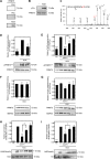
Figure 1. Interaction of MYPT1 with PRMT5.
(A) Immunoprecipitations were carried out using anti-MYPT11-296 and anti-PRMT5 antibodies as well as nonimmune rabbit serum as negative control. Immunoprecipitates and HepG2 total lysate were analysed by Western blots using antibodies specific for MYPT1 and PRMT5. (B) SPR analysis of the interaction of MYPT1 with PRMT5. Full-length GST-MYPT11-1004 was immobilized on anti-GST coupled CM5 sensor chip. FT-PRMT5 was injected over the surfaces in the indicated concentrations. The interaction was monitored using Biacore 3000. The association constant (Ka) value was indicated in the figure.
Figure 2
Figure 2. ROK and MP regulate the methyltransferase activity of PRMT5 through phosphorylation/dephosphorylation at Thr80.
(A) Autoradiograms of PRMT5 phosphorylated in the absence or in the presence of 0.1 μg/ml protein kinase A (PKA, left panel), 0.1 μg/ml protein kinase C (PKC, middle panel) or 0.4 U/ml Rho-associated kinase (ROK, right panel) with 32P-ATP. (B) Western blot analysis of ROK-phosphorylated PRMT5 using antibody specific for phospho-Thr. After stripping the membrane anti-PRMT5 antibody was applied to detect PRMT5 as an input control. (C) Ion trap collision-induced dissociation (CID) spectra of PRMT5 phosphopeptides. CID of m/z: 656.338 (3+) identified as SDLLLSGRDWNpTLIVGK representing [69–85] of the wild type protein. Thr80 was identified as the modification site (see fragment ion y11 (phosphorylated)). Peptide fragments are labeled according to the nomenclature by Biemann. (D) Effect of ROK inhibitor (10 μM H1152) on the phosphorylation level of PRMT5 during in vitro ROK assay. Control samples were prepared in the absence of ROK, positive control samples were prepared in the presence of ROK without ROK inhibitor. Relative phosphorylation level of Thr80 was judged by Western blot using anti- pPRMT5T80 antibody and blots for PRMT5 served as loading control. (E) Effect of 25 nM FT-MYPT1 and 5 nM rPP1cδ or their combination on the phosphorylation level of PRMT5 at Thr8080 as judged by Western blot. Data were compared to ROK-phosphorylated PRMT5. (F,G) Amount of MEP50 bound to FT-PRMT5 during ROK-phosphorylation (F) and dephosphorylation by MP (G) compared to unphosphorylated control samples. MEP50 was detected by anti-MEP50 antibody during Western blot and relative amount was normalized to the level of PRMT5. (H,I) In vitro arginine methyltransferase assay of unphosphorylated and ROK-phosphorylated PRMT5 measured by the symmetric dimethylation level of histone H2A Arg3 (H2AR3me2s, F) or histone H4 Arg3 (H4R3me2s, G) in the presence of 25 nM FT-MYPT1, 5 nM rPP1cδ or their combinations. Gels have been processed under the same experimental conditions. Values represents mean ± SEM; **p < 0.01, ***p < 0.001, ****p < 0.0001, #p < 0.05, one-way ANOVA followed by Tukey’s multiple comparison test, n = 3.
Figure 3
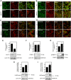
Figure 3. Effect of MYPT1 silencing on the methyltransferase activity of PRMT5 in HepG2 cells.
Immunofluorescent staining of non-target control (Ctrl) and MYPT1-silenced (siMYPT1) cells using antibodies specific for MYPT11-296 (A), PRMT5 (B), histone H2A symmetric dimethyl Arg3 (H2AR3me2s (C) and histone H4 symmetric dimethyl Arg3 (H4R3me2s (D) and actin as inducated in the figures. Scale bars: 50 μM. Enlargement of framed regions in merged images are shown on the right on A, B, C and D panels. Nuclear fractions of non-target control (Ctrl) and MYPT1-silenced (siMYPT1) HepG2 cells were prepared and analysed by Western blot using anti-MYPT11-296 (E), anti-PRMT5 (F), anti-pPRMT5T80 (G), anti-histone H2A symmetric dimethyl Arg3 (H) and anti-histone H4 symmetric dimethyl Arg3 (I) specific antibodies. The relative expression of MYPT1 or PRMT5 and the relative symmetric dimethylation level of H2AR3 or H4R3 were normalized to lamin A/C as internal control. Samples derived from the same experiment and the blots were processed in parallel or assayed after stripping. The relative phosphorylation level of PRMT5T80 was normalized to the expression level of PRMT5 and then to lamin A/C as internal control by densitometry. Mean ± SEM; n = 3; *p < 0.05, **p < 0.01, ***p < 0.001, ****p < 0.0001 by student t-test.
Figure 4
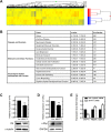
Figure 4. Microarray analysis of MYPT1-silenced HepG2 cells.
(A) Heat map of genes related to MYPT1 silencing in HepG2 cells by microarray analysis. The color code for the signal strength is shown in the box at the bottom in which induced genes are indicated by red and repressed genes are indicated by blue. (B) The onthology of the related genes and their classification by GO terms. Detection of retinoblastoma protein (C) and c-Myc (D) protein expression changes due to MYPT1 silencing from HepG2 whole cell lysates by Western blot. Protein levels were quantified by densitometry normalized to α-tubulin or GAPDH expression level. Samples derived from the same experiment and processed in parallel. RT-PCR analysis of MYPT1, PRMT5 and RAP1A mRNA levels in non-specific siRNA treated and MYPT1 silenced HepG2 cells (E). GAPDH was used as an invariant gene. Values are mean ± SEM from three independent experiments; *p < 0.05, **p < 0.01 by student t-test.
Figure 5
Figure 5. Protein expression profile of HCC and other cancer cell lines.
Reverse phase protein microarray analysis was conducted to study the changes in protein expression and posttranslational modification of PRMT5 (A,B), MYPT1 (C,D) and histone H2A (E,F) proteins in normal and tumor human cell lysates. Human HCC samples (n = 20) were grouped based on their clinically verified stage (middle grey columns) or grade (dark grey columns) classification of tumor. Average of HCC samples irrespectively of grouping and other types of metastatic liver cancer tissues are shown next to each other (light grey columns). Protein microarray contains 15 cancer cell lines and normal tissue lysate of controls of corresponding organs in triplicates (black columns). Value of 1 means average of relative expression, phosphorylation or symmetrical dimethylation of the given protein or residue to the corresponding non-tumor samples. Blots were processed under the same experimental procedure. Datas are mean ± SEM; *p < 0.05, **p < 0.01, ***p < 0.001, ****p < 0.0001 by student t-test.
Figure 6
Figure 6. MP plays a tumorsuppressor role and antagonizes ROK in HCC.
We hypothesize that MP and ROK are involved in gene expression. Under normal conditions MP modulates symmetrical dimethylation of histone core proteins in cell nucleus via dephosphorylation of PRMT5 at its activatory phosphorylation site (Thr80) causing changes in gene expression. In tumor cells inhibitory phosphorylation of MP on Thr850 is increased leading to higher phosphorylation level of PRMT5 at Thr80 by ROK. Activated PRMT5 provokes gene repression by raising symmetrical dimethylation of histone H4 Arg3 that triggers proto-oncogene activation and tumor formation.

Similar articles

Cited by

References

    1. Hou J. et al.. Hepatic RIG-I predicts survival and interferon-alpha therapeutic response in hepatocellular carcinoma. Cancer cell 25, 49–63, doi: 10.1016/j.ccr.2013.11.011 (2014). - DOI - PubMed
    1. Peti W., Nairn A. C. & Page R. Structural basis for protein phosphatase 1 regulation and specificity. The FEBS journal 280, 596–611, doi: 10.1111/j.1742-4658.2012.08509.x (2013). - DOI - PMC - PubMed
    1. Nagao M. et al.. Role of protein phosphatases in malignant transformation. Princess Takamatsu symposia 20, 177–184 (1989). - PubMed
    1. Dombradi V., Axton J. M., Barker H. M. & Cohen P. T. Protein phosphatase 1 activity in Drosophila mutants with abnormalities in mitosis and chromosome condensation. FEBS Lett 275, 39–43 (1990). - PubMed
    1. Berndt N. Protein dephosphorylation and the intracellular control of the cell number. Frontiers in bioscience: a journal and virtual library 4, D22–42 (1999). - PubMed

Publication types

MeSH terms