Review
. 2017 Feb;39(2):243-252.
doi: 10.3892/ijmm.2017.2855. Epub 2017 Jan 11.

The role of sphingolipid signalling in diabetes‑associated pathologies (Review)

Affiliations
Review

The role of sphingolipid signalling in diabetes‑associated pathologies (Review)

Mei Li Ng et al. Int J Mol Med. 2017 Feb.

Abstract

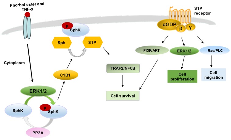
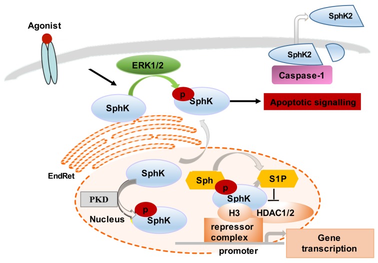
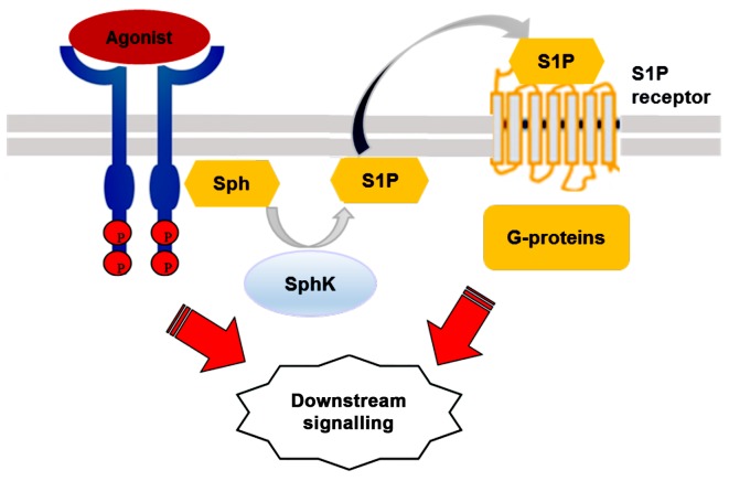
Sphingosine kinase (SphK) is an important signalling enzyme that catalyses the phosphorylation of sphingosine (Sph) to form sphingosine‑1‑phosphate (S1P). The multifunctional lipid, S1P binds to a family of five G protein-coupled receptors (GPCRs). As an intracellular second messenger, S1P activates key signalling cascades responsible for the maintenance of sphingolipid metabolism, and has been implicated in the progression of cancer, and the development of other inflammatory and metabolic diseases. SphK and S1P are critical molecules involved in the regulation of various cellular metabolic processes, such as cell proliferation, survival, apoptosis, adhesion and migration. There is strong evidence supporting the critical roles of SphK and S1P in the progression of diabetes mellitus, including insulin sensitivity and insulin secretion, pancreatic β‑cell apoptosis, and the development of diabetic inflammatory state. In this review, we summarise the current state of knowledge for SphK/S1P signalling effects, associated with the development of insulin resistance, pancreatic β‑cell death and the vascular complications of diabetes mellitus.


Figures

Figure 1
Figure 1
Regulation of SphK1 signalling. SphK1 is located mainly in the cytoplasm. Various agonists (such as phorbol ester and TNF-α) induce activation of SphK1 phosphorylation, activation and translocation to plasma membrane. This relocation is mediated by interaction with CIB1. S1P is exported from the cells and binds to S1P receptors (S1P1–5) to activate classical GPCR signalling pathways, leading to control of cell survival, proliferation and migration. Alternatively, S1P binds to TRAF2 intracellularly and activates the NF-κB pathway and its downstream targets. SphK, sphingosine kinase; CIB1, calcium and integrin-binding protein 1; S1P, sphingosine-1-phosphate; GPCR, G protein-coupled receptor; TRAF2, TNF receptor-associated factor 2; NF-κB, nuclear factor κ-light-chain-enhancer of activated B cells; PLC, phospholipase C; PP2A, protein phosphatase-2A.
Figure 2
Figure 2
Regulation and SphK2 signalling. Several agonists (such as EGF and cross-linking of FcεRI) stimulate SphK2 activation via ERK1/2-mediated phosphorylation. SphK2 in the EndRet induces apoptotic signalling through localised generation of S1P. The release of active SphK2 can also occur via caspase-1-mediated cleavage and allows extracellular generation of S1P. SphK2 localization in the nucleus is regulated via PKD-mediated activation of nuclear export signals. Nuclear SphK2 generates S1P that regulates histone acetylation. S1P specifically binds to the HDAC1/2 and inhibits their enzymatic activity. SphK2 associates with HDAC1/2 in repressor complex at promoters of the genes, where it enhances local H3 acetylation and promotes gene transcription. SphK, sphingosine kinase; EGF, epidermal growth factor; EndRet, endoplasmic reticulum; S1P, sphingosine-1-phosphate; PKD, protein kinase D; HDAC1/2, histone deacetylase 1 and 2; H3, histone 3; Sph, sphingosine.
Figure 3
Figure 3
SphK/S1P pathway 'inside-out' signalling model. Various biologically stimulatory molecules bind and activate receptor tyrosine kinase that, in turn, induces the translocation of SphK1 from the cytosole to the plasma membrane. S1P is then locally produced and secreted from the cells. Extracellular S1P binds and activates S1P receptors to initiate further intracellular downstream signaling cascades such as cell proliferation, migration, and invasion. SphK, sphingosine kinase; S1P, sphingosine-1-phosphate; Sph, sphingosine.

References

    1. Danaei G, Finucane MM, Lu Y, Singh GM, Cowan MJ, Paciorek CJ, Lin JK, Farzadfar F, Khang YH, Stevens GA, et al. Global Burden of Metabolic Risk Factors of Chronic Diseases Collaborating Group (Blood Glucose): National, regional, and global trends in fasting plasma glucose and diabetes prevalence since 1980: Systematic analysis of health examination surveys and epidemiological studies with 370 country-years and 2·7 million participants. Lancet. 2011;378:31–40. doi: 10.1016/S0140-6736(11)60679-X. - DOI - PubMed
    1. Expert Committee on the Diagnosis and Classification of Diabetes Mellitus Report of the expert committee on the diagnosis and classification of diabetes mellitus. Diabetes Care. 2003;26(Suppl 1):S5–S20. doi: 10.2337/diacare.26.2007.S5. - DOI - PubMed
    1. Shao S, Yang Y, Yuan G, Zhang M, Yu X. Signaling molecules involved in lipid-induced pancreatic beta-cell dysfunction. DNA Cell Biol. 2013;32:41–49. doi: 10.1089/dna.2012.1874. - DOI - PMC - PubMed
    1. Newsholme P, Keane D, Welters HJ, Morgan NG. Life and death decisions of the pancreatic beta-cell: The role of fatty acids. Clin Sci (Lond) 2007;112:27–42. doi: 10.1042/CS20060115. - DOI - PubMed
    1. Alemany R, van Koppen CJ, Danneberg K, Ter Braak M, Meyer zu Heringdorf D. Regulation and functional roles of sphingosine kinases. Naunyn Schmiedebergs Arch Pharmacol. 2007;374:413–428. doi: 10.1007/s00210-007-0132-3. - DOI - PubMed

MeSH terms