. 2016 Oct 16;16(1):106.
doi: 10.1093/jisesa/iew089. Print 2016.

Spinosad Induces Antioxidative Response and Ultrastructure Changes in Males of Red Palm Weevil Rhynchophorus ferrugineus (Coleoptera: Dryophthoridae)

Affiliations

Spinosad Induces Antioxidative Response and Ultrastructure Changes in Males of Red Palm Weevil Rhynchophorus ferrugineus (Coleoptera: Dryophthoridae)

Salaheldin A Abdelsalam et al. J Insect Sci. .

Abstract

The red palm weevil, Rhynchophorus ferrugineus, is of great concern worldwide, especially in the Middle East, where dates are a strategic crop. Despite their ecological hazard, insecticides remain the most effective means of control. A bioinsecticide of bacterial origin, spinosad is effective against several pests, and its efficacy against male R. ferrugineus was assessed in the present study. The antioxidative responses of key enzymes including catalase (CAT), superoxide dismutase (SOD), and glutathione-S-transferase (GST) to spinosad were investigated in the midgut and testes, and the effects of this insecticide on the cell ultrastructure of the midgut, Malpighian tubules, and testes were also determined. The lethal concentration 50 of spinosad was measured at 58.8 ppm, and the insecticide inhibited the activities of CAT, SOD, and GST in the midgut. However, no significant changes in the activities of these enzymes were observed in the testes. Spinosad treatment resulted in concentration-dependent changes in the cellular organelles of the midgut, Malpighian tubules, and testes of R. ferrugineus, and some of these effects were similar to those exerted by other xenobiotics. However, specific changes were observed as a result of spinosad treatment, including an increase in the number and size of concretions in Malpighian tubule cells and the occasional absence of the central pair of microtubules in the axonemes of sperm tails. This study introduces spinosad for potential use as an insecticide within an integrated control program against male red palm weevils. Additionally, the study provides biochemical and ultrastructural evidence for use in the development of bioindicators.

Keywords: Malpighian tubule; Rhynchophorus ferrugineus; Spinosad; antioxidant; midgut; testis; ultrastructure.


Figures

Fig. 1.
Fig. 1.
Antioxidant enzyme activities of control and spinosad-treated male R. ferrugineus. Each bar represents the mean ± SE of three replicates. Bar superscripts without a common letter differed significantly from each other; the level of significance was P < 0.05.
Fig. 2.
Fig. 2.
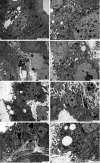
Transmission electron microscopy of R. ferrugineus midgut epithelial cells. Control (A, B): (A) Observe the closely spaced microvilli (mv), oval nucleus (N), abundant mitochondria (M), well-developed cisternae of the ER, intact septate junctions (arrows), and Goblet cells (GC). (B) A high-magnification image of the apical region of control epithelial cells depicts tightly packed mv, a nucleus with homogenous chromatin (N), electron-dense M, arrays of ER, and a normal septate junction (arrow). Spinosad (10 ppm) (C, D): (C) Note the damage to the apical mv, nuclei with chromatin at different condensation levels (arrows), and the increased amount of lysosomal bodies (Ly) passing into the gut lumen. (D) M appear swollen; the cristae are partially disintegrated, and the ER shows dilated cisternae. Fragmented mv can also be seen. (E) Spinosad (50 ppm): Corrugated and indented N with chromatin concentrated at the edge (arrows). The epithelial cells are no longer columnar, and the mv are severely disoriented. (F) Spinosad (100 ppm): marked dilations of junctions (arrows) between epithelial cells. Microvilli are less developed over part of the cell surface. N (at the lower left) is irregular and deformed, and large heterolysosomes (Ly) are discernible in the cytoplasm. Spinosad (200 ppm) (G, H): (G) Note condensed N, extensive lipid accumulation (L) in the cytoplasm, and abnormal dilations of junctions between cells (arrows). Also, the mv and ER arrays are only scanty. (H) Septate cell junctions are disrupted with apparent gaps (arrows) and sign of internalization in the cytoplasm, arrowheads indicate dilated parts of septate junctions, ruptured mv, N, M with indistinct cristae.
Fig. 3.
Fig. 3.
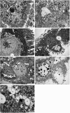
Transmission electron microscopy of Malpighian tubules of R. ferrugineus. Control (A, B): (A) Note the normal appearance of brush border microvilli (mv) containing mitochondria (M), and the laminated concretions (c) in the central cell cytoplasm, N: euchroamatic nucleus. (B) The basal lamina (bl) has branched, irregular infoldings (arrows) forming a well-developed labyrinth associated with M, arrowhead points to convoluted septate junction, mv. (C) Spinosad (10 ppm): Irregular mv, nucleus with several patches of heterochromatin (arrows), and hypertrophied Golgi fields (G). (D) Spinosad (50 ppm): Irregularly arranged mv, numerous concretions (c), N: nucleus with several dark clumps of chromatin. (E) Spinosad (100 ppm): Similar changes (as in D) but at a greater level. Arrow indicates septate junction. Spinosad (200 ppm) (F, G): (F) Lesions include large area of lysed cell cytoplasm (asterisk), chromatin clumping mainly at the nuclear periphery (arrows), enlarged laminated concretions (c), and destroyed mv. (G) Abnormally large M reveal prevalent existence of cristolysis and matrical loss (as compared to A). Note also myelin-like figure (arrow) with complex membrane whorls, large intracellular concretions (c), deformed mv.
Fig. 4.
Fig. 4.
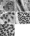
Transmission electron microscopy of normal spermiogenesis of control R. ferrugineus. (A) C.S. (cross section) of early developing spermatids. (B) L.S. (longitudinal section) of late spermatid showing typical chromatin condensation. (C) C.S. of spermatid tails. (D) C.S. of sperm heads and flagella. (E) C.S. of sperm tails (magnified image). N: nucleus, ER: cisternae of ER, nbk: nebenkern, md: mitochondrial derivatives, arrow (in B and D) indicates extension of large md into infloding of the nucleus, ax: axoneme (9 + 9 + 2 arrangement of microtubules), h: head, f: flagellum, ab: accessory bodies, pf: puff-like structure.
Fig. 5.
Fig. 5.
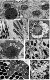
Transmission electron microscopy of testis of spinosad treated R. ferrugineus. Spinosad (10 ppm) (A, B): (A) C.S. of early spermatid showing hypertrophied Golgi body (G), N: nucleus. (B) L.S. of mature sperms. Arrow indicates binucleated sperm head, ac: acrosome. Spinosad (50 ppm) (C, D): (C) C.S. of early spermatids depicting ER-membrane whorls around nucleus (N) and nebenkern (nbk). Arrow points to mitochondrial damage. (D) L.S. of late spermatids. Note extensive swelling of mitochondrial derivatives (md), N: nucleus, ax: spermatid axoneme, c: centriole. (E) Spinosad (100 ppm): C.S. of degenerating early spermatid. Spinosad (200 ppm) (F-I): (F) C.S. of early spermatid showing electron-dense nucleus (N), swollen mitochondria (M), dilated ER cistern, enlarged vesicles of Golgi body (G). (G) L.S. of late-stage spermatids. Note the abnormal chromatin coalescence. (H) C.S. of mature sperms. Axonemes lack central microtubules (arrows), and nuclear envelope of sperm is folded and dilated (arrowheads). (I) C.S. of sperm tails showing marked edematous changes in md.

References

    1. Abarikwu S. O., Duru Q. C., Chinonso O. V., Njoku R. C. 2015. Antioxidant enzymes activity, lipid peroxidation, oxidative damage in the testis and epididymis, and steroidogenesis in rats after co-exposure to atrazine and ethanol. Andrologia. 48(5): 548–557. - PubMed
    1. Abbott W. S. 1925. A method of computing effectiveness of an insecticide. J. Econ. Entomol. 18: 265–267.
    1. Abdelsalam S. A., Korayem A. M., Elsherbini E. A. M., Abdel-Aal A. A., Mohamed D. S. 2014. Photosensitizing Effects of Hematoporphyrin Dihydrochloride against the Flesh Fly Parasarcophaga argyrostoma (Diptera: Sarcophagidae). Florida Entomologist. 97: 1662–1670.
    1. Abouelghar G. E., Sakr H., Ammar H. A., Yousef A., Nassar M. 2013. Sublethal Effects of Spinosad (Tracer®) on the Cotton Leafworm (Lepidoptera: Noctuidae). J. Plant Prot. Res. 53: 275–284.
    1. Aebi H. 1984. Catalase in vitro. Methods Enzymol. 105(C): 121–126. - PubMed

MeSH terms