. 2017 Jan 11;12(1):e0169565.
doi: 10.1371/journal.pone.0169565. eCollection 2017.

Bad to the Bone: On In Vitro and Ex Vivo Microbial Biofilm Ability to Directly Destroy Colonized Bone Surfaces without Participation of Host Immunity or Osteoclastogenesis

Affiliations

Bad to the Bone: On In Vitro and Ex Vivo Microbial Biofilm Ability to Directly Destroy Colonized Bone Surfaces without Participation of Host Immunity or Osteoclastogenesis

Adam Junka et al. PLoS One. .

Abstract

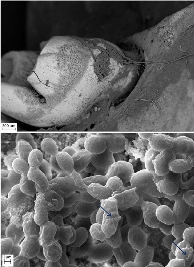
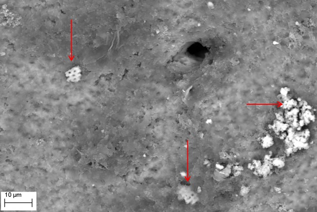
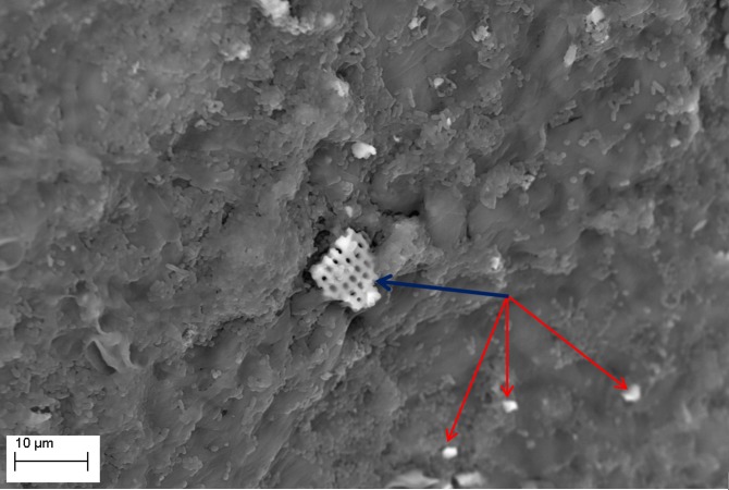
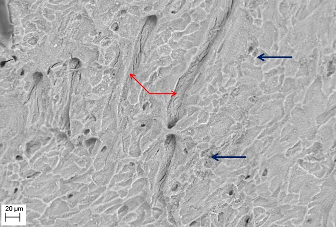
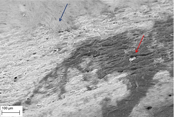
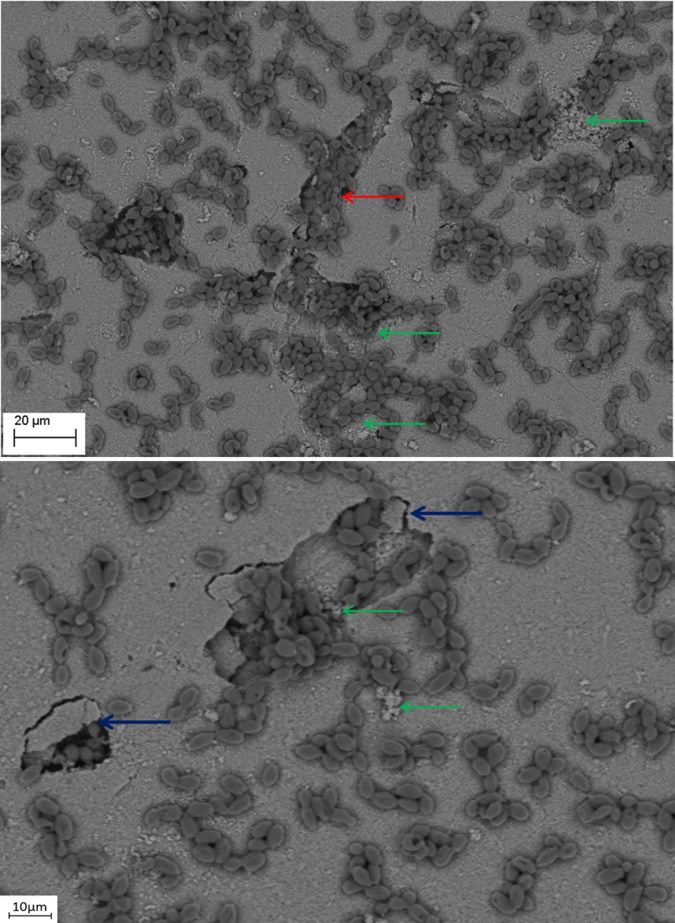
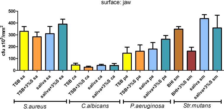
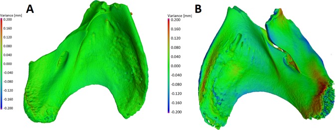
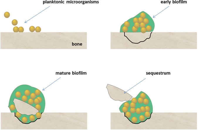
Bone infections are a significant public health burden associated with morbidity and mortality in patients. Microbial biofilm pathogens are the causative agents in chronic osteomyelitis. Research on the pathogenesis of osteomyelitis has focused on indirect bone destruction by host immune cells and cytokines secondary to microbial insult. Direct bone resorption by biofilm pathogens has not yet been seriously considered. In this study, common osteomyelitis pathogens (Staphylococcus aureus, Pseudomonas aeruginosa, Candida albicans, and Streptococcus mutans) were grown as biofilms in multiple in vitro and ex vivo experiments to analyze quantitative and qualitative aspects of bone destruction during infection. Pathogens were grown as single or mixed species biofilms on the following substrates: hydroxyapatite, rat jawbone, or polystyrene wells, and in various media. Biofilm growth was evaluated by scanning electron microscopy and pH levels were monitored over time. Histomorphologic and quantitative effects of biofilms on tested substrates were analyzed by microcomputed tomography and quantitative cultures. All tested biofilms demonstrated significant damage to bone. Scanning electron microscopy indicated that all strains formed mature biofilms within 7 days on all substrate surfaces regardless of media. Experimental conditions impacted pH levels, although this had no impact on biofilm growth or bone destruction. Presence of biofilm led to bone dissolution with a decrease of total volume by 20.17±2.93% upon microcomputed tomography analysis, which was statistically significant as compared to controls (p <0.05, ANOVA). Quantitative cultures indicated that media and substrate did not impact biofilm formation (Kruskall-Wallis test, post-hoc Dunne's test; p <0.05). Overall, these results indicate that biofilms associated with osteomyelitis have the ability to directly resorb bone. These findings should lead to a more complete understanding of the etiopathogenesis of osteomyelitis, where direct bone resorption by biofilm is considered in addition to the well-known osteoclastic and host cell destruction of bone.


Conflict of interest statement

The authors have declared that no competing interests exist.

Figures

Fig 1
Fig 1. Confirmation of absence of eukaryotic live cells in analyzed bone material.
Graph shows live/dead ratio values determined for cells and bones, horizontal bar is median and whiskers represent interquartile range. Differences in live/dead ratio are statistically significant, all medians vary between themselves significantly (Kruskal-Wallis test, p<0.05). Live PDV–positive control sample; green color confirms activity of live eukaryotic cells; Fixed PDV–negative control sample; red color confirms lack of live eukaryotic cells. Bone–analyzed material; red color confirms lack of live eukaryotic cells in analyzed rat jaws.
Fig 2
Fig 2. Native structure of rat jawbone showing characteristic surface morphology and lacunar-canalicular systems (arrows), as viewed under scanning electron microscopy (Magn.811X, SEM Zeiss EVO MA25 microscope).
Right inset shows gross resected rat mandible used in this study and the lingual-bone region (blue) used for biofilm inoculation.
Fig 3
Fig 3. S. aureus ATCC6538 biofilm (upper left inset, magnification 4750X) formed inside a native bone canal (red arrow; Magn.811X).
Higher magnification (blue arrow, lower left inset Magn.14560X) reveals multi-layer composition of staphylococcal biofilm. Media: artificial saliva plus 3% sucrose. SEM Zeiss EVO MA25 microscope.
Fig 4
Fig 4. S. mutans ATCC25175 biofilm (plaque) formed on the surface of bone (Magn.275X).
The dense structure of plaque is seen in the right and central part of the image, whereas plaque disruption is visible in the right portion of the image revealing multilayer composition and biofilm matrix (upper left inset, Magn. 660X). Media: artificial saliva plus 3% sucrose. SEM Zeiss EVO MA25 microscope.
Fig 5
Fig 5. S. mutans ATCC25175 and C. albicans 10231 mixed species biofilm formed on jawbone surface subjacent to a tooth (upper image Magn.95X; lower image Magn.9850X).
Note: streptococci co-aggregation with candida cells (blue arrow). Media: artificial saliva. SEM Zeiss EVO MA25 microscope.
Fig 6
Fig 6. P. aeruginosa ATCC15442 biofilm formed on jawbone surface (Magn.2570X).
Note: fragments of detached bone indicated by red arrows. Elemental composition of these alterations was experimentally confirmed. Media: artificial saliva plus 3% sucrose. SEM Zeiss EVO MA25 microscope.
Fig 7
Fig 7. S. mutans ATCC25175 biofilm formed on jawbone surface.
Note: larger fragment of detached bone indicated by the blue arrow, and smaller fragments indicated by red arrows. Media: artificial saliva. Magn.2990X, SEM Zeiss EVO MA25 microscope.
Fig 8
Fig 8. Removal of P. aeruginosa ATCC15442 biofilm from bone reveals characteristic surface alterations trails (red arrows) and cavities (blue arrows); as compared to Fig 1 representing intact bone structure, and Fig 9 representing the process of cavity and trail formation.
Media: artificial saliva plus 3% sucrose. Magn.645X, SEM Zeiss EVO MA25 microscope.
Fig 9
Fig 9. Removal of C. albicans 10231 biofilm from bone surface.
Note: two types of bone structure are indicated; intact (blue arrow), and altered (red arrow). Media: artificial saliva plus 3% sucrose. SEM Zeiss EVO MA25 microscope.
Fig 10
Fig 10. C. albicans 10231 cells remodeling hydroxyapatite surface.
Magn. 1300X (lower image) and 1010X (upper image). The process of cavity and sequestrum formation is indicated by blue arrows. Formation of trails resembling those seen in Figs 7 and 8 is marked with a red arrow. Destruction of hydroxyapatite is shown with the green arrow. Please see Fig 13 for potential mechanism of sequestrum formation. Media: artificial saliva plus 3% sucrose. SEM Zeiss EVO MA25 microscope.
Fig 11
Fig 11. Number of cells forming biofilm on jaws in TSB, TSB media supplemented with 3% sucrose; artificial saliva, saliva supplemented with 3% sucrose; BHI; BHI supplemented with 3% sucrose.
Fig 12
Fig 12
The geometry of control bone sample (A, left side of Figure), incubated for 30 days in sterile media and geometry of treated bone sample (B, right side of Figure), which was used as a surface for S. aureus biofilm growth. The green surface represents position points which did not change significantly from day 0 to day 30. The colors from light blue to violet represent points which collapsed during 30 days in comparison to day 0, and colors from orange to red represent points which were higher after 30 days of incubation than the same points in the beginning of experiment (day 0).
Fig 13
Fig 13. This schematic illustrates the development of sequestrum over time as planktonic organisms attach to bone and form mature biofilms, leading to bone cavitation and eventual detachment of infected fragments.

References

    1. Boody BS, Jenkins TJ, Maslak J, Hsu WK, Patel AA. Vertebral Osteomyelitis and Spinal Epidural Abscess: An Evidence-based Review. J Spinal Disord Tech. 2015. July;28(6):E316–27. 10.1097/BSD.0000000000000294 - DOI - PubMed
    1. Sanders J, Mauffrey C. Long bone osteomyelitis in adults: fundamental concepts and current techniques. Orthopedics. 2013. May;36(5):368–75. 10.3928/01477447-20130426-07 - DOI - PubMed
    1. Maffulli N, Papalia R, Zampogna B, Torre G, Albo E, Denaro V. The management of osteomyelitis in the adult. Surgeon. 2016. January 21. pii: S1479-666X(15)00124-9. - PubMed
    1. Stoodley P, Ehrlich GD, Sedghizadeh PP, Hall-Stoodley L, Baratz ME, Altman DT, et al. Orthopaedic biofilm infections. Curr Orthop Pract. 2011. November;22(6):558–563. 10.1097/BCO.0b013e318230efcf - DOI - PMC - PubMed
    1. Donlan RM, Costerton JW. Biofilms: survival mechanisms of clinically relevant microorganisms. Clin Microbiol Rev. 2002. April;15(2):167–93. 10.1128/CMR.15.2.167-193.2002 - DOI - PMC - PubMed

MeSH terms