

Effect of Trp53 gene deficiency on brain injury after neonatal hypoxia-ischemia
- PMID: 28076846
- PMCID: PMC5355327
- DOI: 10.18632/oncotarget.14518
Effect of Trp53 gene deficiency on brain injury after neonatal hypoxia-ischemia
Abstract
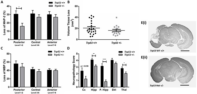
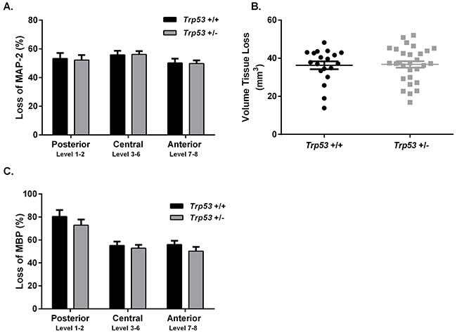
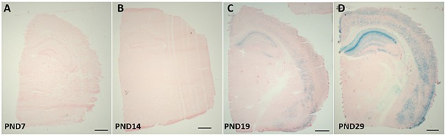
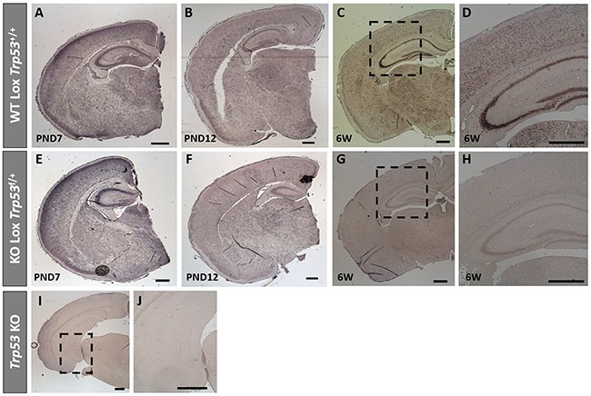
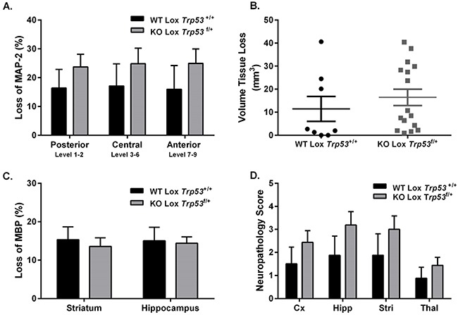
Hypoxia-ischemia (HI) can result in permanent life-long injuries such as motor and cognitive deficits. In response to cellular stressors such as hypoxia, tumor suppressor protein p53 is activated, potently initiating apoptosis and promoting Bax-dependent mitochondrial outer membrane permeabilization. The aim of this study was to investigate the effect of Trp53 genetic inhibition on injury development in the immature brain following HI. HI (50 min or 60 min) was induced at postnatal day 9 (PND9) in Trp53 heterozygote (het) and wild type (WT) mice. Utilizing Cre-LoxP technology, CaMK2α-Cre mice were bred with Trp53-Lox mice, resulting in knockdown of Trp53 in CaMK2α neurons. HI was induced at PND12 (50 min) and PND28 (40 min). Extent of brain injury was assessed 7 days following HI. Following 50 min HI at PND9, Trp53 het mice showed protection in the posterior hippocampus and thalamus. No difference was seen between WT or Trp53 het mice following a severe, 60 min HI. Cre-Lox mice that were subjected to HI at PND12 showed no difference in injury, however we determined that neuronal specific CaMK2α-Cre recombinase activity was strongly expressed by PND28. Concomitantly, Trp53 was reduced at 6 weeks of age in KO-Lox Trp53 mice. Cre-Lox mice subjected to HI at PND28 showed no significant difference in brain injury. These data suggest that p53 has a limited contribution to the development of injury in the immature/juvenile brain following HI. Further studies are required to determine the effect of p53 on downstream targets.
Keywords: brain injury; cell death; hypoxia-ischemia; mitochondria; p53.
Conflict of interest statement
The authors declare no conflicts of interest.
Figures
References
-
- Evans K, Rigby AS, Hamilton P, Titchiner N, Hall DM. The relationships between neonatal encephalopathy and cerebral palsy: a cohort study. J Obstet Gynaecol. 2001;21:114–20. - PubMed
-
- Pappas A, Shankaran S, McDonald SA, Vohr BR, Hintz SR, Ehrenkranz RA, Tyson JE, Yolton K, Das A, Bara R, Hammond J, Higgins RD. Hypothermia Extended Follow-up Subcommittee of the Eunice Kennedy Shriver NNRN. Cognitive outcomes after neonatal encephalopathy. Pediatrics. 2015;135:e624–34. - PMC - PubMed
-
- Azzopardi D, Strohm B, Marlow N, Brocklehurst P, Deierl A, Eddama O, Goodwin J, Halliday HL, Juszczak E, Kapellou O, Levene M, Linsell L, Omar O, et al. Effects of hypothermia for perinatal asphyxia on childhood outcomes. N Engl J Med. 2014;371:140–9. - PubMed
-
- Hagberg H, Mallard C, Rousset CI, Thornton C. Mitochondria: hub of injury responses in the developing brain. Lancet Neurol. 2014;13:217–32. - PubMed
MeSH terms
Substances
Grants and funding
LinkOut - more resources
Full Text Sources
Other Literature Sources
Molecular Biology Databases
Research Materials
Miscellaneous
