Review
. 2016 Nov 28;60(4):2725.
doi: 10.4081/ejh.2016.2725.

In vivo imaging techniques: a new era for histochemical analysis

Affiliations
Review

In vivo imaging techniques: a new era for histochemical analysis

A Busato et al. Eur J Histochem. .

Abstract

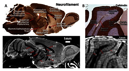
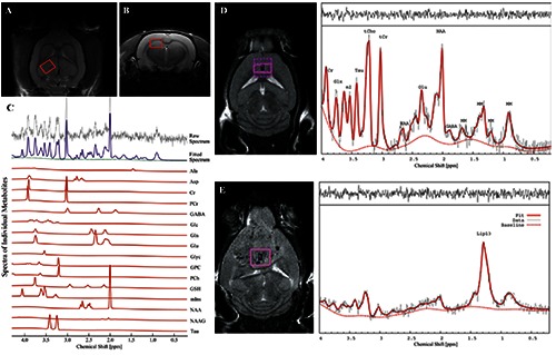
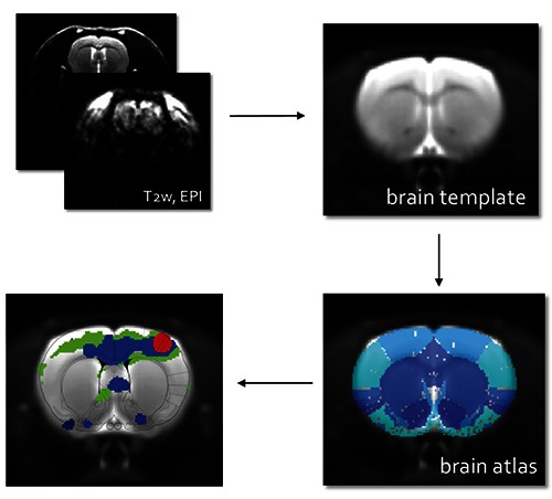
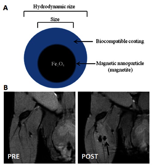
In vivo imaging techniques can be integrated with classical histochemistry to create an actual histochemistry of water. In particular, Magnetic Resonance Imaging (MRI), an imaging technique primarily used as diagnostic tool in clinical/preclinical research, has excellent anatomical resolution, unlimited penetration depth and intrinsic soft tissue contrast. Thanks to the technological development, MRI is not only capable to provide morphological information but also and more interestingly functional, biophysical and molecular. In this paper we describe the main features of several advanced imaging techniques, such as MRI microscopy, Magnetic Resonance Spectroscopy, functional MRI, Diffusion Tensor Imaging and MRI with contrast agent as a useful support to classical histochemistry.


Conflict of interest statement

The authors declare that they have no conflict of interest.

Figures

Figure 1.
Figure 1.
A) Sagittal sections of MRI microscopy and a similar neurofilament histology section in the same brain, showing visible white-matter anatomy. B) Detail through the cerebellum showing the correspondence of structures between histology and MRI microscopy. Adapted from Cleary et al., Neuroimage 2011;56:974-83 with permission.
Figure 2.
Figure 2.
In vivo T2-weighted MR images A) coronal and B) axial showing regions of interest (ROI) for 1H-MRS experiments. Here, the ROI is centered in the right dorsal hippocampal region of the rat brain. C) Quantification of neuro-metabolite signals from in vivo 1H-MRS in the right dorsal hippocampal region of the Stress-induced Sleep Perturbation (SSP) model. D) 1H-MRS voxel location on a T2-weighted MRI and the corresponding 1H-MRS spectra from a control mouse and E) from a mouse with large a glioblastoma. 1HMRS spectra from glioblastoma are characterized by increased lipid signals. tCr, total creatine; Glx, glutamate + glutamine; Ins, myo-Inositol; Tau, Taurine; tCho, total choline; NAA, N-acetyl-aspartate; Glu, glutamate; MM, macromolecules; Lac, Lactate; GABA, gamma-aminobutyric acid. A, B and C panels were adapted from Lee et al., PLoS One 2016;11:e0153346 with permission; D and E panels were adapted from Park et al., PLoS One 2014;9:e94755 with permission.
Figure 3.
Figure 3.
Procedures to analyze fMRI data: acquisition of Echo Planar Images (EPI), registration of EPI to a common MRI brain template, overlapping to brain atlas to correctly label different brain region and addition of the activation maps; the final result is shown in the last panel.
Figure 4.
Figure 4.
A) Basic schematic diagram of a magnetic nanoparticle’s structure; a magnetic nanoparticle consists of magnetic core and biocompatible coating. B) In vivo MR Images of USPIO-labeled stem cells (2.5x10³) PRE and POST intra-muscle injection.

References

    1. Medhi B, Misra S, Avti PK, Kumar P, Kumar H, Singh B. Role of neuroimaging in drug development. Rev Neurosci 2014;25:663-73. - PubMed
    1. Garcia-Figueiras R, Baleato-Gonzalez S, Padhani AR, Oleaga L, Vilanova JC, Luna A, et al. Proton magnetic resonance spectroscopy in oncology: the fingerprints of cancer? Diagn Interv Radiol 2016;22:75-89. - PMC - PubMed
    1. Mansfield P. MRI imaging in biomedicine. Advances in magnetic resonance. Academic Press, New York: 1982.
    1. Callaghan PC. Principles of nuclear magnetic resonance microscopy. Clarendon Press, Oxford: 1993.
    1. Blackband SJ, Buckley DL, Bui JD, Phillips MI. NMR microscopy—beginnings and new directions. Magn Reson Mater Phy 1999;9:112-6. - PubMed