. 2017 Jan 11;18(1):4.
doi: 10.1186/s12865-016-0186-4.

Antigen presenting capacity of murine splenic myeloid cells

Affiliations

Antigen presenting capacity of murine splenic myeloid cells

Ying-Ying Hey et al. BMC Immunol. .

Abstract

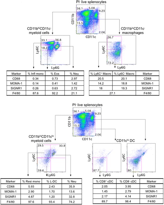
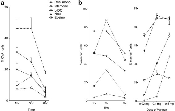
Background: The spleen is an important site for hematopoiesis. It supports development of myeloid cells from bone marrow-derived precursors entering from blood. Myeloid subsets in spleen are not well characterised although dendritic cell (DC) subsets are clearly defined in terms of phenotype, development and functional role. Recently a novel dendritic-like cell type in spleen named 'L-DC' was distinguished from other known dendritic and myeloid cells by its distinct phenotype and developmental origin. That study also redefined splenic eosinophils as well as resident and inflammatory monocytes in spleen.

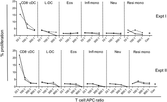
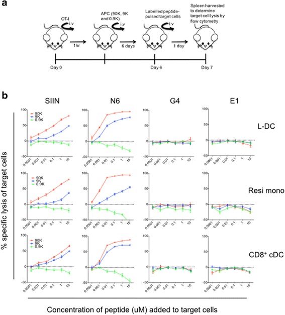
Results: L-DC are shown to be distinct from known splenic macrophages and monocyte subsets. Using a new flow cytometric procedure, it has been possible to identify and isolate L-DC in order to assess their functional competence and ability to activate T cells both in vivo and in vitro. L-DC are readily accessible to antigen given intravenously through receptor-mediated endocytosis. They are also capable of CD8+ T cell activation through antigen cross presentation, with subsequent induction of cytotoxic effector T cells. L-DC are MHCII- cells and unable to activate CD4+ T cells, a property which clearly distinguishes them from conventional DC. The myeloid subsets of resident monocytes, inflammatory monocytes, neutrophils and eosinophils, were found to have varying capacities to take up antigen, but were uniformly unable to activate either CD4+ T cells or CD8+ T cells.

Conclusion: The results presented here demonstrate that L-DC in spleen are distinct from other myeloid cells in that they can process antigen for CD8+ T cell activation and induction of cytotoxic effector function, while both L-DC and myeloid subsets remain unable to activate CD4+ T cells. The L-DC subset in spleen is therefore distinct as an antigen presenting cell.

Keywords: Antigen presentation/processing; Dendritic cells; Myeloid cells; Spleen.


Figures

Fig. 1
Fig. 1
Expression of macrophage specific markers. Splenocytes were clear of red blood cells by lysis and enriched for myeloid and DC subsets via T and B cell depletion. Cells were then stained with fluorochrome-conjugated antibodies specific for CD11b (PE-Cy7), CD11c (APC), Ly6C (FITC), Ly6G (PE), along with biotinylated antibodies to CD68, MOMA-1, SIGNR1 and F4/80. APC-Cy7-streptavidin was used as a secondary conjugate. L-DC, dendritic and myeloid subsets were gated as described in Table 1 and Hey et al., (2016) [10]. a Expression of CD68, MOMA-1, SIGNR1 and F4/80 on inflammatory monocytes (Infl mono), eosinophils (Eos), neutrophils (Neu) and macrophages (Macro). b Expression of CD68, MOMA-1, SIGNR1 and F4/80 on resident monocytes (Resi mono), L-DC and cDC subsets. Data are reflective of three independent analyses
Fig. 2
Fig. 2
Comparison of endocytic ability of myeloid and dendritic subsets. The ability of cells to endocytose antigen was measured by uptake of OVA-FITC and mannan-FITC. Spleens were collected for analysis at the same time, and splenocytes prepared by lysis of red blood cells with enrichment for dendritic and myeloid cells via T and B cell depletion. Cells were stained with antibodies to identify L-DC and myeloid subsets as shown in Table 1. Uptake of antigen was assessed in terms of % FITC staining cells. C57BL/6 J mice were given: a OVA-FITC at 1, 3, and 6 h prior to euthanasia for spleen collection (intravenously; 1 mg per mouse). Data reflect mean ± SE (n = 4); b mannan-FITC (intravenously; 0.5 mg per mouse) at 1, 3 and 6 h prior to euthanasia for spleen collection. Single mice only were analysed tin a pilot study to determibe optimal time of 3 h used in a subsequent dose response experiment. Data reflect mean ± SE (n = 2). Control mice were given PBS
Fig. 3
Fig. 3
Activation of CD4+ T cells by splenic dendritic and myeloid subsets. Antigen presenting ability of myeloid subsets purified from spleens of Act-mOVA mice was assessed. L-DC, eosinophils (Eos), inflammatory monocytes (Infl mono), neutrophils (Neu), resident monocytes (Resi mono) and CD8 cDC (as a control), were sorted as described in Table 1 following enrichment of splenocytes by depletion of red blood cells and T and B lymphocytes using magnetic bead technology. Diluting numbers of APC were plated following treatment with LPS (10 μg/ml) (solid line) and without LPS (dotted line) for 2 h. This was followed by addition of 105 CFSE-labelled OT-II (TCR-tg) CD4+ T cells purified from mouse spleen through depletion of B cells, CD8+ T cells, DC and myeloid cells using magnetic bead protocols. Cells were cultured at T cell:APC ratios of 33:1, 100:1, 300:1 and 900:1 for 72 h. CD4+ OT-II T cells were then gated as PIThy1.2+Vα2+CD8 cells, and assessed flow cytometrically for CFSE dilution as an indicator of T cell proliferation. OT-II T cells cultured alone served as controls (con). Graphs show % proliferating OT-II cells. Two independent replicate experiments were conducted
Fig. 4
Fig. 4
Ability of splenic APC to induce a cytotoxic T cell response. The ability of APC subsets to induce cytotoxic effector function in CD8+ T cells was assessed by measuring lysis of OVA peptide-pulsed target cells in a fluorescent target assay (FTA). a The experimental procedure is shown as a timeline. On Day 0, CD8+ T cells from OT-I TCR-Tg mice were prepared by red blood cell lysis of splenocytes and sorting for PIThy1.2+Vα2+CD4 cells. OT-I T cells (3.5 × 106) were delivered intravenously into host mice (C57BL/6). An hour later, several APC subsets sorted from Act-mOVA mice were also delivered into host mice. These were sorted as described in Table 1 and three cell doses (90 K, 9 K or 0.9 K) given intravenously. In order to measure the effector function of activated CD8+ T cells after 7 days, B6. SJL splenocytes were prepared as targets and adoptively transferred intravenously on Day 6. Target cells were labelled with several concentrations of CFSE, CTV and CPD for later identification. Overall, labelled target cells were then pulsed with 6 different concentrations of 4 distinct OVA peptides: SIINFEKL (SIIN), GLEQLESIINFEKL (N6), SIIGFEKL (G4) and EIINFEKL (E1). Specific killing of the distinctly labelled, antigen-pulsed target cells was determined by flow cytometric analysis to determine the number of target cells remaining in the test mouse compared with the control mouse given OT-I T cells only. Calculation of target lysis involved the formula described in Materials and Methods. b Data shows % specific lysis of target cells pulsed with different concentrations of peptides by OT-I T cells primed with three different APC types. Data is expressed as mean ± SE (n = 6)

References

    1. Cesta MF. Normal structure, function, and histology of the spleen. Toxicol Pathol. 2006;34(5):455–65. doi: 10.1080/01926230600867743. - DOI - PubMed
    1. Kurotaki D, Uede T, Tamura T. Functions and development of red pulp macrophages. Microbiol Immunol. 2015;59(2):55–62. doi: 10.1111/1348-0421.12228. - DOI - PubMed
    1. Nolte MA, Hoen ENM T, Van Stijn A, Kraal G, Mebius RE. Isolation of the intact white pulp. Quantitative and qualitative analysis of the cellular composition of the splenic compartments. Eur J Immunol. 2000;30(2):626–34. doi: 10.1002/1521-4141(200002)30:2<626::AID-IMMU626>3.0.CO;2-H. - DOI - PubMed
    1. Akashi K, Traver D, Miyamoto T, Weissman IL. A clonogenic common myeloid progenitor that gives rise to all myeloid lineages. Nature. 2000;404(6774):193–7. doi: 10.1038/35004599. - DOI - PubMed
    1. Yona S, Kim KW, Wolf Y, Mildner A, Varol D, Breker M, Strauss-Ayali D, Viukov S, Guilliams M, Misharin A, et al. Fate mapping reveals origins and dynamics of monocytes and tissue macrophages under homeostasis. Immunity. 2013;38:79–91. doi: 10.1016/j.immuni.2012.12.001. - DOI - PMC - PubMed

Publication types