. 2017 Jan 5;18(1):1.
doi: 10.1186/s13059-016-1139-1.

Distinct 5-methylcytosine profiles in poly(A) RNA from mouse embryonic stem cells and brain

Affiliations

Distinct 5-methylcytosine profiles in poly(A) RNA from mouse embryonic stem cells and brain

Thomas Amort et al. Genome Biol. .

Abstract

Background: Recent work has identified and mapped a range of posttranscriptional modifications in mRNA, including methylation of the N6 and N1 positions in adenine, pseudouridylation, and methylation of carbon 5 in cytosine (m5C). However, knowledge about the prevalence and transcriptome-wide distribution of m5C is still extremely limited; thus, studies in different cell types, tissues, and organisms are needed to gain insight into possible functions of this modification and implications for other regulatory processes.

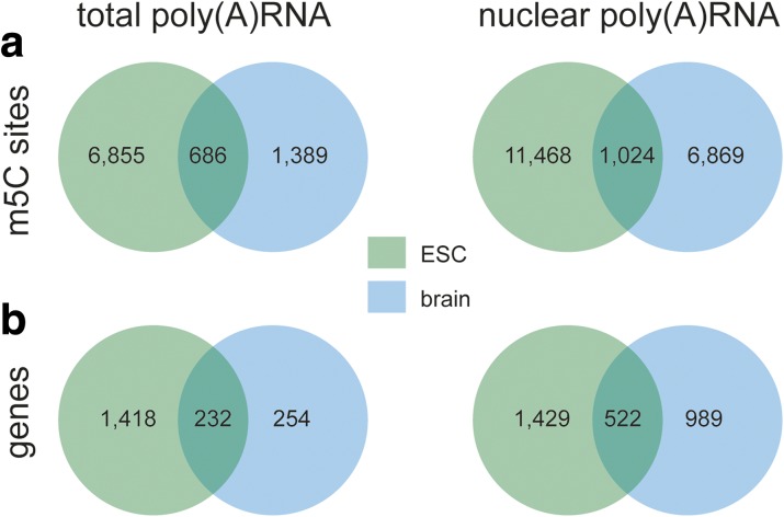
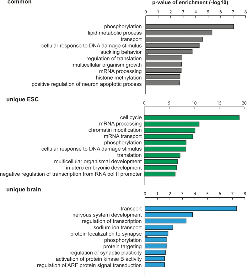
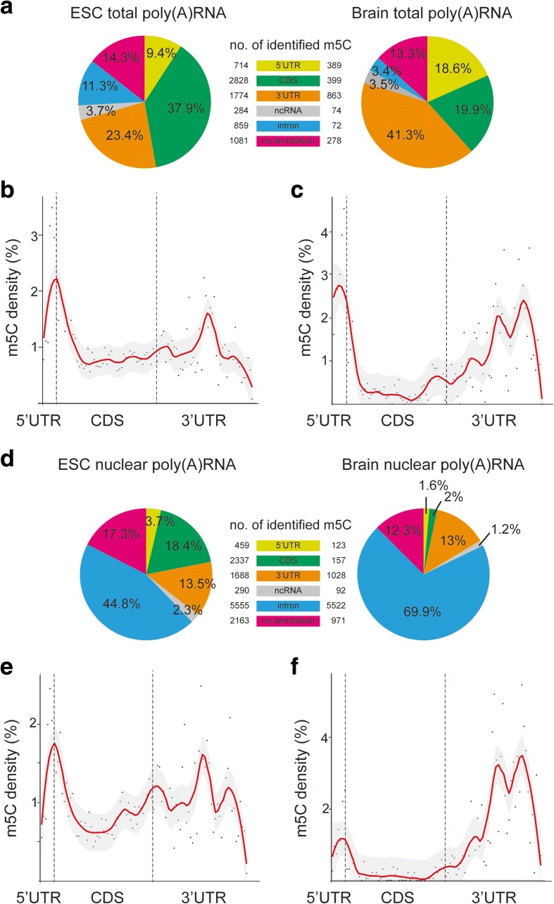
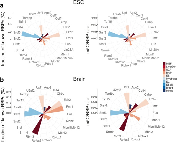
Results: We have carried out an unbiased global analysis of m5C in total and nuclear poly(A) RNA of mouse embryonic stem cells and murine brain. We show that there are intriguing differences in these samples and cell compartments with respect to the degree of methylation, functional classification of methylated transcripts, and position bias within the transcript. Specifically, we observe a pronounced accumulation of m5C sites in the vicinity of the translational start codon, depletion in coding sequences, and mixed patterns of enrichment in the 3' UTR. Degree and pattern of methylation distinguish transcripts modified in both embryonic stem cells and brain from those methylated in either one of the samples. We also analyze potential correlations between m5C and micro RNA target sites, binding sites of RNA binding proteins, and N6-methyladenosine.

Conclusion: Our study presents the first comprehensive picture of cytosine methylation in the epitranscriptome of pluripotent and differentiated stages in the mouse. These data provide an invaluable resource for future studies of function and biological significance of m5C in mRNA in mammals.

Keywords: 5-Methylcytosine; Bisulfite sequencing; Embryonic stem cells; Epitranscriptome; Mouse brain; RNA binding proteins; RNA methylation; m5C; m6A; meRIP.


Figures

Fig. 1
Fig. 1
BS-seq of total and nuclear poly(A) RNA samples from ESCs and brain reveals shared and sample-specific methylation sites. a Venn diagrams of methylation sites identified in total poly(A) RNA (left) or nuclear poly(A) RNA (right) from mouse ESC and brain. b Venn diagrams of number of genes to which identified m5Cs were mapped
Fig. 2
Fig. 2
Verification of candidate methylated transcripts by meRIP. a Graphical depiction of the meRIP approach. RNA was extracted from cells, chemically fragmented, incubated with an anti-5-methylcytosine antibody or IgG, and antigen-antibody complexes were captured with protein A beads. Specific candidate RNAs (blue bars in b) were analyzed by qPCR of immunoprecipitated material, and enrichment relative to the IgG control (black bar in b) was calculated. b MeRIP shows significant enrichment of 13 out of 16 candidate transcripts. The Tbp transcript (white bar) served as a negative control, since it was not detected in our m5C dataset. Data are shown as mean ± standard error of the mean (SEM) of three independent experiments. Statistical significance was determined by unpaired t test, significance threshold p < 0.05 (*)
Fig. 3
Fig. 3
The majority of uniquely methylated cytosines in ESC total poly(A) RNA are due to differential methylation rather than differential expression between ESC and brain. a The expression levels and methylation rates of m5Cs identified as unique to ESCs were analyzed in the brain samples. b The expression levels and methylation rates of m5Cs identified as unique to brain were analyzed in the ESC samples. Multi-level pie charts display the numbers of sites on annotated and non-annotated transcripts in the innermost ring, the numbers of sites on transcripts with a mean normalized count of more (dark green) or fewer (light green) than 10 reads in the middle ring, and the numbers of sites with sequence coverage <10 reads (blue) or sequence coverage >10 reads but methylation rate lower than 0.2 (yellow) in the outer ring. Positions in which the mean values for coverage and non-conversion were skewed towards methylation by an individual replicate were classified as biased mean
Fig. 4
Fig. 4
GO term enrichment analysis reveals distinct predominance of different gene categories in transcripts methylated in both ESCs and brain (common) versus transcripts methylated uniquely in one of the samples (unique). GO terms were analyzed with DAVID and further clustered using REVIGO. The ten most significantly enriched categories are shown
Fig. 5
Fig. 5
Methylated cytosines are preferentially located around the translational start codon of mRNAs. a The percentages of m5Cs detected in ESC (left) or brain (right) total poly(A) RNA mapping to the indicated transcript classes are shown. b Meta-gene profiles of all m5C locations detected in total poly(A) RNA of ESCs along the rescaled segments 5′ UTR, coding sequence (CDS), and 3′ UTR of a normalized mRNA are shown and indicate a peak of m5C at the translational start codon. Red line represents the loess smoothed conditional mean and gray areas the 0.95 confidence interval. Dashed lines separate the different mRNA segments at the translational start and stop codons. c Same as in b for brain total poly(A) RNA. d Pie chart of the percentages of m5Cs detected in the indicated transcript classes in ESC (left) or brain (right) nuclear poly(A) RNA. e, f Meta-gene analysis as in b reveals accumulation of m5C sites around the start codon in ESC (e) and brain (f) nuclear poly(A) RNA as well as in the 3′ UTR of brain nuclear RNA transcripts (f)
Fig. 6
Fig. 6
Radar plots show an overlap of m5C sites with binding sites of several RNA binding proteins (RBPs) available in the CLIPdb. a Left panel, fraction of binding sites overlapping with an m5C site for each particular RBP. Right panel, number of m5Cs overlapping with binding sites for a particular protein was normalized against the total number of binding sites of the respective RBP. Cell/tissue types in which the RBP binding sites had been detected are color coded and explained in the legend (MEF mouse embryonic fibroblasts, Liver36h liver partial hepatectomy 36 h, N2A Neuro2a, ES embryonic stem cells, EC embryonal carcinoma, ESdN ES-derived neuronal). b Same as in a for brain total and nuclear poly(A) RNA

Similar articles

Cited by

References

    1. Machnicka MA, Olchowik A, Grosjean H, Bujnicki JM. Distribution and frequencies of post-transcriptional modifications in tRNAs. RNA Biol. 2014;11:1619–29. doi: 10.4161/15476286.2014.992273. - DOI - PMC - PubMed
    1. Motorin Y, Helm M. RNA nucleotide methylation. Wiley Interdiscip Rev RNA. 2011;2:611–31. doi: 10.1002/wrna.79. - DOI - PubMed
    1. Sibbritt T, Patel HR, Preiss T. Mapping and significance of the mRNA methylome. Wiley Interdiscip Rev RNA. 2013;4:397–422. doi: 10.1002/wrna.1166. - DOI - PubMed
    1. Meyer KD, Jaffrey SR. The dynamic epitranscriptome: N6-methyladenosine and gene expression control. Nat Rev Mol Cell Biol. 2014;15:313–26. doi: 10.1038/nrm3785. - DOI - PMC - PubMed
    1. He C. Grand challenge commentary: RNA epigenetics? Nat Chem Biol. 2010;6:863–5. doi: 10.1038/nchembio.482. - DOI - PubMed

Publication types