Review
. 2017 Feb:312:1-14.
doi: 10.1016/j.cellimm.2016.12.003. Epub 2016 Dec 23.

Interplay of extracellular matrix and leukocytes in lung inflammation

Affiliations
Review

Interplay of extracellular matrix and leukocytes in lung inflammation

Thomas N Wight et al. Cell Immunol. 2017 Feb.

Abstract

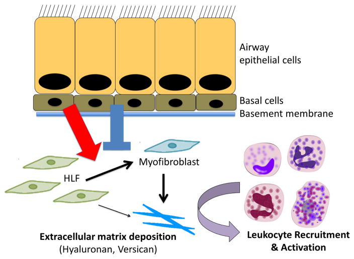
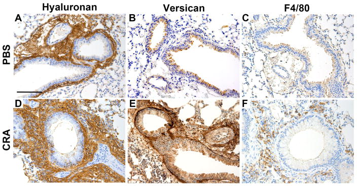
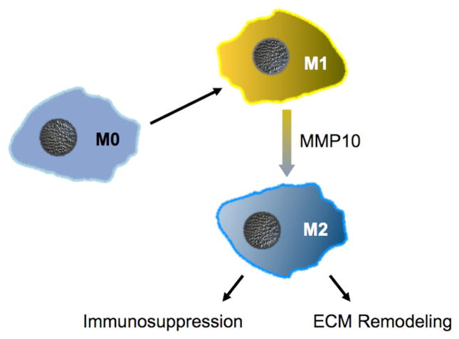
During inflammation, leukocytes influx into lung compartments and interact with extracellular matrix (ECM). Two ECM components, versican and hyaluronan, increase in a range of lung diseases. The interaction of leukocytes with these ECM components controls leukocyte retention and accumulation, proliferation, migration, differentiation, and activation as part of the inflammatory phase of lung disease. In addition, bronchial epithelial cells from asthmatic children co-cultured with human lung fibroblasts generate an ECM that is adherent for monocytes/macrophages. Macrophages are present in both early and late lung inflammation. Matrix metalloproteinase 10 (MMP10) is induced in alveolar macrophages with injury and infection and modulates macrophage phenotype and their ability to degrade collagenous ECM components. Collectively, studies outlined in this review highlight the importance of specific ECM components in the regulation of inflammatory events in lung disease. The widespread involvement of these ECM components in the pathogenesis of lung inflammation make them attractive candidates for therapeutic intervention.

Keywords: Asthma; Extracellular matrix; Fibrosis; Hyaluronan; Immunity; Inflammation; Macrophage; Matrix metalloproteinase 10; Versican.


Figures

Figure 1
Figure 1
Extravasation of leukocytes across the endothelium and/or epithelium (E) into the interstitium of the tissue during an inflammatory response. The leukocytes interact with specific ECM components, such as versican and hyaluronan, generated by resident cells of the tissue, such as endothelia and epithelia, and stromal cells, such as fibroblasts and smooth muscle cells. This interaction involves receptor-mediated interactions with hyaluronan and versican via cell surface receptors such as PSGL-1, TLR2, and CD44. These interactions in turn influence leukocyte phenotype by stimulating intracellular signals that promote their adhesion, proliferation, migration, differentiation, and activation. Furthermore, the leukocytes themselves may produce versican and hyaluronan in response to inflammatory stimuli to further enrich the matrix with these specific components. Such matrices, depending on their interactive partners, may exhibit either pro-inflammatory or anti-inflammatory properties. Figure from: Thomas N. Wight, Inkyung Kang, Mervyn J. Merrilees, Matrix Biology. 35:152–161, 2014, http://dx.doi.org/10.1016/j.matbio.2014.01.015. Reuse permitted under Creative Commons Attribution-NonCommercial-No Derivatives License (CC BY NC ND).
Figure 2
Figure 2
Versican accumulation during embryonic mouse development and in lungs of a mouse with Pseudomonas aeruginosa lung infection. (A) Versican accumulation in fetal lung tissue at E14.5 days. (B) Versican accumulation in the lung of a 16-week-old mouse treated with PBS as a vehicle control. (C) Versican accumulation from a 16-week-old mouse infected with live Pseudomonas aeruginosa for 5 days. Brown indicates positive staining for versican β-GAG; blue, hematoxylin counterstain. Br, bronchiole; Di, diaphragm; Ri, rib; PV, postcapillary Vein; TB, terminal bronchiole. Arrows indicate versican staining in the alveolar septa; * marks an area of positive staining of the alveolar septa; cells in alveolar space makes it difficult to distinguish these two anatomical compartments. (D) The amount of versican-stained lung tissue as a percentage of total lung tissue in control mice (PBS) and those exposed to live P. aeruginosa for up to 5 days. Values are the mean β SEM (n = 3–6). asignificantly different from PBS, bsignificantly different from 4 hr, csignificantly different from 24 hr. p<00001 using a one-way ANOVA with Tukey’s multiple comparison test. Scale (A–C) 100 μm; (C inset) 50 μm. Figure reused with permission from: Jessica M. Snyder, Ida M. Washington, Timothy Birkland, Mary Y. Chang, Charles W. Frevert, Journal of Histochemistry & Cytochemistry (Volume 63 Issue 12) pp. 952–967, copyright c 2015 by The Authors. Reprinted by Permission of SAGE Publications, Inc.
Figure 3
Figure 3
Working hypothesis of leukocyte/ECM interaction in asthma suggesting that ongoing injury, irritation, and/or bacterial/viral infection of the epithelial cells results in signals that promote disordered wound repair resulting in altered ECM remodeling and the formation of a versican-/hyaluronan-rich ECM that promotes leukocyte recruitment and activation. Blue arrow indicates agonists that promote ECM accumulation and red arrow indicates antagonists the prevent ECM accumulation.
Figure 4
Figure 4
Hyaluronan (A,D), versican (B,E ) and macrophage (C,F) involvement (brown color) in control- and CRA-treated mouse lungs showing increases in hyaluronan and versican content in the subepithelial region of airway bronchioles in the CRA-treated lungs. These areas were also enriched in F4/80 positive macrophages. Figure adapted with permission from: Stephen R. Reeves, Gernot Kaber, Alyssa Sheih, Georgiana Cheng, Mark A. Aronica, Mervyn J. Merrilees, Jason S. Debley, Charles W. Frevert, Steven F. Ziegler, Thomas N. Wight. Journal of Histochemistry & Cytochemistry (Volume 64, Issue 6) pp. 364–380, copyright c 2016 by The Histochemical Society. Reprinted by Permission of SAGE Publications, Inc.
Figure 5
Figure 5
MMP10 functions in a cell-autonomous manner to control the state of macrophage activation. Likely via shedding of a yet-to-be-identified surface protein, MMP10 drives the conversion of pro-inflammatory M1-biased macrophages towards immunosuppressive M2-biased cells. In addition, MMP10 controls the activation of ECM degrading activity in M2 macrophages, such as by promoting expression of MMP13, a collagenolytic proteinase.

Similar articles

Cited by

References

    1. Booth AJ, Hadley R, Cornett AM, Dreffs AA, Matthes SA, Tsui JL, Weiss K, Horowitz JC, Fiore VF, Barker TH, Moore BB, Martinez FJ, Niklason LE, White ES. Acellular normal and fibrotic human lung matrices as a culture system for in vitro investigation. Am J Respir Crit Care Med. 2012;186:866–76. - PMC - PubMed
    1. Weibel ER, Crystal RG. Structural organization of the pulmonary interstitium. In: Crystal RG, West JB, editors. The Lung: Scientific Foundations. Raven Press; New York: 1991. pp. 369–380.
    1. Gill S, Wight TN, Frevert CW. Proteoglycans: key regulators of pulmonary inflammation and the innate immune response to lung infection. Anat Rec (Hoboken) 2010;293:968–81. - PMC - PubMed
    1. Nastase MV, Iozzo RV, Schaefer L. Key roles for the small leucine-rich proteoglycans in renal and pulmonary pathophysiology. Biochim Biophys Acta. 2014;1840:2460–70. - PMC - PubMed
    1. Andersson-Sjoland A, Hallgren O, Rolandsson S, Weitoft M, Tykesson E, Larsson-Callerfelt AK, Rydell-Tormanen K, Bjermer L, Malmstrom A, Karlsson JC, Westergren-Thorsson G. Versican in inflammation and tissue remodeling: The impact on lung disorders. Glycobiology. 2014 - PMC - PubMed

Publication types