. 2017 Jun 1;312(6):F982-F991.
doi: 10.1152/ajprenal.00569.2016. Epub 2017 Jan 11.

Extracellular microRNA signature in chronic kidney disease

Affiliations

Extracellular microRNA signature in chronic kidney disease

Jagdeesan Muralidharan et al. Am J Physiol Renal Physiol. .

Abstract

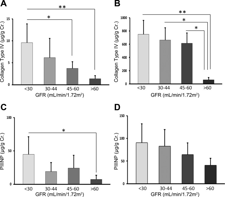
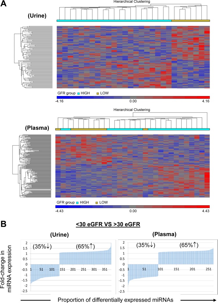
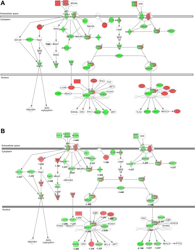
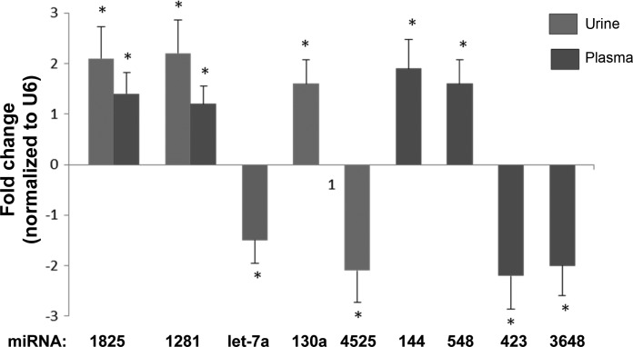
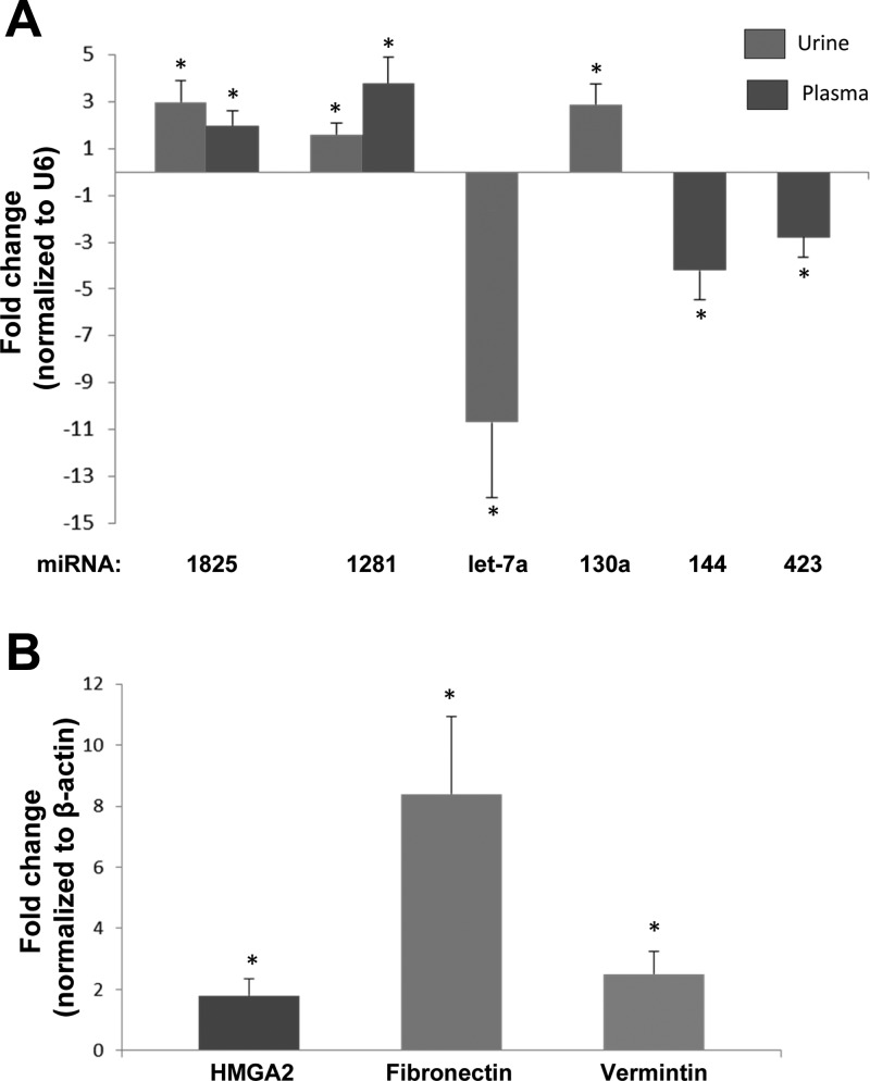
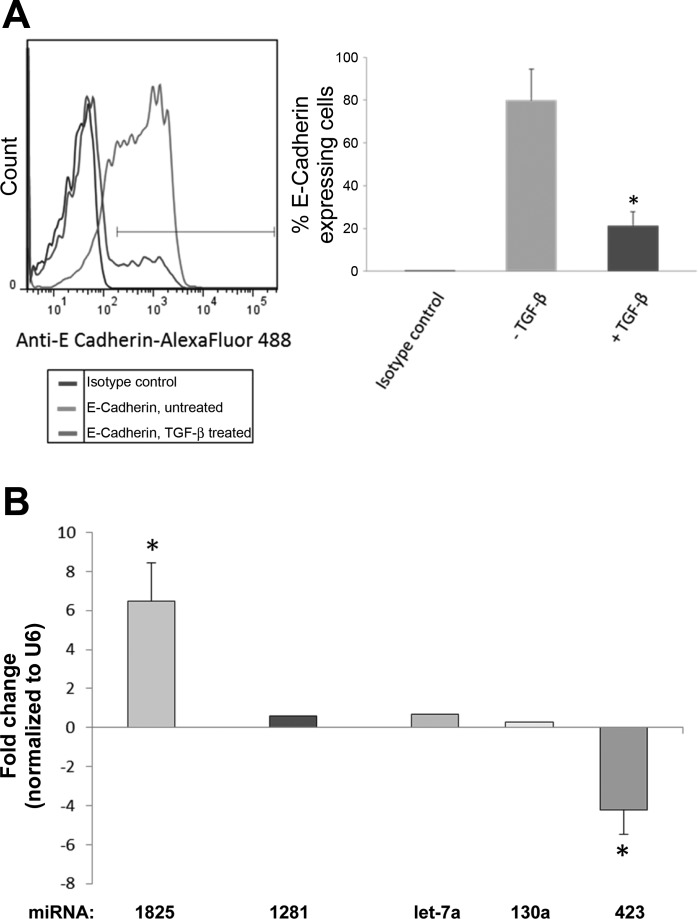
MicroRNAs (miRNAs) are noncoding RNAs that regulate posttranscriptional gene expression. In this study we characterized the circulating and urinary miRNA pattern associated with reduced glomerular filtration rate, using Affymetrix GeneChip miR 4.0 in 28 patients with chronic kidney disease (CKD). Top miRNA discoveries from the human studies were validated in an Alb/TGFβ mouse model of CKD, and in rat renal proximal tubular cells (NRK52E) exposed to TGFβ1. Plasma and urinary levels of procollagen III N-terminal propeptide and collagen IV were elevated in patients with decreased estimated glomerular filtration rate (eGFR). Expression of 384 urinary and 266 circulatory miRNAs were significantly different between CKD patients with eGFR ≥30 vs. <30 ml·min-1·1.73 m-2 Pathway analysis mapped multiple miRNAs to TGFβ signaling-related mRNA targets. Specifically, Let-7a was significantly downregulated, and miR-130a was significantly upregulated, in urine of patients with eGFR <30; miR-1825 and miR-1281 were upregulated in both urine and plasma of patients with decreased eGFR; and miR-423 was significantly downregulated in plasma of patients with decreased eGFR. miRNA expression in urine and plasma of Alb/TGFβ mice generally resembled and confirmed most, although not all, of the observations from the human studies. In response to TGFβ1 exposure, rat renal proximal tubular cells overexpressed miR-1825 and downregulated miR-423. Thus, miRNA are associated with kidney fibrosis, and specific urinary and plasma miRNA profile may have diagnostic and prognostic utility in CKD.

Keywords: TGFβ; chronic kidney disease; fibrosis.


Figures

Fig. 1.
Fig. 1.
Urine and plasma levels of collagen type IV (A and B) and procollagen III NH2-terminal propeptide (PIIINP; C and D) in chronic kidney disease (CKD) patients in relation to eGFR levels. The urinary excretion levels of type IV collagen and PIIINP are expressed as micrograms per gram of creatinine. *P < 0.05, ** P < 0.01.
Fig. 2.
Fig. 2.
Comparative analysis of miRNA expression levels in plasma and urine samples obtained from patients grouped by eGFR. A: heatmap showing unsupervised hierarchical clustering of miRNAs (Pearson correlation, average linkage). Colors represent relative miRNA expression as indicated in the color key for each panel. Brackets on the top margins indicate samples from the same cohort. Samples are in columns, miRNAs in rows. Only miRNAs that survived multiple testing (FDR), and had a fold change >3 or <−3 and P < 0.05 are shown. B: miRNAs with significantly dysregulated expression are graphed for plasma and urine samples. Percentages of up- or downregulated miRNAs are shown with corresponding up or down arrows on each graph.
Fig. 3.
Fig. 3.
Pathway analysis of miRNA discovery. The TGFβ canonical pathway was among the top ranked as determined by Ingenuity Pathways Analysis (IPA) software (Ingenuity, Redwood City, CA). Green indicates predicted downregulation of target transcripts by differentially expressed miRNAs; red indicates predicted upregulation. A, urine; B, plasma. Among the altered miRNAs, expression levels of miR-1825, miR-1281, let-7a, and miR-130a are experimentally-validated.
Fig. 4.
Fig. 4.
qRT-PCR confirmation of the microarray discoveries in CKD patients. Error bars are the SE for each analysis. Selected miRNAs were verified using single-well TaqMan qRT-PCR. The 2−ΔΔCt method was used to calculate relative changes in miRNA expression. Results were normalized to levels of U6 snRNA. Each data point represents mean ± SE. *P < 0.05.
Fig. 5.
Fig. 5.
A: qRT-PCR verification of microarray discoveries in urine and plasma samples of Alb/TGFβ mice compared with wild-type mice. Error bars are SE for each analysis. Selected miRNAs were verified using single-well TaqMan qRT-PCR. The 2−ΔΔCt method was used to calculate relative changes in miRNA expression. Results were normalized to levels of U6 snRNA. Each data point represents the mean ± SE, P < 0.05. B: qRT-PCR verification of mRNA targets of fibrosis-associated miRNAs. The 2−ΔΔCt method was used to calculate relative changes in mRNA expression. Results were normalized to levels of endogenous β-actin. Each data point represents the mean ± SE. *P < 0.05.
Fig. 6.
Fig. 6.
A: flow cytometry analysis showing expression of E-cadherin in rat renal proximal tubular cell line (NRK52E) with or without TGFβ stimulation. B: real-time PCR of fibrotic miRNAs in NRK52E cells stimulated by TGFβ. Selected miRNAs were verified using single-well TaqMan qRT-PCR. The 2−ΔΔCt method was used to calculate relative changes in miRNA expression. Results were normalized to levels of U6 snRNA. Each data point represents the mean ± SE. *P < 0.05.

References

    1. Araki S, Haneda M, Koya D, Isshiki K, Kume S, Sugimoto T, Kawai H, Nishio Y, Kashiwagi A, Uzu T, Maegawa H. Association between urinary type IV collagen level and deterioration of renal function in type 2 diabetic patients without overt proteinuria. Diabetes Care 33: 1805–1810, 2010. doi:10.2337/dc10-0199. - DOI - PMC - PubMed
    1. Butz H, Rácz K, Hunyady L, Patócs A. Crosstalk between TGFβ signaling and the microRNA machinery. Trends Pharmacol Sci 33: 382–393, 2012. doi:10.1016/j.tips.2012.04.003. - DOI - PubMed
    1. Chang CJ, Hsu CC, Chang CH, Tsai LL, Chang YC, Lu SW, Yu CH, Huang HS, Wang JJ, Tsai CH, Chou MY, Yu CC, Hu FW. Let-7d functions as novel regulator of epithelial-mesenchymal transition and chemoresistant property in oral cancer. Oncol Rep 26: 1003–1010, 2011. doi:10.3892/or.2011.1360. - DOI - PubMed
    1. Chen PY, Qin L, Barnes C, Charisse K, Yi T, Zhang X, Ali R, Medina PP, Yu J, Slack FJ, Anderson DG, Kotelianski V, Wang F, Tellides G, Simons M. FGF regulates TGFβ signaling and endothelial-to-mesenchymal transition via control of let-7 miRNA expression. Cell Reports 2: 1684–1696, 2012. doi:10.1016/j.celrep.2012.10.021. - DOI - PMC - PubMed
    1. Chung AC, Huang XR, Meng X, Lan HY. miR-192 mediates TGF-beta/Smad3-driven renal fibrosis. J Am Soc Nephrol 21: 1317–1325, 2010. doi:10.1681/ASN.2010020134. - DOI - PMC - PubMed

Publication types

MeSH terms