. 2017 Apr;16(4 suppl 1):S200-S214.
doi: 10.1074/mcp.M116.064741. Epub 2017 Jan 11.

Human Antiviral Protein IFIX Suppresses Viral Gene Expression during Herpes Simplex Virus 1 (HSV-1) Infection and Is Counteracted by Virus-induced Proteasomal Degradation

Affiliations

Human Antiviral Protein IFIX Suppresses Viral Gene Expression during Herpes Simplex Virus 1 (HSV-1) Infection and Is Counteracted by Virus-induced Proteasomal Degradation

Marni S Crow et al. Mol Cell Proteomics. 2017 Apr.

Abstract

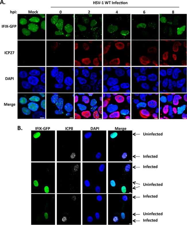
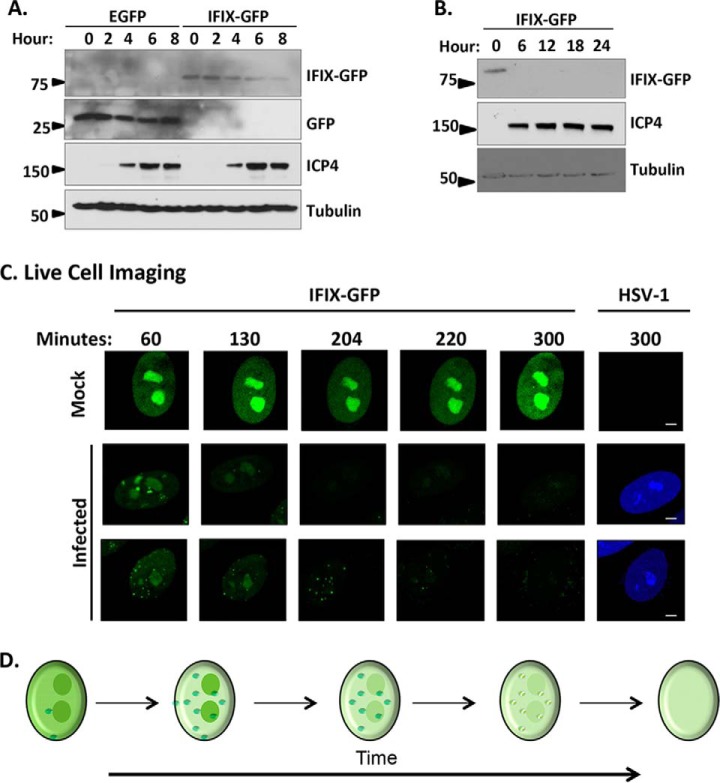
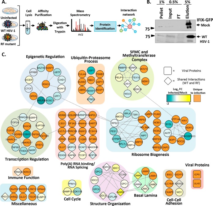
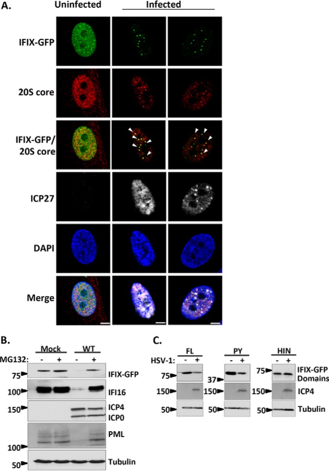
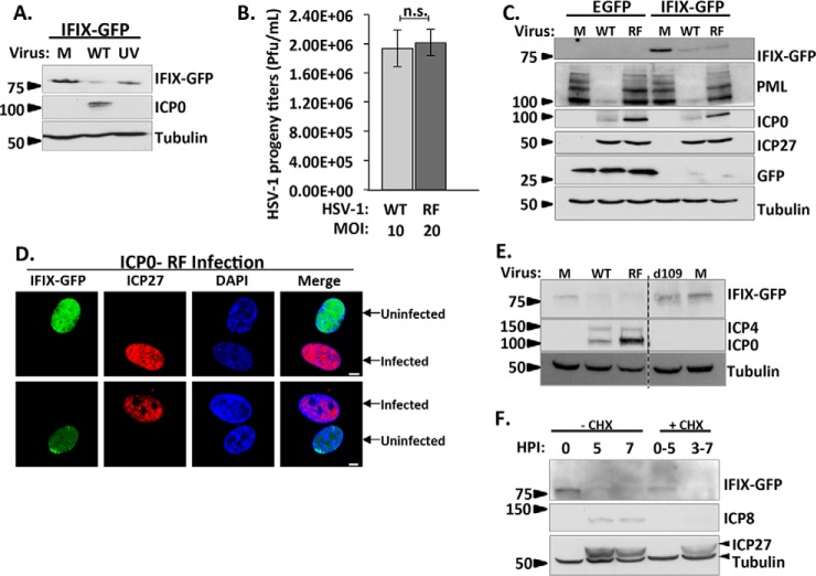
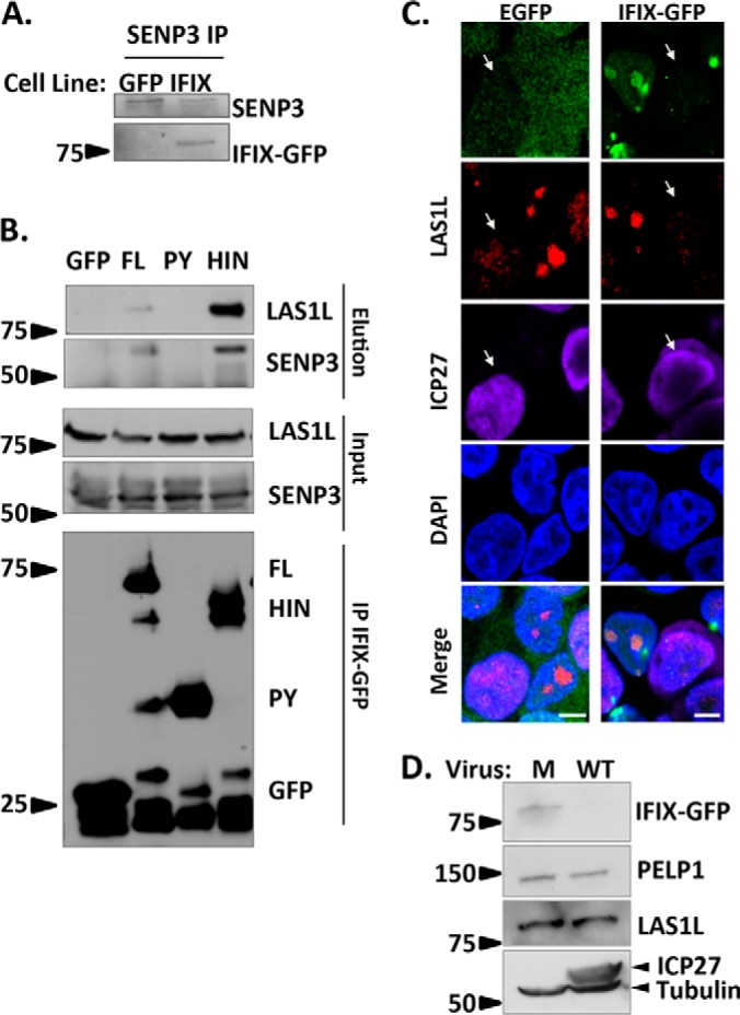
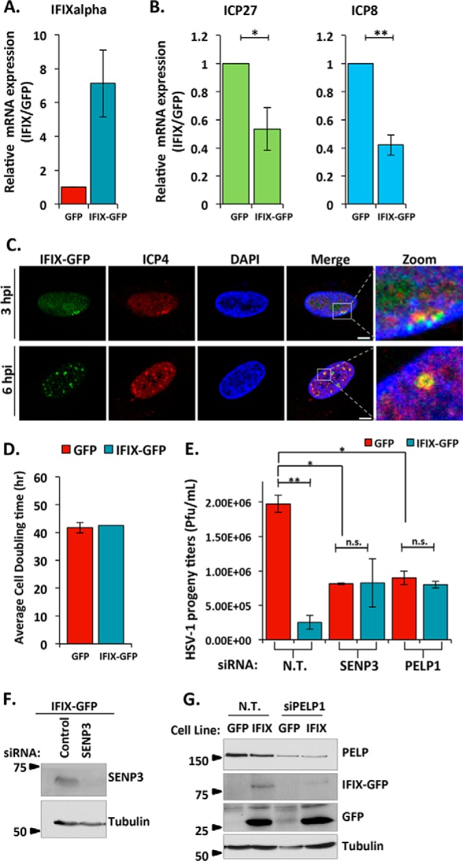
The interferon-inducible protein X (IFIX), a member of the PYHIN family, was recently recognized as an antiviral factor against infection with herpes simplex virus 1 (HSV-1). IFIX binds viral DNA upon infection and promotes expression of antiviral cytokines. How IFIX exerts its host defense functions and whether it is inhibited by the virus remain unknown. Here, we integrated live cell microscopy, proteomics, IFIX domain characterization, and molecular virology to investigate IFIX regulation and antiviral functions during HSV-1 infection. We find that IFIX has a dynamic localization during infection that changes from diffuse nuclear and nucleoli distribution in uninfected cells to discrete nuclear puncta early in infection. This is rapidly followed by a reduction in IFIX protein levels. Indeed, using immunoaffinity purification and mass spectrometry, we define IFIX interactions during HSV-1 infection, finding an association with a proteasome subunit and proteins involved in ubiquitin-proteasome processes. Using synchronized HSV-1 infection, microscopy, and proteasome-inhibition experiments, we demonstrate that IFIX co-localizes with nuclear proteasome puncta shortly after 3 h of infection and that its pyrin domain is rapidly degraded in a proteasome-dependent manner. We further demonstrate that, in contrast to several other host defense factors, IFIX degradation is not dependent on the E3 ubiquitin ligase activity of the viral protein ICP0. However, we show IFIX degradation requires immediate-early viral gene expression, suggesting a viral host suppression mechanism. The IFIX interactome also demonstrated its association with transcriptional regulatory proteins, including the 5FMC complex. We validate this interaction using microscopy and reciprocal isolations and determine it is mediated by the IFIX HIN domain. Finally, we show IFIX suppresses immediate-early and early viral gene expression during infection. Altogether, our study demonstrates that IFIX antiviral functions work in part via viral transcriptional suppression and that HSV-1 has acquired mechanisms to block its functions via proteasome-dependent degradation.


Conflict of interest statement

The authors declare that they have no conflicts of interest with the contents of this article

Figures

Fig. 1.
Fig. 1.
IFIX displays altered nuclear localization and protein levels during infection with WT HSV-1. A, time course showing IFIX-GFP localization in Flp-In 293s at the indicated hpi. m.o.i.: 5, ICP27 is marker of infection. B, IFIX-GFP localization upon infection of primary HFFs stably expressing IFIX-GFP. m.o.i.: 3 was taken at 4 hpi; ICP8 is marker of infection. A and B, bar, 5 μm.
Fig. 2.
Fig. 2.
Protein levels of IFIX-GFP decrease by 4 h post-infection and remain diminished over the course of HSV-1 infection in primary fibroblasts. A, time course of EGFP or IFIX-GFP HFF cells during infection with BFP-HSV-1 analyzed by Western blotting. The blot reveals a decrease in IFIX-GFP levels over time that is not due to GFP being targeted or cleaved. B, late times of WT HSV-1 infection in IFIX-GFP stable HFFs analyzed by Western blotting showing the lack of IFIX-GFP signal late in infection. C, still images extracted from live cell microscopy movies, taken with ×60 objective, monitoring IFIX-GFP in uninfected (mock) and BFP-tagged HSV-1 infected stable HFF cells (see also supplemental Movie S1). Bar, 5 μm. D, schematic of the changes in IFIX sub-nuclear localization and levels during HSV-1 infection. Small green dots represent diffuse IFIX recruitment to distinct puncta that are dispersed over time.
Fig. 3.
Fig. 3.
IFIX protein interactions early in HSV-1 infection. A, experimental workflow. B, immunoblot showing the efficiency of IFIX isolations in uninfected and WT infected cells. FT, flow-through. C, IFIX interactions during HSV-1 infection. Diamond-shaped nodes indicate proteins found to be specific in both WT and RF infections. The blue-to-yellow gradient depicts the fold change of WT infected/mock relative quantification of spectral counts. Orange nodes represent proteins found to be unique in infected cells. Proteins are grouped according to their GO annotations assigned by ClueGO.
Fig. 4.
Fig. 4.
IFIX is targeted for proteasome-dependent degradation during infection with HSV-1. A, direct- and immunofluorescence microscopy during WT HSV-1 infection displaying IFIX-GFP puncta localizing to nuclear proteasomes. The 20S core subunit of the proteasome is in the red channel, and ICP27 as a marker for infection is in white. m.o.i.: 10, 3.5 hpi. Bar, 5 μm. B, Western blot of IFIX-GFP stable HFFs during WT HSV-1 infection with and without proteasome inhibitor MG132 treatment. ICP4 and ICP0 are markers for infection. IFI16 and PML are positive controls. IFIX levels decrease during infection but are rescued by MG132 treatment. m.o.i.: 10, 6 hpi. C, Western blot of 293Ts transfected with IFIX-GFP domain constructs reveals the pyrin domain of IFIX is the target for degradation. FL, full length; PY, pyrin domain; HIN, HIN200 domain. ICP4 is marker for infection. m.o.i.: 10, 6 hpi. See supplemental Fig. S1 for exposures of the intact Western blot membranes.
Fig. 5.
Fig. 5.
Decrease in IFIX levels requires viral gene expression, yet is not dependent on the E3 ligase activity of the viral protein ICP0. A, UV-induced inactivation of viral genes rescues IFIX protein levels. Cells were mock-infected (M) or infected with WT or UV-treated HSV-1 at m.o.i. 10, harvested at 6 hpi, and analyzed by Western blotting. B, testing equivalence of infection progression of wild type HSV-1 and ICP0 RING finger mutant (RF) HSV-1 by comparing HSV-1 progeny titers in HFF cells stably expressing EGFP. Cells were infected with WT at m.o.i. 10 or RF at m.o.i. 20. Cells and supernatants were collected at 22 hpi (95% cytopathic effect) and titered on U2OS cells. n.s., not significant. Error bars represent S.D. of two biological replicates run in technical duplicates. C, Western blot in HFFs stably expressing EGFP or IFIX-GFP comparing the indicated proteins during mock, WT (m.o.i. 10), or RF (m.o.i. 20) infection was at 6 hpi. IFIX levels decrease during infection with WT and RF viruses. PML is blotted to show ICP0 ligase activity is inhibited in the RF virus. ICP0 is marker for infection; ICP27 displays equal levels in both viruses. D, microscopy during RF infection displays decrease in IFIX-GFP in primary fibroblasts stably expressing IFIX-GFP. m.o.i.: 3, 6hpi; ICP27 is marker of infection. Bar, 5 μm. E, Western blot in HFFs stably expressing IFIX-GFP demonstrate IFIX levels do not decrease during d109 virus infection. Mock (M), WT (m.o.i. 10), RF (m.o.i. 20), or d109 (m.o.i. 20) infection was at 6 hpi. ICP4 and ICP0 are markers for infection and a validation for d109, which lacks all immediate-early genes. F, time course analyzed by Western blotting comparing IFIX-GFP levels in fibroblasts treated with cycloheximide (CHX) at 10 μg/ml to inhibit immediate-early viral protein expression (0–5 hpi) or delayed-early viral protein expression (3–7 hpi) and IFIX-GFP levels in fibroblasts not treated with CHX during WT HSV-1 infection.
Fig. 6.
Fig. 6.
HIN domain of IFIX mediates its interaction with the 5FMC complex. A, validating IFIX co-interaction with SENP3 by reciprocal IP. B, IFIX interacts with 5FMC components LAS1L and SENP3 through its HIN domain. Forward IPs using GFP antibody were performed in cells transfected with IFIX constructs full-length (FL), IFIX Pyrin domain (PY), and IFIX HIN200 domain (HIN) in pEGFP. Inputs (1.5%), elutions (20%), and isolated IFIX constructs (IP, 20%) were blotted for LAS1L, SENP3, and GFP. C, IF microscopy in IFIX-GFP and EGFP control 293 cells showing a redistribution of 5FMC protein LAS1L in infected cells (white arrows), m.o.i.: 5, 4 hpi. Co-localization of IFIX and LAS1L is pronounced in uninfected cells (for PELP1, see supplemental Fig. S3). D, Levels of PELP1 and LAS1L are not reduced during HSV-1 infection. PELP1 and LAS1L levels were monitored by western blotting at 6hpi. ICP27 is marker for infection. M, mock. Microscopy images were taken at ×60 oil objective. Bar, 5 μm.
Fig. 7.
Fig. 7.
IFIX inhibits viral gene transcription. A, confirmation of IFIX is overexpressed in stable HFFs. B, relative mRNA levels of viral genes ICP27 and ICP8 determined by quantitative PCR. Cells were infected with WT HSV-1 at an m.o.i. of 10 and collected at 6 hpi. Error bars represent S.D. of three biological replicates. Significance was determined by t test. *, p ≤ 0.05; **, p ≤ 0.005. C, IFIX-GFP localizes to viral genomes when its degradation is inhibited. Cells were treated with MG132 to block the proteasome during the course of 3- or 6-h infections. ICP4 is marker for viral genomes. Size bars, 5 μm. D, doubling times of HFFs stably expressing EGFP or IFIX-GFP. E, siRNA-mediated knockdown of 5FMC components PELP1 and SENP3 impact HSV-1 progeny titers in human fibroblasts. EGFP or IFIX-GFP HFFs were transfected with the indicated siRNAs. Cells were infected with WT HSV-1 at an m.o.i. of 10 at 24 h post-transfection. N.T., non-targeted. Error bars indicate S.D. of two biological replicates in technical duplicate; significance was determined by t test. *, p ≤ 0.05; **, p ≤ 0.005. n.s., not significant. F and G, validation of knockdown efficiency of PELP1 and SENP3 by Western blotting.

References

    1. Brubaker S. W., Bonham K. S., Zanoni I., and Kagan J. C. (2015) Innate immune pattern recognition: a cell biological perspective. Annu. Rev. Immunol. 33, 257–290 - PMC - PubMed
    1. Diner B. A., Li T., Greco T. M., Crow M. S., Fuesler J. A., Wang J., and Cristea I. M. (2015) The functional interactome of PYHIN immune regulators reveals IFIX is a sensor of viral DNA. Mol. Syst. Biol. 11, 787. - PMC - PubMed
    1. Fairbrother W. J., Gordon N. C., Humke E. W., O'Rourke K. M., Starovasnik M. A., Yin J.-P., and Dixit V. M. (2001) The PYRIN domain: a member of the death domain-fold superfamily. Protein Sci. 10, 1911–1918 - PMC - PubMed
    1. Jin T., Perry A., Smith P., Jiang J., and Xiao T. S. (2013) Structure of the absent in melanoma 2 (AIM2) pyrin domain provides insights into the mechanisms of AIM2 autoinhibition and inflammasome assembly. J. Biol. Chem. 288, 13225–13235 - PMC - PubMed
    1. Li T., Chen J., and Cristea I. M. (2013) Human cytomegalovirus tegument protein pUL83 inhibits IFI16-mediated DNA sensing for immune evasion. Cell Host Microbe 14, 591–599 - PMC - PubMed

Publication types

MeSH terms

LinkOut - more resources