. 2017 Feb;216(2):393-408.
doi: 10.1083/jcb.201606126. Epub 2017 Jan 11.

Regulating chromosomal movement by the cochaperone FKB-6 ensures timely pairing and synapsis

Affiliations

Regulating chromosomal movement by the cochaperone FKB-6 ensures timely pairing and synapsis

Benjamin Alleva et al. J Cell Biol. 2017 Feb.

Abstract

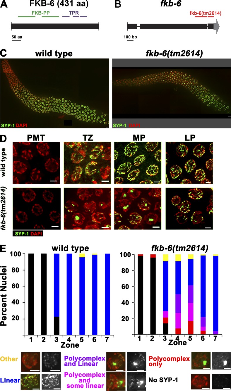
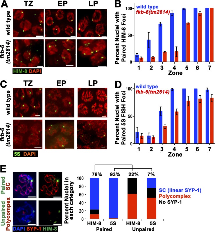
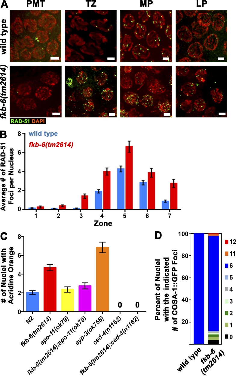
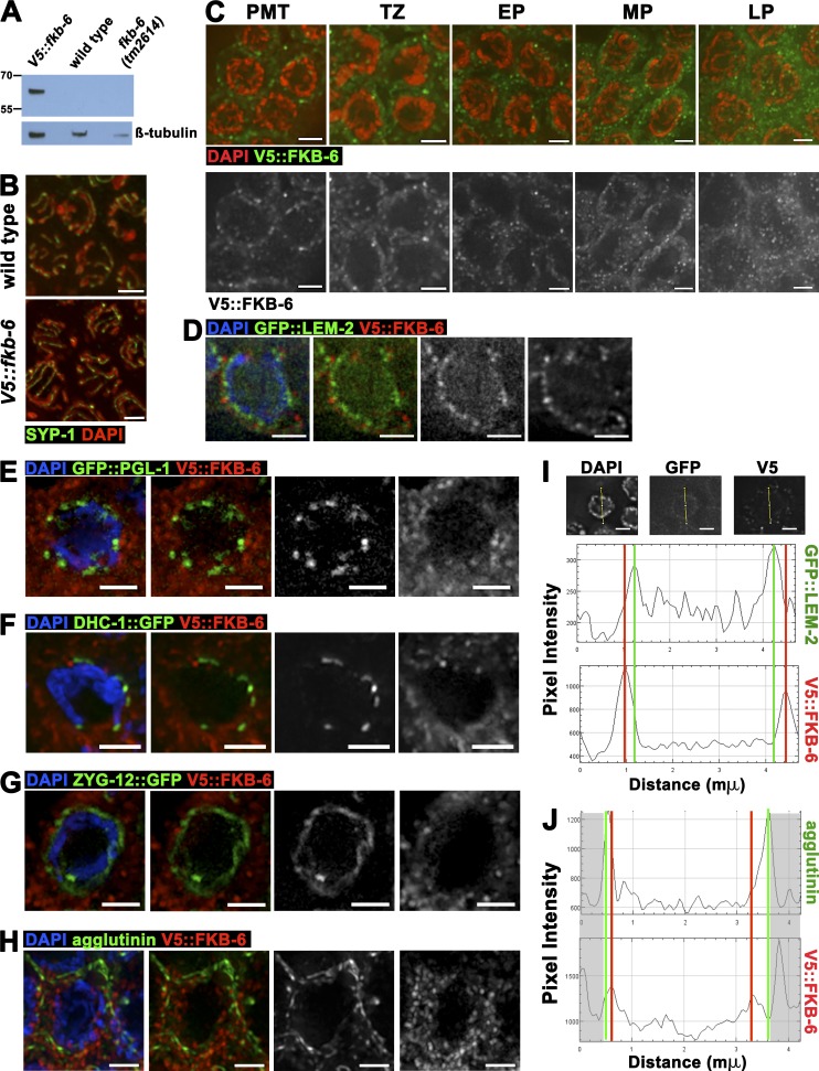
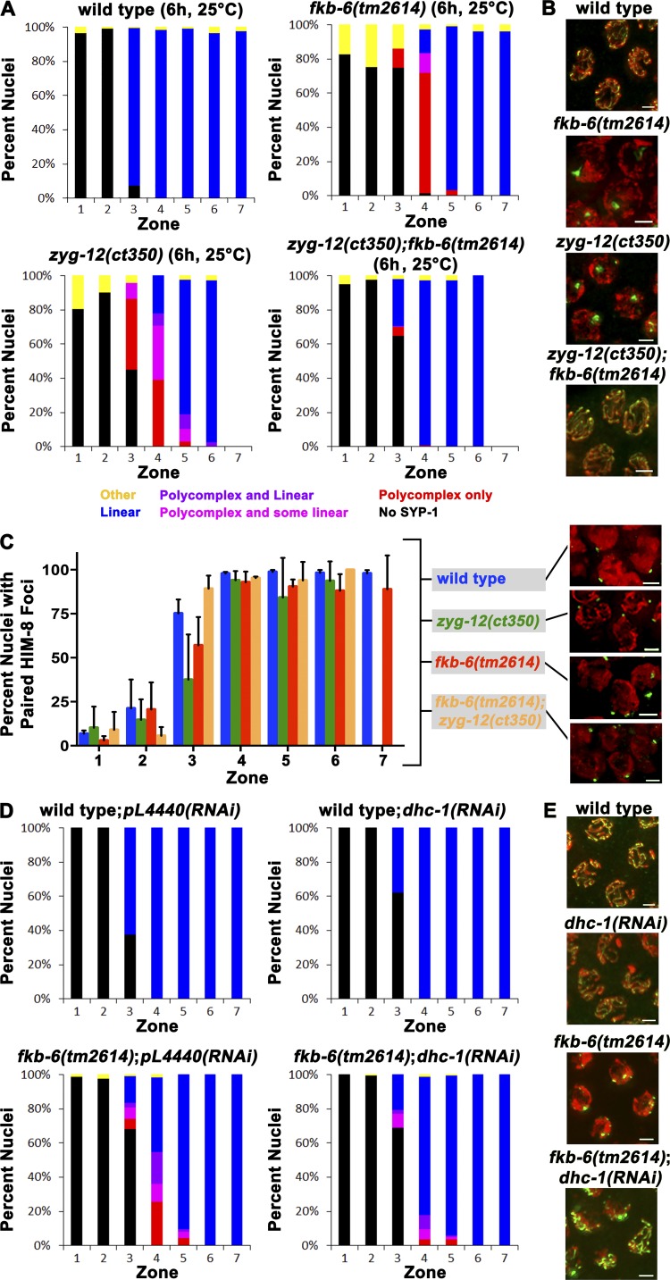
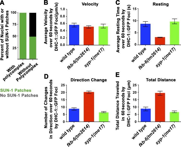
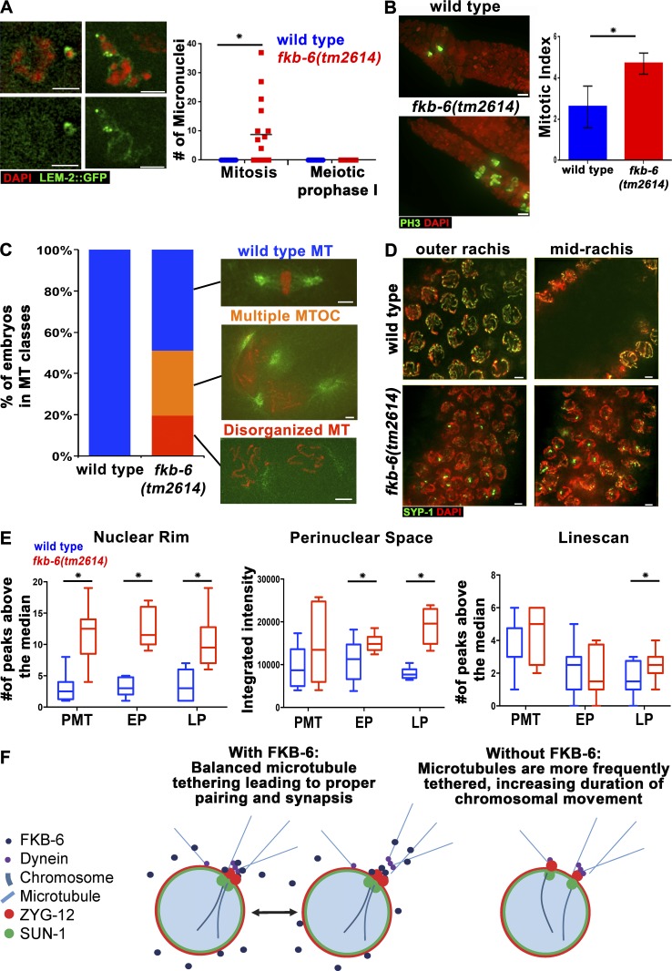
In meiotic prophase I, homologous chromosome pairing is promoted through chromosome movement mediated by nuclear envelope proteins, microtubules, and dynein. After proper homologue pairing has been established, the synaptonemal complex (SC) assembles along the paired homologues, stabilizing their interaction and allowing for crossing over to occur. Previous studies have shown that perturbing chromosome movement leads to pairing defects and SC polycomplex formation. We show that FKB-6 plays a role in SC assembly and is required for timely pairing and proper double-strand break repair kinetics. FKB-6 localizes outside the nucleus, and in its absence, the microtubule network is altered. FKB-6 is required for proper movement of dynein, increasing resting time between movements. Attenuating chromosomal movement in fkb-6 mutants partially restores the defects in synapsis, in agreement with FKB-6 acting by decreasing chromosomal movement. Therefore, we suggest that FKB-6 plays a role in regulating dynein movement by preventing excess chromosome movement, which is essential for proper SC assembly and homologous chromosome pairing.


Figures

Figure 1.
Figure 1.
PC formation of SYP-1 proteins in fkb-6(tm2614) mutant germline. (A) C. elegans FKB-6 protein mapped with its protein domains. (B) Schematics of the fkb-6 gene with the position of the tm2614 deletion. (C) Images of full-length gonad stained with SYP-1 (central region, green) and DAPI (false-colored red) of the genotypes indicated. Images are z-stack projections of half the germline. (D) Representative images of SYP-1 staining in PMT, TZ, MP, and LP. Images are projections of a z-stack halfway through the nuclei. (E) Quantification of SYP-1 staining of wild-type and fkb-6 mutants. Scoring was divided into six categories, as indicated at the bottom. Representation of zones in terms of meiotic stage are as follows: zones 1 and 2, PMT (mitotic nuclei); zone 3, mostly TZ and some PMT; zone 4, mostly EP; zones 5 and 6, MP; and zone 7, LP. n nuclei: wild type = 657 and fkb-6 = 621. Bars: (A) 50 aa; (B) 100 bp; (C) 5 µm; (D and E) 2 µm.
Figure 2.
Figure 2.
fkb-6(tm2614) mutants show defects in pairing of the X-chromosome and chromosome V. Representative images of HIM-8 (A) antibody staining or 5S FISH (C) in the genotypes indicated in TZ/zone 3, EP/zone 4, and LP/zone 7. Stained with HIM-8 or FISH (green) and DAPI (red). Images are z-stack projections halfway through the nuclei. Bars, 2 µm. (B) Quantification of percentage of nuclei with paired HIM-8 foci. n nuclei: wild type = 790 and fkb-6 = 614. (D) Quantification of percentage of nuclei with paired 5S foci. n nuclei: wild type = 1,113 and fkb-6 = 1,277. Zones in B and D in terms of meiotic stage are as indicated in Fig. 1 D. Error bars are SD. (E) Representative images of common classes and division into categories of SYP-1 staining with HIM8 or 5S FISH probe (n = 743, 1,028 nuclei). Bars, 2 μm.
Figure 3.
Figure 3.
fkb-6 mutants accumulate recombination intermediates. (A) Representative images of RAD-51 antibody staining in wild-type and fkb-6 mutants in PMT/zone 1, TZ/zone 3, EP/zone 4, and LP/zone 7. Stained with RAD-51 (green) and DAPI (red). Images are a z-stack projection halfway through the nuclei. Bars, 2 µm. (B) Quantification of RAD-51 immunostaining. Representation of zones in terms of meiotic stage are as indicated in Fig. 1 E. Error bars are SEM. n nuclei: wild type = 718 and fkb-6 = 610. (C) Apoptotic nuclei per gonadal arm in the genotypes indicated. Error bars are SEM. n gonads: wild type = 125, fkb-6 = 74, syp3 = 57, spo-11 = 42, fkb-6;spo-11 = 56, ced-4 = 22, and fkb-6;ced-4 = 21. (D) Distribution of the number of COSA-1 foci per gonad in LP nuclei. n nuclei: wild type = 168 and fkb-6 = 123.
Figure 4.
Figure 4.
FKB-6 localizes to the cytoplasmic space throughout the germline. (A) Western blot of V5 antigen (top) and β-tubulin (bottom) as loading control of the genotypes indicated. (B) Projections of a z-stack halfway through the nuclei of V5::fkb-6 transgenic worms. Gonad stained with SYP-1 (green) and DAPI (red). (C) Representative images of V5 antibody staining in V5::fkb-6 strain in PMT, TZ, EP, MP, and LP. FKB-6 (via V5; green) and DAPI (red). Images on the bottom are V5::FKB-6 channel (white) of the images shown on the left. (D–H) Early pachytene images of V5::fkb-6 strain stained for V5 (red); in green, GFP::LEM-2 (D), GFP::PGL-1 (E), DHC-1::GFP (F), ZYG-12::GFP (G), and agglutinin (H). (I and J) Intensity plots of the LEM-2::GFP (I) or agglutinin (H) and V5::fkb-6 strain. Shaded boxes are part of the adjacent nucleus. Bars, 2 µm.
Figure 5.
Figure 5.
fkb-6(tm2614) suppresses pairing and SC assembly defects in mutants with defective chromosomal movement. (A and D) Quantification of SYP-1 staining of the indicated genotypes and conditions. Representation of zones in terms of meiotic stage and classes of scoring are as indicated in Fig. 1 E. fkb-6(tm2614) suppresses the SC assembly defects in zyg-12(ct350) and dhc-1(RNAi). (B, E, and C inset) Representative images of zone 4 of A, D, and C, respectively. SYP-1/HIM-8 (green) and DAPI (red). (C) Quantification of HIM-8 pairing. Representation of zones in terms of meiotic stage are as indicated in Fig. 1 E. Error bars are SD. n nuclei: (A and C) wild type = 1,261, fkb-6 = 1,162, zyg-12 = 628, fkb-6;zyg-12 = 545, wild type;pl4440(RNAi) = 834, wild type;dhc-1(RNAi) = 1,126, fkb-6;pl4440(RNAi) = 899, and fkb-6;dhc-1(RNAi) = 1,832; (B) wild type = 818, fkb-6 = 706, zyg-12 = 533, and fkb-6;zyg-12 = 415. Bars, 2 μm.
Figure 6.
Figure 6.
fkb-6(tm2614) mutants show decreased resting time of DHC-1 foci between movements. (A) Percentage of nuclei with SUN-1 patches in nuclei that do and do not show PC formation in fkb-6(tm2614) mutants. n nuclei: fkb-6(tm2614) mutants = 387. (B) Mean velocity of DHC-1 patches. (C) Resting time of DHC-1 patches. (D) Change in direction of DHC-1 patches. (E) Total distance traveled by DHC-1 patches in wild type, fkb-6(tm2614), and syp-1 mutants. Error bars in B–E are SEM; n nuclei = 10 for all.
Figure 7.
Figure 7.
fkb-6(tm2614) mutants show defects typical to microtubule organization defects in the germline. (A) Micronuclei in fkb-6(tm2614) mutants (grown at 25°C for 6 h). LEM-2 (nuclear envelope, green) and DAPI (red). Images are a z-stack projection halfway through the nuclei. n gonads: wild type = 16 and fkb-6(tm2614) mutants = 16. (B) Metaphase nuclei are found in higher proportion in fkb-6(tm2614) mutants. PH-3 to mark metaphase nuclei (green) and DAPI (red). Images are a z-stack projection through the nuclei at the PMT. Error bars are SD. n gonads: wild type = 8 and fkb-6(tm2614) mutants = 8. (C) Microtubule defects found in embryos, as indicated in the images, distributed to categories in wild-type (n = 68) and fkb-6(tm2614) mutants (n = 31). (D) Nuclear positioning defects in fkb-6(tm2614) mutants, SYP-1 (red) and DAPI (blue). Images are a z-stack projection of the top and bottom layer. Bars, 2 μm. (E) Quantification of microtubule density around the nucleus by three methods (ImageJ). Box-and-whisker plots are made with minimum to maximum values. n nuclei: wild type = 12 and fkb-6 = 12 for all. *, P < 0.05. (F) Model.

References

    1. Arribere J.A., Bell R.T., Fu B.X.H., Artiles K.L., Hartman P.S., and Fire A.Z.. 2014. Efficient marker-free recovery of custom genetic modifications with CRISPR/Cas9 in Caenorhabditis elegans. Genetics. 198:837–846. 10.1534/genetics.114.169730 - DOI - PMC - PubMed
    1. Bilgir C., Dombecki C.R., Chen P.F., Villeneuve A.M., and Nabeshima K.. 2013. Assembly of the synaptonemal complex is a highly temperature-sensitive process that is supported by PGL-1 during Caenorhabditis elegans meiosis. G3 (Bethesda). 3:585–595. 10.1534/g3.112.005165 - DOI - PMC - PubMed
    1. Birnby D.A., Link E.M., Vowels J.J., Tian H., Colacurcio P.L., and Thomas J.H.. 2000. A transmembrane guanylyl cyclase (DAF-11) and Hsp90 (DAF-21) regulate a common set of chemosensory behaviors in Caenorhabditis elegans. Genetics. 155:85–104. - PMC - PubMed
    1. Boateng K.A., Bellani M.A., Gregoretti I.V., Pratto F., and Camerini-Otero R.D.. 2013. Homologous pairing preceding SPO11-mediated double-strand breaks in mice. Dev. Cell. 24:196–205. 10.1016/j.devcel.2012.12.002 - DOI - PMC - PubMed
    1. Brockway H., Balukoff N., Dean M., Alleva B., and Smolikove S.. 2014. The CSN/COP9 signalosome regulates synaptonemal complex assembly during meiotic prophase I of Caenorhabditis elegans. PLoS Genet. 10:e1004757 10.1371/journal.pgen.1004757 - DOI - PMC - PubMed

Publication types

MeSH terms

LinkOut - more resources