Review
. 2017 Feb 1;9(1):3-15.
doi: 10.1093/jmcb/mjx002.

Anatomy of Mdm2 and Mdm4 in evolution

Affiliations
Review

Anatomy of Mdm2 and Mdm4 in evolution

Ban Xiong Tan et al. J Mol Cell Biol. .

Abstract

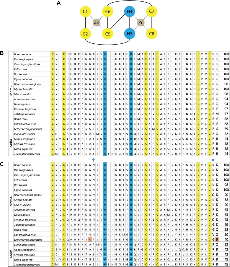
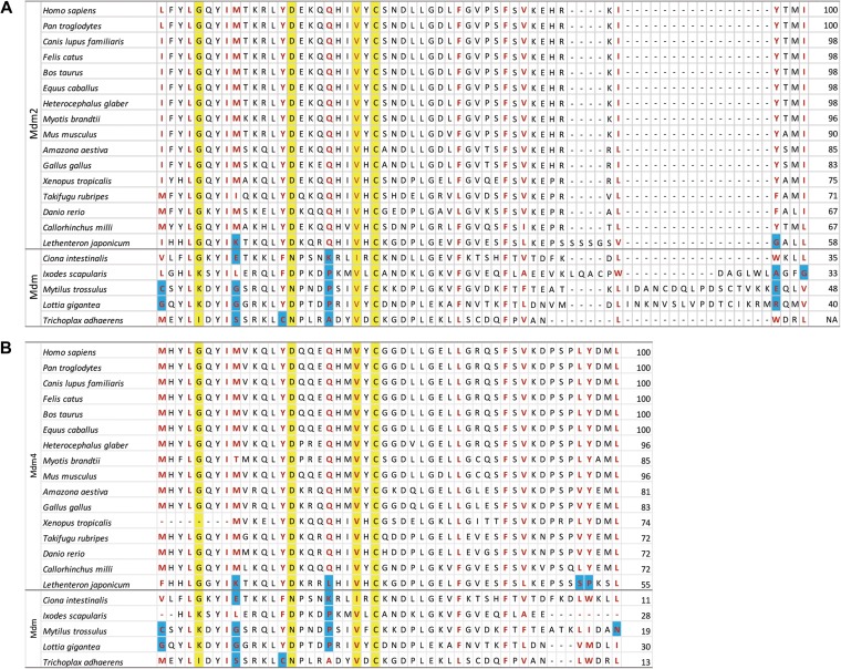
Mouse double minute (Mdm) genes span an evolutionary timeframe from the ancient eukaryotic placozoa Trichoplax adhaerens to Homo sapiens, implying a significant and possibly conserved cellular role throughout history. Maintenance of DNA integrity and response to DNA damage involve many key regulatory pathways, including precise control over the tumour suppressor protein p53. In most vertebrates, degradation of p53 through proteasomal targeting is primarily mediated by heterodimers of Mdm2 and the Mdm2-related protein Mdm4 (also known as MdmX). Both Mdm2 and Mdm4 have p53-binding regions, acidic domains, zinc fingers, and C-terminal RING domains that are conserved throughout evolution. Vertebrates typically have both Mdm2 and Mdm4 genes, while analyses of sequenced genomes of invertebrate species have identified single Mdm genes, suggesting that a duplication event occurred prior to emergence of jawless vertebrates about 550-440 million years ago. The functional relationship between Mdm and p53 in T. adhaerens, an organism that has existed for 1 billion years, implies that these two proteins have evolved together to maintain a conserved and regulated function.

Keywords: Mdm2; Mdm4; evolution; p53.


Figures

Figure 1
Figure 1
The RING finger domains of Mdm2 and Mdm4. (A) The Mdm2/Mdm4 RING finger motif consists of an octet of two cysteines (C1 and C2), two histidines (H3 and H4) and four cysteines (C5–C8). Two zinc ions are bound in coordinate bonds (dotted lines) by half of the motif (C1+C2+C5+C6 and H3+H4+C7+C8). (B) This C2H2C4 cross-brace motif is conserved in vertebrate Mdm2 and invertebrate Mdm. The cysteine and histidine residues involved are numbered and highlighted in yellow and blue, respectively. (C) The blue arrowheads indicate the asparagines (N448 in humans) that can be substituted to cysteines, as well as lysines (K478 in humans) to arginines, to make the Mdm4 functional E3 ligases. The residues highlighted in orange present in L. japonicum Mdm4 potentially make them functional E3 ligases. The numbers on the right of the table indicate the percentage identity compared to H. sapiens Mdm2 or Mdm4.
Figure 2
Figure 2
Alignments of the p53-binding domains of Mdm2 and Mdm4. (A) The residues in red form the hydrophobic cleft in which an α helix from the p53 transactivation domain binds to Mdm2, while the residues highlighted in yellow represent the genetically determined amino acids that are crucial in p53 interaction. The blue highlight indicates amino acids that differ in physical properties from the consensus, for instance a charged lysine in place of a hydrophobic methionine. (B) The homologous residues on Mdm4 are similarly highlighted. The numbers on the right of the table indicate the percentage identity compared to H. sapiens Mdm2 or Mdm4.
Figure 3
Figure 3
m dm2 knockout in p53-morphant zebrafish is not embryonic lethal if tp53 is temporarily repressed during embryogenesis. (A) mdm2−/− fish grow to adulthood, albeit physically smaller, and lack developed reproductive organs. (B) Immunohistochemical staining reveals that wild-type p53 does not accumulate in mdm2−/− adult fish while, in contrast, mutant p53 protein is stabilized in mdm2−/−p53M214K/M214K fish.

References

    1. Barak Y., Juven T., Haffner R., et al. (1993). mdm2 expression is induced by wild type p53 activity. EMBO J. 12, 461–468. - PMC - PubMed
    1. Bennett-Lovsey R., Hart S.E., Shirai H., et al. (2002). The SWIB and the MDM2 domains are homologous and share a common fold. Bioinformatics 18, 626–630. - PubMed
    1. Bernardi R., Scaglioni P.P., Bergmann S., et al. (2004). PML regulates p53 stability by sequestering Mdm2 to the nucleolus. Nat. Cell Biol. 6, 665–672. - PubMed
    1. Bista M., Petrovich M., and Fersht A.R (2013). MDMX contains an autoinhibitory sequence element. Proc. Natl Acad. Sci. USA 110, 17814–17819. - PMC - PubMed
    1. Blackford A.N., and Grand R.J.A (2009). Adenovirus E1B 55-kilodalton protein: multiple roles in viral infection and cell transformation. J. Virol. 83, 4000–4012. - PMC - PubMed

Publication types

Substances