. 2017 Feb 28;91(6):e02154-16.
doi: 10.1128/JVI.02154-16. Print 2017 Mar 15.

Chikungunya, Influenza, Nipah, and Semliki Forest Chimeric Viruses with Vesicular Stomatitis Virus: Actions in the Brain

Affiliations

Chikungunya, Influenza, Nipah, and Semliki Forest Chimeric Viruses with Vesicular Stomatitis Virus: Actions in the Brain

Anthony N van den Pol et al. J Virol. .

Abstract

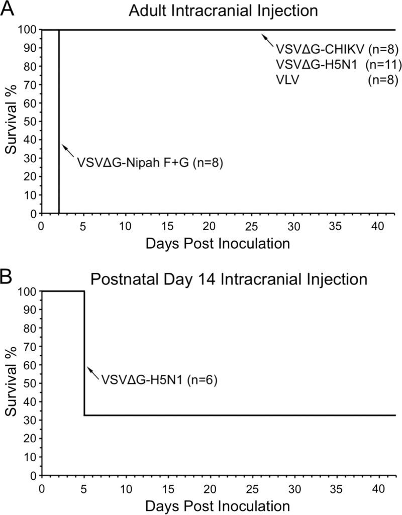
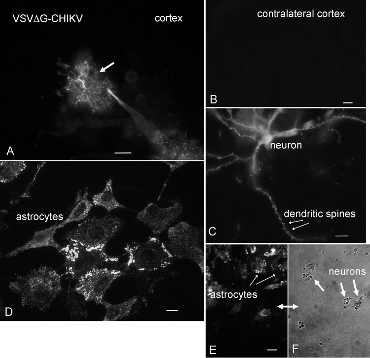
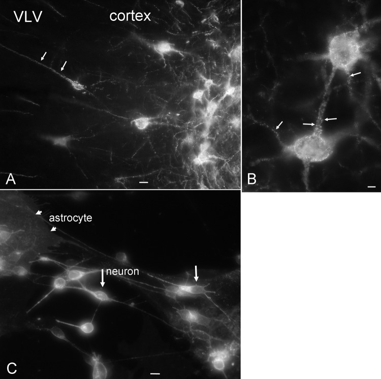
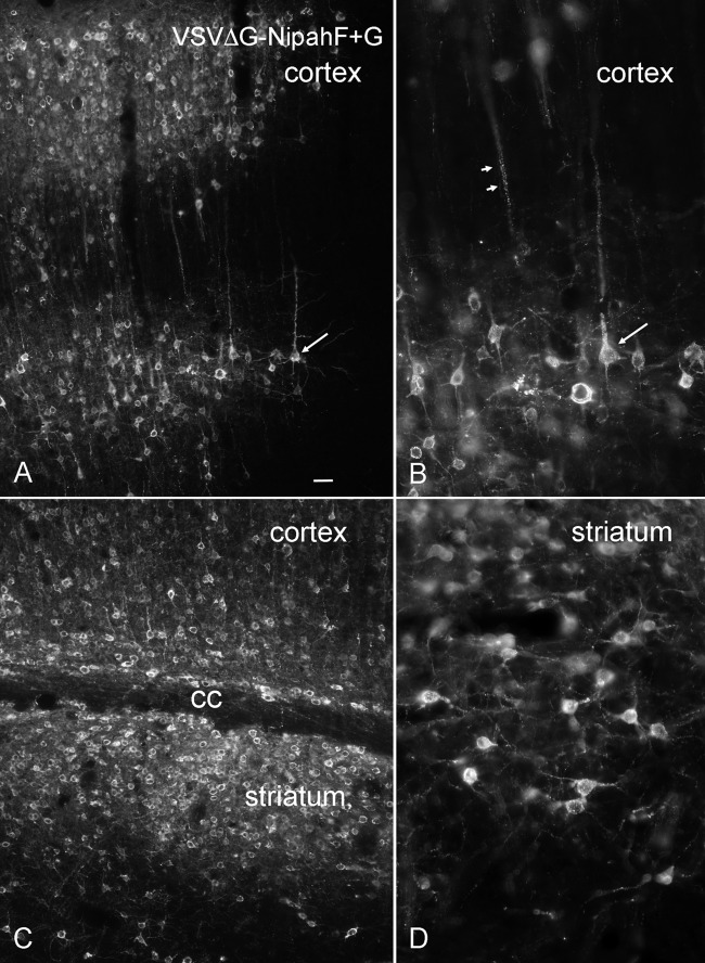
Recombinant vesicular stomatitis virus (VSV)-based chimeric viruses that include genes from other viruses show promise as vaccines and oncolytic viruses. However, the critical safety concern is the neurotropic nature conveyed by the VSV glycoprotein. VSVs that include the VSV glycoprotein (G) gene, even in most recombinant attenuated strains, can still show substantial adverse or lethal actions in the brain. Here, we test 4 chimeric viruses in the brain, including those in which glycoprotein genes from Nipah, chikungunya (CHIKV), and influenza H5N1 viruses were substituted for the VSV glycoprotein gene. We also test a virus-like vesicle (VLV) in which the VSV glycoprotein gene is expressed from a replicon encoding the nonstructural proteins of Semliki Forest virus. VSVΔG-CHIKV, VSVΔG-H5N1, and VLV were all safe in the adult mouse brain, as were VSVΔG viruses expressing either the Nipah F or G glycoprotein. In contrast, a complementing pair of VSVΔG viruses expressing Nipah G and F glycoproteins were lethal within the brain within a surprisingly short time frame of 2 days. Intranasal inoculation in postnatal day 14 mice with VSVΔG-CHIKV or VLV evoked no adverse response, whereas VSVΔG-H5N1 by this route was lethal in most mice. A key immune mechanism underlying the safety of VSVΔG-CHIKV, VSVΔG-H5N1, and VLV in the adult brain was the type I interferon response; all three viruses were lethal in the brains of adult mice lacking the interferon receptor, suggesting that the viruses can infect and replicate and spread in brain cells if not blocked by interferon-stimulated genes within the brain.IMPORTANCE Vesicular stomatitis virus (VSV) shows considerable promise both as a vaccine vector and as an oncolytic virus. The greatest limitation of VSV is that it is highly neurotropic and can be lethal within the brain. The neurotropism can be mostly attributed to the VSV G glycoprotein. Here, we test 4 chimeric viruses of VSV with glycoprotein genes from Nipah, chikungunya, and influenza viruses and nonstructural genes from Semliki Forest virus. Two of the four, VSVΔG-CHIKV and VLV, show substantially attenuated neurotropism and were safe in the healthy adult mouse brain. VSVΔG-H5N1 was safe in the adult brain but lethal in the younger brain. VSVΔG Nipah F+G was even more neurotropic than wild-type VSV, evoking a rapid lethal response in the adult brain. These results suggest that while chimeric VSVs show promise, each must be tested with both intranasal and intracranial administration to ensure the absence of lethal neurotropism.

Keywords: Nipah virus; blood-brain barrier; brain; chikungunya; influenza virus; neurotropic viruses; vesicular stomatitis virus; viral vaccine.


Figures

FIG 1
FIG 1
Schematic diagram of recombinant chimeric VSVs. The top diagram, VSV, shows the gene sequence and relative gene size of normal VSV. Below that are schematics of the chimeric virus genomes with the VSV glycoprotein gene (green) deleted and replaced with the glycoprotein genes from chikungunya virus (yellow), influenza virus H5N1 (orange), and Nipah virus F (red) and G (blue) and VLV, in which only the VSV glycoprotein gene is used and all other VSV genes are deleted and replaced by the Semliki Forest virus nonstructural protein genes (pink). VSVΔG-Nipah F+G is the combination of VSVΔG-Nipah F and VSVΔG-Nipah G.
FIG 2
FIG 2
Identification of neurons and astrocytes. (A) Neuronal nuclei are immunostained red for the neuronal antigen NeuN (short arrows). (B) Neurons with 2 to 4 thin dendritic or axonal processes contain NeuN-positive nuclei. (A and B) Bar, 20 μm. (C) Neurons are labeled by NeuN immunostaining (red), and astrocytes are labeled green by GFAP immunostaining. (D) In the same field shown in panel C, phase contrast shows that phase-bright neurons and astrocyte immunostaining overlap. (C and D) Bar, 40 μm. (E) Astrocytes 2 days after inoculation with VSVΔG-H5N1. Astrocyte processes have withdrawn, and GFAP immunoreactivity is no longer fiber-like, as seen in normal astrocytes (C and F), but has become more globular, indicating cell cytolysis. (F) Normal astrocytes immunolabeled with GFAP show long, thin GFAP staining. (E and F) Bar, 40 μm.
FIG 3
FIG 3
Immunostaining for chimeric VSVs in mouse brain cultures containing neurons and glia. (A and B) VSVΔG-Nipah F+G and VSVΔG-H5N1 showed immunostaining in small neurons (arrows). Scale bar, 30 μm (all panels). (C) VSVΔG-CHIKV showed immunoreactivity in the plasma membranes of large astrocytes (long arrows) and also staining in some small neuron-like cells (short arrows). (D) VLV showed staining in neurons (arrows). (E) Control cultures with no virus showed no immunoreactivity.
FIG 4
FIG 4
Survival and infection after intranasal inoculation with chimeric VSVs. (A) In postnatal day 14 mice, viruses were applied to the external nares. VSV-G/GFP was lethal, as previously reported. VSVΔG-CHIKV and VLV were without lethal effect. VSV-IFN showed no adverse effect. Similarly, VSVΔG-LCMV, used as a negative control for safety, showed no lethal effect. In contrast, VSVΔG-Nipah F+G was lethal in many mice. VSVΔG-H5N1 showed a very strong lethal effect, resulting in a fatal response in almost all of the 23 mice tested. (B) VSVΔG-H5N1 at 5 dpi showed strong infection of the left cortex with minimal infection of the adjacent right cortex. (C) VSVΔG-H5N1 in the same brain strongly infected cells of the hypothalamus in the region of the arcuate nucleus (ARC). (D and E) Hippocampus (D) and lungs (E) showed no detectable infection in the same mouse. (F) VSVΔG-Nipah F+G showed strong infection of periglomerular cells and mitral cells in the olfactory bulb at 3 dpi.
FIG 5
FIG 5
VSVΔG-CHIKV, VSVΔG-H5N1, and VLV are safe in the adult brain, whereas VSVΔG-Nipah F+G is lethal. (A) Of the 4 viruses tested, only VSVΔG-Nipah F+G was rapidly lethal when injected into the adult mouse brain. VSVΔG-CHIKV, VSVΔG-H5N1, and VLV were all completely without lethal effect in the adult brain. (B) In contrast to its lack of adverse action in the adult brain, VSVΔG-H5N1 showed lethal actions when injected into the brains of younger, postnatal day 14 mice.
FIG 6
FIG 6
VSVΔG-CHIKV in brain and culture. (A) Injection of VSVΔG-CHIKV into the brain infected both neurons and astrocytes at 3 dpi. The arrow points to an astrocyte with many radiating processes. Bar, 35 μm. (B) No infection was seen in the contralateral cortex. Bar, 25 μm. (C) Some neurons also showed VSVΔG-CHIKV infection. Bar, 25 μm. (D) In culture, large, flat astrocytes are preferentially infected at 1 dpi. (E) Astrocytes are infected in vitro at 1 dpi. Bar, 12 μm. (F) The arrows indicate cells with neuronal morphology in the same field shown in panel E. Whereas many glial cells were infected, neurons were not infected. Bar, 30 μm.
FIG 7
FIG 7
VLV in brain and culture. (A) After intracranial inoculation, VLV infects mostly neurons. The arrows show a dendrite stretching toward the outer cortex filled with small vesicles, a typical sign of early infection. Bar, 10 μm. (B) Two infected cells in the cortex, with vesicle-like labeling of the cell body and processes (arrows). Bar, 4 μm. (C) In vitro, both astrocytes and neurons are infected by VLV. Bar, 9 μm.
FIG 8
FIG 8
VSVΔG-H5N1 in the brain. (A) Cortical neurons were infected by VSVΔG-H5N1 at 3 dpi in the adult mouse brain. Bar, 18 μm. (B) High magnification of a neuronal dendrite with dendritic spines (arrows). Bar, 4 μm. Although the cells were infected at the injection site, the virus showed little spread. (C) In vitro, neurons and astrocytes show infection. Bar, 14 μm.
FIG 9
FIG 9
VSVΔG-Nipah F+G in the brain. (A) At 1 day after a small, 0.4-μl intracortical injection, VSVΔG-Nipah F+G had spread to a large number of neurons. The arrow indicates a neuron with pyramidal cell morphology. Bar, 22 μm. (B) Higher magnification of panel A. Arrows point to the same neuron in panels A and B. (C) Virus spread across the corpus callosum (cc) into the striatum. (D) Higher magnification of infected striatal cells. In both the striatum, as shown, and the cortex (B), most infected cells had the morphology of neurons. No infection was seen in the contralateral cortex.
FIG 10
FIG 10
Type I IFN blocks chimeric VSVs in human brain cultures. (A) Primary cultures of human brain astrocytes were pretreated with 0 (control), 1, or 10 U/ml of type I IFN for 18 h and then inoculated with the indicated chimeric VSVs (final concentration, 2 × 10⁵ PFU/ml) or medium control and allowed to incubate for a further 24 h. The cells were then fixed and immunostained. The phase contrast images show typical cell density. The VSV-immunostained cultures show virus infection. Scale bar, 50 μm. (B) Percent infected cells in cultures without IFN pretreatment or with 1 U/ml IFN; 1 U/ml IFN was sufficient to completely block chimeric VSV infection, and 10 U/ml IFN (not shown) also completely blocked infection. The error bars indicate standard errors of the mean (SEM) (n = 3). (C) Three viruses, VSVΔG-H5N1, VSVΔG-CHIKV, and VLV, were injected intracranially into adult mice lacking type I IFN receptors. All three viruses were lethal within 1 week in all IFNR-KO mice tested.

Similar articles

Cited by

References

    1. Kurup D, Wirblich C, Feldmann H, Marzi A, Schnell MJ. 2015. Rhabdovirus-based vaccine platforms against henipaviruses. J Virol 89:144–154. doi:10.1128/JVI.02308-14. - DOI - PMC - PubMed
    1. Rose NF, Marx PA, Luckay A, Nixon DF, Moretto WJ, Donahoe SM, Montefiori D, Roberts A, Buonocore L, Rose JK. 2001. An effective AIDS vaccine based on live attenuated vesicular stomatitis virus recombinants. Cell 106:539–549. doi:10.1016/S0092-8674(01)00482-2. - DOI - PubMed
    1. Clarke DK, Cooper D, Egan MA, Hendry RM, Parks CL, Udem SA. 2006. Recombinant vesicular stomatitis virus as an HIV-1 vaccine vector. Springer Semin Immunopathol 28:239–253. doi:10.1007/s00281-006-0042-3. - DOI - PMC - PubMed
    1. Chattopadhyay A, Park S, Delmas G, Suresh R, Senina S, Perlin DS, Rose JK. 2008. Single-dose, virus-vectored vaccine protection against Yersinia pestis challenge: CD4+ cells are required at the time of challenge for optimal protection. Vaccine 26:6329–6337. doi:10.1016/j.vaccine.2008.09.031. - DOI - PMC - PubMed
    1. Geisbert TW, Daddario-Dicaprio KM, Lewis MG, Geisbert JB, Grolla A, Leung A, Paragas J, Matthias L, Smith MA, Jones SM, Hensley LE, Feldmann H, Jahrling PB. 2008. Vesicular stomatitis virus-based Ebola vaccine is well-tolerated and protects immunocompromised nonhuman primates. PLoS Pathog 4:e1000225. doi:10.1371/journal.ppat.1000225. - DOI - PMC - PubMed

Publication types

MeSH terms