. 2017 Mar 13;91(7):e02079-16.
doi: 10.1128/JVI.02079-16. Print 2017 Apr 1.

Expression of Ifnlr1 on Intestinal Epithelial Cells Is Critical to the Antiviral Effects of Interferon Lambda against Norovirus and Reovirus

Affiliations

Expression of Ifnlr1 on Intestinal Epithelial Cells Is Critical to the Antiviral Effects of Interferon Lambda against Norovirus and Reovirus

Megan T Baldridge et al. J Virol. .

Abstract

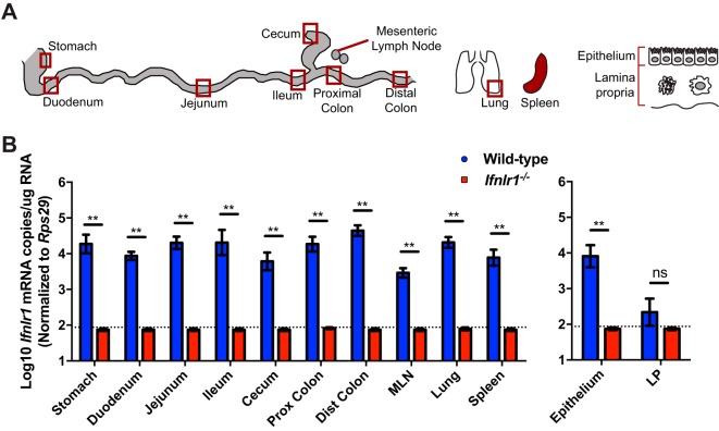
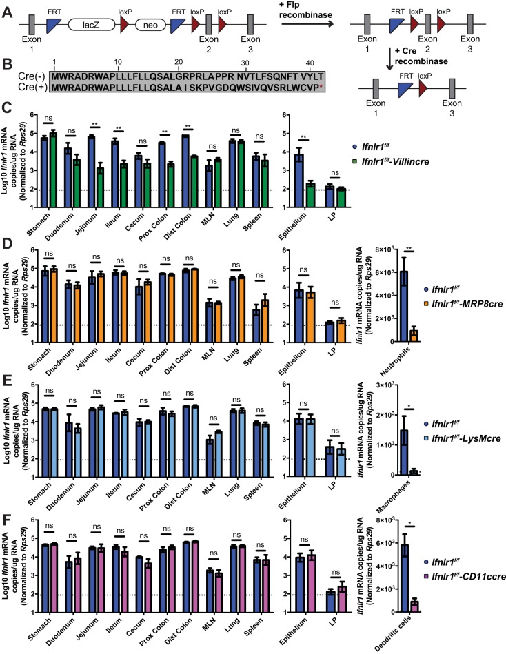
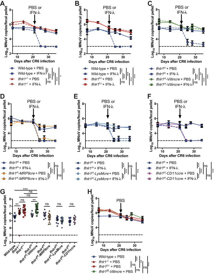
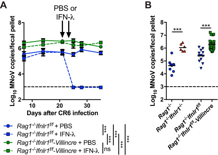
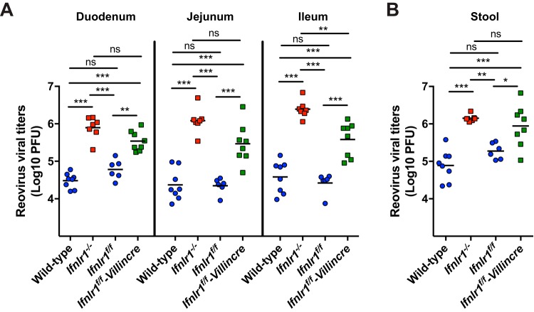
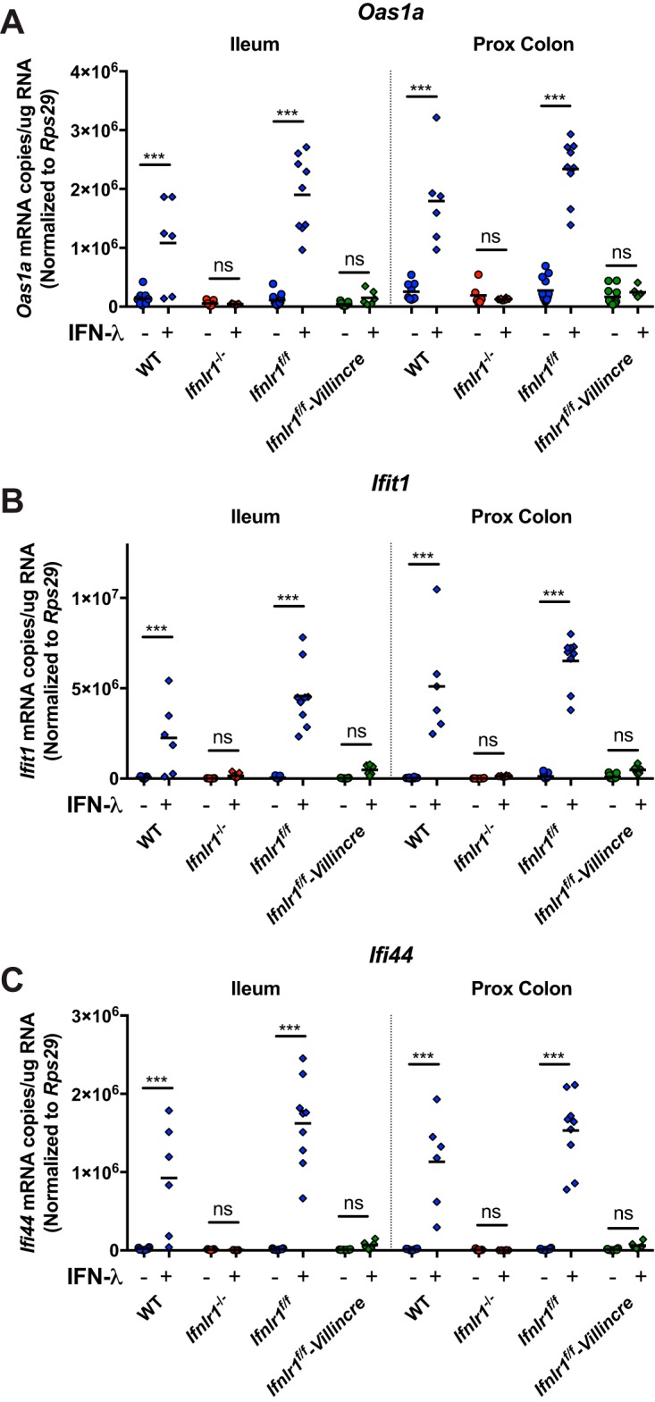
Lambda interferon (IFN-λ) has potent antiviral effects against multiple enteric viral pathogens, including norovirus and rotavirus, in both preventing and curing infection. Because the intestine includes a diverse array of cell types, however, the cell(s) upon which IFN-λ acts to exert its antiviral effects is unclear. Here, we sought to identify IFN-λ-responsive cells by generation of mice with lineage-specific deletion of the receptor for IFN-λ, Ifnlr1 We found that expression of IFNLR1 on intestinal epithelial cells (IECs) in the small intestine and colon is required for enteric IFN-λ antiviral activity. IEC Ifnlr1 expression also determines the efficacy of IFN-λ in resolving persistent murine norovirus (MNoV) infection and regulates fecal shedding and viral titers in tissue. Thus, the expression of Ifnlr1 by IECs is necessary for the response to both endogenous and exogenous IFN-λ. We further demonstrate that IEC Ifnlr1 expression is required for the sterilizing innate immune effects of IFN-λ by extending these findings in Rag1-deficient mice. Finally, we assessed whether our findings pertained to multiple viral pathogens by infecting mice specifically lacking IEC Ifnlr1 expression with reovirus. These mice phenocopied Ifnlr1-null animals, exhibiting increased intestinal tissue titers and enhanced reovirus fecal shedding. Thus, IECs are the critical cell type responding to IFN-λ to control multiple enteric viruses. This is the first genetic evidence that supports an essential role for IECs in IFN-λ-mediated control of enteric viral infection, and these findings provide insight into the mechanism of IFN-λ-mediated antiviral activity.IMPORTANCE Human noroviruses (HNoVs) are the leading cause of epidemic gastroenteritis worldwide. Type III interferons (IFN-λ) control enteric viral infections in the gut and have been shown to cure mouse norovirus, a small-animal model for HNoVs. Using a genetic approach with conditional knockout mice, we identified IECs as the dominant IFN-λ-responsive cells in control of enteric virus infection in vivo Upon murine norovirus or reovirus infection, Ifnlr1 depletion in IECs largely recapitulated the phenotype seen in Ifnlr1-/- mice of higher intestinal tissue viral titers and increased viral shedding in the stool. Moreover, IFN-λ-mediated sterilizing immunity against murine norovirus requires the capacity of IECs to respond to IFN-λ. These findings clarify the mechanism of action of this cytokine and emphasize the therapeutic potential of IFN-λ for treating mucosal viral infections.

Keywords: innate immunity; interferons; mucosal immunity; norovirus; reovirus.


Figures

FIG 1
FIG 1
Ifnlr1 is expressed in the epithelial fraction along the length of the intestine. (A) RNA was isolated from sites along the intestine and the lung, indicated by red boxes, whole mesenteric lymph node (MLN) and spleen, and epithelial and lamina propria (LP) fractions of the small intestine from Ifnlr1-sufficient and -deficient mice. (B) Ifnlr1 expression was quantified by quantitative real-time PCR of RNA from sites depicted in panel A. n = 4 to 6 samples per group, from two independent experiments, analyzed by Mann-Whitney test. **, P < 0.01; ns, not significant. Prox, proximal; dist, distal.
FIG 2
FIG 2
Ifnlr1 expression is decreased in the small and large intestines of Ifnlr1f/f-Villincre mice. (A) Schematic depicting the Ifnlr1 gene locus in Ifnlr1tm1a(EUCOMM)Wtsi mice. After crossing with mice expressing Flp recombinase (+Flp recombinase), the region between the two FRT sites was deleted, leaving conditional-ready Ifnlr1f/f mice. In the absence of Cre, all exons are present. With the addition of Cre recombinase, the floxed exon 2 is deleted. (B) In the absence of Cre [Cre(−)], the IFNLR1 protein is expressed. In the presence of Cre [Cre(+)], the protein sequence is altered at amino acid 20 and a premature stop codon is introduced at amino acid 42. (C to F) Ifnlr1 expression was assessed by quantitative real-time PCR of sites along the intestine and the lung, MLN and spleen, and epithelial and LP fractions from Ifnlr1f/f-Villincre (C), Ifnlr1f/f-MRP8cre (D), Ifnlr1f/f-LysMcre (E), and Ifnlr1f/f-CD11ccre (F) mice compared to their Ifnlr1f/f littermates. Ifnlr1 expression was also assessed by quantitative real-time PCR of isolated bone marrow neutrophils from Ifnlr1f/f-MRP8cre (D), splenic macrophages from Ifnlr1f/f-LysMcre (E), and splenic dendritic cells from Ifnlr1f/f-CD11ccre (F) mice compared to their Ifnlr1f/f littermates. n = 4 to 7 samples per group, from two independent experiments, analyzed by Mann-Whitney test. *, P < 0.05; **, P < 0.01; ns, not significant.
FIG 3
FIG 3
Expression of Ifnlr1 on intestinal epithelial cells is required for the antiviral effects of endogenous and exogenous IFN-λ against MNoV. (A to E) Time course of MNoV genome copies shed into fecal pellets with time points at 7, 14, 21, 24, 28, and 35 days after CR6 infection. PBS or recombinant IFN-λ was injected intraperitoneally on day 21 into wild-type and Ifnlr1−/− (A), wild-type and Ifnlr1f−/− (B), Ifnlr1f/f-Villincre (C), Ifnlr1f/f-MRP8cre (D), Ifnlr1f/f-LysMcre (E), or Ifnlr1f/f-CD11ccre (F) mice and their Ifnlr1f/f littermates. n = 6 to 12 mice per group, from two to three independent experiments, analyzed by two-way ANOVA followed by Tukey's multiple-comparison test; a P value of <0.001 by ANOVA column factor was found for panels A to F. (G) Individual data points depicting MNoV genome copies shed into fecal pellets on day 7 from panels A to F. n = 9 to 21 mice per group, from two to three independent experiments, analyzed by one-way ANOVA followed by Tukey's multiple-comparison test; a P value of <0.001 was determined by ANOVA. (H) Fecal shedding data from PBS-treated mice in panels A to C is shown superimposed to facilitate comparison between strains. n = 8 to 11 mice per group, from two to three independent experiments, analyzed by two-way ANOVA followed by Tukey's multiple-comparison test; a P value of <0.001 was determined by ANOVA column factor. *, P < 0.05; **, P < 0.01; ***, P < 0.001; ns, not significant.
FIG 4
FIG 4
Expression of Ifnlr1 on intestinal epithelial cells is required for the antiviral effects of IFN-λ against MNoV in the absence of adaptive immunity. (A) Time course of MNoV genome copies shed into fecal pellets with time points at 7, 14, 21, 24, 28, and 35 days after CR6 infection. PBS or recombinant IFN-λ was injected intraperitoneally on day 21 and day 23 into Rag1−/− Ifnlr1f/f-Villincre or Rag1−/− Ifnlr1f/f mice. n = 6 to 14 mice per group, combined from three independent experiments, analyzed by two-way ANOVA followed by Tukey's multiple-comparison test; a P value of <0.001 was found by ANOVA column factor. (B) Individual data points depicting MNoV genome copies shed into fecal pellets on day 7 from Rag1−/−, Rag1−/− Ifnlr1−/− double knockouts or mice depicted in panel A. n = 6 to 22 mice per group, combined from two to three independent experiments, analyzed by Mann-Whitney test. ***, P < 0.001; ns, not significant.
FIG 5
FIG 5
Ifnlr1 expression on intestinal epithelial cells limits reovirus infection. (A and B) Titers of reovirus strain T1L were assessed at day 4 postinoculation in the different compartments of the small intestine (A) and stool (B) from wild-type, Ifnlr1−/−, Ifnlr1f/f-Villincre, and Ifnlr1f/f littermate control mice. The small intestine was resected from the pylorus to the cecum and sectioned into three equal parts, representing the duodenum, jejunum, and ileum. Titers are expressed as PFU per milliliter of tissue homogenate or gram of stool. n = 6 to 8 mice per group, combined from two independent experiments, analyzed by one-way ANOVA followed by Tukey's multiple-comparison test; a P value of <0.001 was determined by ANOVA column factor for all tissues and stool. *, P < 0.05; **, P < 0.01; ***, P < 0.001; ns, not significant.
FIG 6
FIG 6
Ifnlr1 expression on intestinal epithelial cells is necessary for induction of interferon-stimulated genes. Oas1a (A), Ifit1 (B), and Ifi44 (C) expression was assessed by quantitative real-time PCR of RNA from distal ileum and proximal colon tissue from wild-type (WT), Ifnlr1−/−, Ifnlr1f/f, and Ifnlr1f/f-Villincre mice at 1 day posttreatment with PBS or recombinant IFN-λ. n = 5 to 9 mice per group, combined from two independent experiments, analyzed by one-way ANOVA followed by Tukey's multiple-comparison test; a P value of <0.001 was determined by ANOVA column factor for all tissues. ***, P < 0.001; ns, not significant.

Similar articles

Cited by

References

    1. Karst SM. 2010. Pathogenesis of noroviruses, emerging RNA viruses. Viruses 2:748–781. doi:10.3390/v2030748. - DOI - PMC - PubMed
    1. Glass RI, Parashar UD, Estes MK. 2009. Norovirus gastroenteritis. N Engl J Med 361:1776–1785. doi:10.1056/NEJMra0804575. - DOI - PMC - PubMed
    1. Koo HL, Ajami N, Atmar RL, DuPont HL. 2010. Noroviruses: the leading cause of gastroenteritis worldwide. Discov Med 10:61–70. - PMC - PubMed
    1. Scallan E, Hoekstra RM, Angulo FJ, Tauxe RV, Widdowson MA, Roy SL, Jones JL, Griffin PM. 2011. Foodborne illness acquired in the United States–major pathogens. Emerg Infect Dis 17:7–15. doi:10.3201/eid1701.P11101. - DOI - PMC - PubMed
    1. Lazear HM, Nice TJ, Diamond MS. 2015. Interferon-lambda: immune functions at barrier surfaces and beyond. Immunity 43:15–28. doi:10.1016/j.immuni.2015.07.001. - DOI - PMC - PubMed

Publication types

MeSH terms

Substances

LinkOut - more resources