. 2017 Jan 11;37(2):258-268.
doi: 10.1523/JNEUROSCI.2251-16.2016.

Connexin-Mediated Signaling in Nonsensory Cells Is Crucial for the Development of Sensory Inner Hair Cells in the Mouse Cochlea

Affiliations

Connexin-Mediated Signaling in Nonsensory Cells Is Crucial for the Development of Sensory Inner Hair Cells in the Mouse Cochlea

Stuart L Johnson et al. J Neurosci. .

Abstract

Mutations in the genes encoding for gap junction proteins connexin 26 (Cx26) and connexin 30 (Cx30) have been linked to syndromic and nonsyndromic hearing loss in mice and humans. The release of ATP from connexin hemichannels in cochlear nonsensory cells has been proposed to be the main trigger for action potential activity in immature sensory inner hair cells (IHCs), which is crucial for the refinement of the developing auditory circuitry. Using connexin knock-out mice, we show that IHCs fire spontaneous action potentials even in the absence of ATP-dependent intercellular Ca2+ signaling in the nonsensory cells. However, this signaling from nonsensory cells was able to increase the intrinsic IHC firing frequency. We also found that connexin expression is key to IHC functional maturation. In Cx26 conditional knock-out mice (Cx26Sox10-Cre), the maturation of IHCs, which normally occurs at approximately postnatal day 12, was partially prevented. Although Cx30 has been shown not to be required for hearing in young adult mice, IHCs from Cx30 knock-out mice exhibited a comprehensive brake in their development, such that their basolateral membrane currents and synaptic machinery retain a prehearing phenotype. We propose that IHC functional differentiation into mature sensory receptors is initiated in the prehearing cochlea provided that the expression of either connexin reaches a threshold level. As such, connexins regulate one of the most crucial functional refinements in the mammalian cochlea, the disruption of which contributes to the deafness phenotype observed in mice and DFNB1 patients.

Significance statement: The correct development and function of the mammalian cochlea relies not only on the sensory hair cells, but also on the surrounding nonsensory cells. Although the nonsensory cells have been largely implicated in the general homeostasis in the mature cochlea, their involvement in the initial functional differentiation of the sensory inner hair cells is less clear. Using mutant mouse models for the most common form of congenital deafness in humans, which are knock-outs for the gap-junction channels connexin 26 and connexin 30 genes, we show that defects in nonsensory cells prevented the functional maturation of inner hair cells. In connexin knock-outs, inner hair cells remained stuck at a prehearing stage of development and, as such, are unable to process sound information.

Keywords: cochlea; connexin; deafness; development; gap-junction; inner hair cells.


Figures

Figure 1.
Figure 1.
Connexins do not alter the biophysical properties of immature IHCs. A, Spontaneous APs recorded from IHCs under whole-cell current-clamp configuration from P3 Cx30(−/−) mice and control littermates (+/+). In this and the following figures, black represents control (wild-type or heterozygous) and gray represents mutant or knock-out mice. B, APs in cell-attached voltage clamp recorded from a P4 control (top) and P4 Cx30(−/−) (bottom) IHC. C, Coefficient of variation from each IHC against their firing rate. Open symbols represent data from single IHCs. Closed symbols represent averages. D, E, Potassium currents elicited from P3 IHCs by applying depolarizing voltage steps in 10 mV nominal increments from −144 mV, starting from the holding potential of −84 mV. Recordings were performed at body temperature.
Figure 2.
Figure 2.
Spontaneous APs in IHCs are present in the absence of Ca2+ signals from nonsensory cells in Cx30(−/−) mice. A, B, Representative false-color images of Fluo-4 fluorescence changes (ΔF/F0), encoded as shown by the color scale bar (top) and obtained as maximal projection rendering of all frames recorded in 200 s (10 frame/s). The images show a small part of the GER in the proximity of the patched IHC (arrows) from P6 Cx30(−/−) mice. Note the absence (A) or some residual (B) Ca2+ signals from nonsensory cells in the GER. IHCs were patched from the pillar side to prevent damage to the GER. Scale bar, 10 μm. C, D, Simultaneous recording of Ca2+ transients in the nonsensory cells present in the GER using fluorescence imaging (ΔF/F0; see Materials and Methods) from white ROI delineated by the dashed white line in panel A and B, respectively. Middle panels, IHC firing activity using cell-attached patch clamp. Bottom panels, Changes in AP frequency during the recordings. APs were present even when Ca2+ transients in the GER were absent (A; C, top), but their frequency increases during the residual Ca2+ transients in Cx30(−/−) mice (B; D, top). Recordings were performed at body temperature.
Figure 3.
Figure 3.
Current and voltage responses recorded from IHCs of Cx30(−/−) mice. A, B, Potassium currents recorded from P18 IHCs of wild-type (A) and littermate Cx30(−/−) (B) mice using depolarizing voltage steps in 10 mV nominal increments from the holding potential of −84 mV to the various test potentials shown by some of the traces. The adult-type currents (IK,f and IK,n) were only present in IHCs from wild-type mice (A). IHCs from Cx30(−/−) mice retained the currents characteristic of immature cells (IK,s and IK1). The presence of the rapidly activating IK,f in control IHCs is evident when comparing the activation time course of the total outward currents shown in the insets on an expanded time scale. C–E, Voltage responses elicited by applying hyperpolarizing and depolarizing current injections to control (C) and Cx30(−/−) adult IHCs (D, E) from their respective membrane potentials. In some IHCs, depolarizing current injections caused slow APs at the onset of responses. Recordings were performed at room temperature.
Figure 4.
Figure 4.
Efferent activity is still present in mature Cx30(−/−) IHCs. A, Outward currents obtained by using a 4 s depolarizing step to 0 mV from the holding potential of −84 mV in control and Cx30(−/−) P16 IHCs (Marcotti et al., 2004). Although the SK2 current (ISK2) is normally downregulated after the onset of hearing (Glowatzki and Fuchs, 2000), it was still expressed in mature Cx30(−/−) IHCs. B, Spontaneous IPSCs recorded from a P16 Cx30(−/−) IHC indicate that these IHCs retain the efferent endings that normally make only transient axosomatic synaptic contacts with IHCs during immature stages (Simmons et al., 1996; Katz et al., 2004). Recordings were performed at body temperature.
Figure 5.
Figure 5.
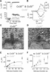
Exocytosis and ribbon morphology in Cx30(−/−) IHCs. A, ICa and corresponding ΔCm recorded from adult control and Cx30(−/−) IHCs obtained in response to 50 ms voltage steps, in 10 mV increments, from −81 mV. For clarity, only maximal responses at −11 mV are shown. B, Average peak ICa (bottom) and ΔCm (top) curve from control (P17–P25, n = 15) and Cx30(−/−) (P18–P24, n = 5) IHCs. C, D, Typical cross-sectional profiles of synaptic ribbons obtained from a control (C) and a Cx30(−/−) (D) IHC. Some of the synaptic vesicles are missing around the ribbon of the Cx30(−/−) IHC (arrow). Scale bar, 200 nm. E, F, Average ΔCm from 12 control and 14 Cx30(−/−) IHCs in response to voltage steps from 2 ms to 2 s (to ∼−11 mV) showing the RRP (E) and SRP (F). E, The points at 100 ms represent the recruitment of the SRP. Recordings were performed at body temperature.
Figure 6.
Figure 6.
IHCs from Cx30Δ/Δ knock-out mice develop normally. A, Potassium currents recorded from mature IHCs (P25) of Cx30Δ/Δ knock-out mice using the same voltage protocol described in Figure 3. Inset, The presence of IK,f is evident. B, Current-voltage curves measured from IHCs at 2 ms from the stimulus onset of Cx30Δ/Δ (n = 6, P25), wild-type (Cx30+/+: n = 10, P16–P18), and Cx30(−/−) (Cx30−/−: n = 9, P16–P18) mice. C, Voltage responses recorded from a mature IHC of a Cx30Δ/Δ mouse, which were elicited by applying hyperpolarizing and depolarizing current injections. Recordings were performed at room temperature.
Figure 7.
Figure 7.
Current and voltage responses recorded from IHCs of Cx26Sox10-Cre mice. A, B, Potassium currents recorded from P25 IHCs of wild-type (A) and littermate Cx26Sox10-Cre (B) mice using the same voltage protocol as described in Figure 3. Both the rapidly activating IK,f and the negatively activated IK,n were present in the Cx26Sox10-Cre IHC, although reduced in size compared with the control cell. C, Current-voltage curves obtained as in Figure 6B from IHCs of wild-type (Cx26+/+: n = 3, P25) and Cx26Sox10-Cre (Cx26−/−: n = 8, P25) mice. D, E, Voltage responses elicited as described in Figure 3 from control (D) and knock-out (E) adult IHCs from the same cells shown in A and B, respectively. Recordings were performed at room temperature.

References

    1. Ahmad S, Chen S, Sun J, Lin X. Connexins 26 and 30 are co-assembled to form gap junctions in the cochlea of mice. Biochem Biophys Res Commun. 2003;307:362–368. doi: 10.1016/S0006-291X(03)01166-5. - DOI - PubMed
    1. Ahmad S, Tang W, Chang Q, Qu Y, Hibshman J, Li Y, Söhl G, Willecke K, Chen P, Lin X. Restoration of connexin26 protein level in the cochlea completely rescues hearing in a mouse model of human connexin30-linked deafness. Proc Natl Acad Sci U S A. 2007;104:1337–1341. doi: 10.1073/pnas.0606855104. - DOI - PMC - PubMed
    1. Anselmi F, Hernandez VH, Crispino G, Seydel A, Ortolano S, Roper SD, Kessaris N, Richardson W, Rickheit G, Filippov MA, Monyer H, Mammano F. ATP release through connexin hemichannels and gap junction transfer of second messengers propagate Ca2+ signals across the inner ear. Proc Natl Acad Sci U S A. 2008;105:18770–18775. doi: 10.1073/pnas.0800793105. - DOI - PMC - PubMed
    1. Beltramello M, Piazza V, Bukauskas FF, Pozzan T, Mammano F. Impaired permeability to Ins(1,4,5)P3 in a mutant connexin underlies recessive hereditary deafness. Nat Cell Biol. 2005;7:63–69. doi: 10.1038/ncb1205. - DOI - PubMed
    1. Beutner D, Moser T. The presynaptic function of mouse cochlear inner hair cells during development of hearing. J Neurosci. 2001;21:4593–4599. - PMC - PubMed