. 2017 Jan 11;37(2):362-370.
doi: 10.1523/JNEUROSCI.2714-16.2016.

Excitatory Hindbrain-Forebrain Communication Is Required for Cisplatin-Induced Anorexia and Weight Loss

Affiliations

Excitatory Hindbrain-Forebrain Communication Is Required for Cisplatin-Induced Anorexia and Weight Loss

Amber L Alhadeff et al. J Neurosci. .

Abstract

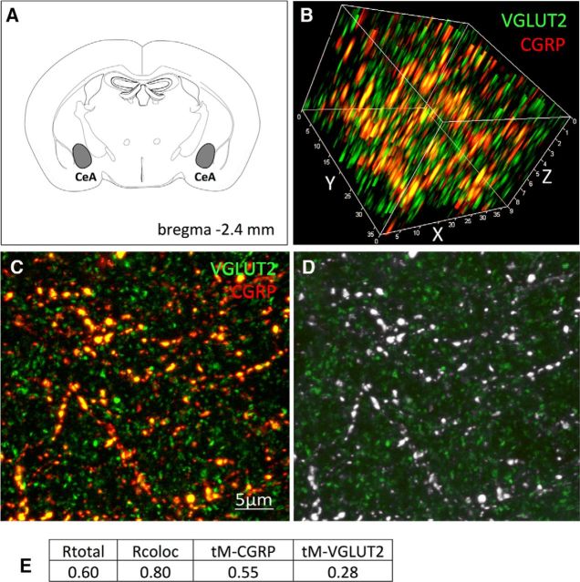
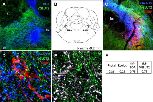
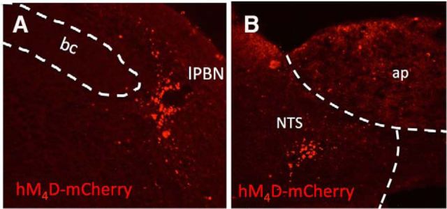
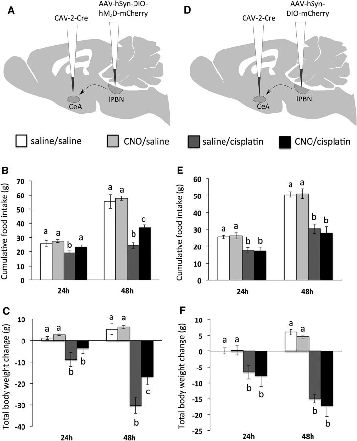
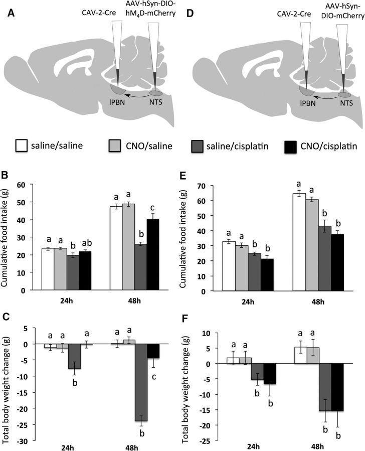
Cisplatin chemotherapy is commonly used to treat cancer despite severe energy balance side effects. In rats, cisplatin activates nucleus tractus solitarius (NTS) projections to the lateral parabrachial nucleus (lPBN) and calcitonin-gene related peptide (CGRP) projections from the lPBN to the central nucleus of the amygdala (CeA). We demonstrated previously that CeA glutamate receptor signaling mediates cisplatin-induced anorexia and body weight loss. Here, we used neuroanatomical tracing, immunofluorescence, and confocal imaging to demonstrate that virtually all NTS→lPBN and lPBN→CeA CGRP projections coexpress vesicular glutamate transporter 2 (VGLUT2), providing evidence that excitatory projections mediate cisplatin-induced energy balance dysregulation. To test whether lPBN→CeA projection neurons are required for cisplatin-induced anorexia and weight loss, we inhibited these neurons chemogenetically using a retrograde Cre-recombinase-expressing canine adenovirus-2 in combination with Cre-dependent inhibitory Designer Receptors Exclusive Activated by Designer Drugs (DREADDs) before cisplatin treatment. Inhibition of lPBN→CeA neurons attenuated cisplatin-induced anorexia and body weight loss significantly. Using a similar approach, we additionally demonstrated that inhibition of NTS→lPBN neurons attenuated cisplatin-induced anorexia and body weight loss significantly. Together, our data support the view that excitatory hindbrain-forebrain projections are necessary for cisplatin's untoward effects on energy intake, elucidating a key neuroanatomical circuit driving pathological anorexia and weight loss that accompanies chemotherapy treatment.

Significance statement: Chemotherapy treatments are commonly used to treat cancers despite accompanying anorexia and weight loss that may limit treatment adherence and reduce patient quality of life. Strikingly, we lack a neural understanding of, and effective treatments for, chemotherapy-induced anorexia and weight loss. The current data characterize the excitatory nature of neural projections activated by cisplatin in rats and reveal the necessity of specific hindbrain-forebrain projections for cisplatin-induced anorexia and weight loss. Together, these findings help to characterize the neural mechanisms mediating cisplatin-induced anorexia, advancing opportunities to develop better-tolerated chemotherapies and adjuvant therapies to prevent anorexia and concurrent nutritional deficiencies during cancer treatment.

Keywords: amygdala; anorexia; chemotherapy; cisplatin; glutamate; hindbrain.


Figures

Figure 1.
Figure 1.
Virtually all CGRP-positive fibers within the CeA coexpress VGLUT2. A, Location of CeA region analyzed. B, 3D rotated confocal z-stack image of VGLUT2 (green)- and CGRP (red)-immunopositive axon terminals within the CeA. C, 2D flattened confocal image of CeA immunolabeling; orange/yellow profiles are double labeled for both VGLUT2 (green) and CGRP (red) immunofluorescence. D, Confocal grayscale image of CeA immunolabeling: colocalized pixels (white) are those in which both VGLUT2 and CGRP signals are above their respective thresholds. Because all CGRP-positive profiles within the field also are VGLUT2 positive, they appear gray/white. E, Table of Pearson's and Mander's correlation coefficients for colocalization of CGRP and VGLUT2. Rtotal, Pearson's correlation coefficient for all pixels above background (no thresholds applied); Rcoloc, Pearson's correlation coefficient for all pixels in which both CGRP and VGLUT2 channels are above their respective thresholds; tM-CGRP, tM-VGLUT2, Mander's split channel colocalization coefficient using threshold.
Figure 2.
Figure 2.
Virtually all NTS→lPBN fiber terminals in close apposition to lPBN CGRP neurons coexpress VGLUT2. A, Image of caudal NTS indicating BDA tracer injection placement (blue), with VGLUT2 (green) also imaged to reveal tissue structure and landmarks. ap, Area postrema; cc, central canal; dmnx, dorsal motor nucleus of the vagus; ts, solitary tract. B, Location of lPBN region analyzed. C, Epifluorescence micrograph showing triple labeling of BDA+ axons (blue), VGLUT2 (green), and CGRP+ neurons (red) within the lPBN; 20× objective. bc, Brachium conjunctivum. #Region of the lPBN analyzed for triple labeling. D, Higher-magnification confocal image showing triple labeling of BDA+ axons and terminals (blue), VGLUT2 (green), and CGRP (red) within the lPBN; 100× oil objective and 3× digital zoom. E, Confocal grayscale image: colocalized pixels (white) are those in which both BDA and VGLUT2 signals are above their respective thresholds. Because all BDA-positive profiles within the field also are VGLUT2 positive, they appear gray/white. F, Table of Pearson's and Mander's correlation coefficients for colocalization of BDA and VGLUT2. Rtotal, Pearson's correlation coefficient for all pixels above background (no thresholds applied); Rcoloc, Pearson's correlation coefficient for all pixels in which both BDA and VGLUT2 channels are above their respective threshold; tM-BDA, tM-VGLUT2, Mander's split channel colocalization coefficient using threshold.
Figure 3.
Figure 3.
Representative images of inhibitory DREADD (hM4D-mCherry) virus expression within the lPBN (A) and caudal medial NTS (B). ap, Area postrema; bc, brachium conjunctivum.
Figure 4.
Figure 4.
lPBN→CeA projections are required for cisplatin-induced anorexia and weight loss. A, Surgical setup for lPBN→CeA neuron inhibition: bilateral injection of CAV-2-Cre in the CeA and the inhibitory Cre-dependent DREADD hM4D in the lPBN. B, lPBN→CeA neuron inhibition by CNO attenuated cisplatin-induced anorexia. C, lPBN→CeA neuron inhibition by CNO attenuated cisplatin-induced weight loss. D, Surgical setup for lPBN→CeA experiment in control rats: bilateral injection of CAV-2-Cre in the CeA and AAV-mCherry in the lPBN. E, CNO injection in control rats did not alter cisplatin-induced anorexia significantly. F, CNO injection in control rats did not alter cisplatin-induced weight loss significantly. Data are expressed as mean ± SEM; different letters denote significant differences between groups within time points (p < 0.05, B and C: n = 5–6/group, E and F: n = 5/group).
Figure 5.
Figure 5.
NTS→lPBN projections are required for cisplatin-induced anorexia and weight loss. A, Surgical setup for NTS→lPBN neuron inhibition: bilateral injection of CAV-2-Cre in the lPBN and the inhibitory Cre-dependent DREADD hM4D in the caudal medial NTS. B, NTS→lPBN neuron inhibition by CNO attenuated cisplatin-induced anorexia. C, NTS→lPBN neuron inhibition by CNO attenuated cisplatin-induced weight loss. D, Surgical setup for NTS→lPBN experiment in control rats: bilateral injection of CAV-2-Cre in the lPBN and AAV-mCherry in the NTS. E, CNO injection in control rats did not alter cisplatin-induced anorexia significantly. F, CNO injection in control rats did not alter cisplatin-induced weight loss significantly. Data are expressed as mean ± SEM; different letters denote significant differences between groups within time points (p < 0.05, B and C: n = 6–7 per group, E and F: n = 4–5/group).

References

    1. Alhadeff AL, Baird JP, Swick JC, Hayes MR, Grill HJ. Glucagon-like peptide-1 receptor signaling in the lateral parabrachial nucleus contributes to the control of food intake and motivation to feed. Neuropsychopharmacology. 2014;39:2233–2243. doi: 10.1038/npp.2014.74. - DOI - PMC - PubMed
    1. Alhadeff AL, Holland RA, Nelson A, Grill HJ, De Jonghe BC. Glutamate receptors in the central nucleus of the amygdala mediate cisplatin-induced malaise and energy balance dysregulation through direct hindbrain projections. J Neurosci. 2015;35:11094–11104. doi: 10.1523/JNEUROSCI.0440-15.2015. - DOI - PMC - PubMed
    1. Andrews PL, Horn CC. Signals for nausea and emesis: Implications for models of upper gastrointestinal diseases. Auton Neurosci. 2006;125:100–115. doi: 10.1016/j.autneu.2006.01.008. - DOI - PMC - PubMed
    1. Armbruster BN, Li X, Pausch MH, Herlitze S, Roth BL. Evolving the lock to fit the key to create a family of G protein-coupled receptors potently activated by an inert ligand. Proc Natl Acad Sci U S A. 2007;104:5163–5168. doi: 10.1073/pnas.0700293104. - DOI - PMC - PubMed
    1. Boender AJ, de Jong JW, Boekhoudt L, Luijendijk MC, van der Plasse G, Adan RA. Combined use of the canine adenovirus-2 and DREADD-technology to activate specific neural pathways in vivo. PLoS One. 2014;9:e95392. doi: 10.1371/journal.pone.0095392. - DOI - PMC - PubMed

Publication types

MeSH terms