. 2017 Jan 19;541(7637):365-370.
doi: 10.1038/nature20794. Epub 2017 Jan 11.

Integration of temporal and spatial patterning generates neural diversity

Affiliations

Integration of temporal and spatial patterning generates neural diversity

Ted Erclik et al. Nature. .

Abstract

In the Drosophila optic lobes, 800 retinotopically organized columns in the medulla act as functional units for processing visual information. The medulla contains over 80 types of neuron, which belong to two classes: uni-columnar neurons have a stoichiometry of one per column, while multi-columnar neurons contact multiple columns. Here we show that combinatorial inputs from temporal and spatial axes generate this neuronal diversity: all neuroblasts switch fates over time to produce different neurons; the neuroepithelium that generates neuroblasts is also subdivided into six compartments by the expression of specific factors. Uni-columnar neurons are produced in all spatial compartments independently of spatial input; they innervate the neuropil where they are generated. Multi-columnar neurons are generated in smaller numbers in restricted compartments and require spatial input; the majority of their cell bodies subsequently move to cover the entire medulla. The selective integration of spatial inputs by a fixed temporal neuroblast cascade thus acts as a powerful mechanism for generating neural diversity, regulating stoichiometry and the formation of retinotopy.


Figures

Figure 1
Figure 1. The OPC NE is patterned along its dorsal/ventral axis
a. Lateral view of a three-dimensional reconstruction of a third instar larval brain. The dotted line outlines one arm of the OPC. Dac labels the lamina (red), E-Cad marks the neuroepithelium (blue) and neuroblasts are marked by deadpan-lacZ (green). b. Cartoon schematic of the compartmentalization of the OPC neuroepithelium. c. Vsx1 (red) labels the central region (cOPC) and Optix>GFP (green) marks the adjacent ventral and dorsal main regions (v-mOPC and d-mOPC). d. Rx (blue) marks the pOPC and Optix>GFP (green) marks the mOPC. e and e′. Rx (red) labels the pOPC. The pOPC is subdivided into wg-positive (wg>GFP in green) and dpp-positive (dpp-lacZ in blue) domains (e′). f and g. E-cad labels the NE (blue) and hh-lineage trace is detected with βgal (green). hh is active in only the ventral half of the OPC, including the cOPC marked by Vsx1 (red). h. A MARCM GFP clone (green) located in the d-mOPC extends to, but cannot invade, the cOPC compartment, which is marked with Vsx1 (red). I and j. A lineage trace experiment with Vsx1-Gal4 (i) and Optix-Gal4 (j) (GFP in green) marks only the cOPC (i) or the mOPC (j) and its progeny in the lamina and medulla. The OPC (in blue) is labeled with DE-Cadherin (i) or with Eya (j). k. Cartoon schematic depicting the sequence of transcription factors expressed during the temporal progression of medulla neuroblasts. l and l′. The progression of temporal neuroblast transcription factors is the same throughout the OPC. The cOPC is marked with the Vsx1 lineage trace (cyan in l and an outline in l′). m. Cartoon schematic of the NB progression. The sequence is the same throughout the crescent.
Figure 2
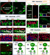
Figure 2. Distinct neuronal cell types are generated along the D/V axis of the OPC
a. Lateral view of the third instar medulla cortex with Bsh (red) and Svp (green). b. Hth+ neurons (blue) in the cOPC can be divided into mutually exclusive Bsh (red) and Svp (green) populations. c. Svp (green) and Vsx1 (red) are co-expressed in a subset of cOPC-derived Hth+ neurons (blue). d. Hth+ neurons in the cOPC (blue) can be divided into NotchON (Ap in red) and NotchOFF (Hth alone) populations. e. Vsx1 (red) is not expressed in the NotchON (ap-lacZ in green) Hth+ neurons of the cOPC. f. A Pm3 neuron (GFP in green) generated by flip-out with Vsx1-Gal4. Photoreceptors are labeled with 24B10 (red). g. Cross-section view of the Svp-positive neurons of the pOPC (green). Only the ventral population of pOPC neurons expresses Tsh (red). h–i. Single cell flip-out clones using 27b-Gal4 label Pm2 (h) and Pm1 (i) neurons. j. Tsh (red) is not expressed in the NotchON (ap-lacZ in green) Hth+ neurons of the pOPC. k. Cleaved-caspase-3 (green) marks dying cells that are intermingled with NON Hth+ in the mOPC. l–n. Schematic models summarizing neurogenesis in Hth+ neuroblasts of different compartments along the D/V axis of the OPC.
Figure 3
Figure 3. Temporal and spatial inputs are required for neuronal specification in the medulla
a. In hth mutant MARCM clone, Svp expression (green) is lost but Vsx1 (red) is unaffected in the cOPC. b. Svp expression (green) in lost in the pOPC in hth mutant MARCM clone. c and d. Bsh (red) is ectopically expressed in the progeny of older neuroblasts in hth gain-of-function (GOF) clones in the cOPC (c) or in the pOPC (d). Svp expression (green) is unaffected in both case (c and d). e–j. Bsh (red) and Svp (green) expression in different LOF and GOF. e. Vsx1 RNAi loss-of-function (LOF) clone in the cOPC. f. Vsx1 GOF clone in the mOPC. g. Rx LOF mutant MARCM clone in the pOPC. h. Rx GOF clones in the mOPC. i. Optix LOF mutant MARCM clone in the mOPC,. j. Optix GOF clone in the cOPC. k. Lateral view of the third instar medulla cortex. Svp (green) is ectopically expressed in Rx GOF clones. The Pm1 marker Tsh (red) is ectopically expressed in ventral mOPC clones (solid arrow) but is not in clones in the dorsal mOPC or ventral cOPC (outlined arrows). l. Wild-type adult optic lobe with 27b-Gal4 driving GFP expression (green) in Pm1 and Pm2 neurons in the medulla rim and Tm1 neurons in the cortex. The neuropil is labeled in red with N-Cadherin. m. In Rx mutant adult optic lobe Pm1 and Pm2 neurons are absent from the medulla rim (boxed region). Tm1 neurons are unaffected.
Figure 4
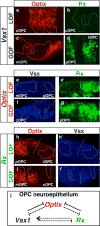
Figure 4. Spatial genes cross-regulate each other in the OPC neuroepithelium
a–i′. Expression of Optix (red), Rx (green), and Vsx1 (blue) in different clones. a. and b. Expression of Optix and Rx in Vsx1 RNAi LOF clones in the cOPC neuroepithelium. c. and d. Expression of Optix and Rx in Vsx1 GOF clones in the OPC NE. e. and e′. Expression of Vsx and Rx in Optix mutant MARCM clones in the OPC NE. f. and g. Expression of Vsx and Rx in Optix GOF clones in the OPC NE. h. and h′. Expression of Optix and Vsx in Rx mutant MARCM clones in the OPC NE. i. and i′. Expression of Optix and Vsx in Rx GOF clones in the cOPC NE. j. The spatial genes negatively cross-regulate each other in the OPC NE. Filled line indicates that genes are necessary and sufficient while dotted line indicates sufficiency only.
Figure 5
Figure 5. Neuronal movement during medulla development is restricted to multi-columnar cell-types
a–d. Lineage trace of mOPC-derived neurons using Optix-Gal4 (a and c) or cOPC-derived neurons using Vsx1-Gal4 (b and d) in larva (a and b) or adult (c and d). The dotted lines indicate the enrichment of GFP+ neurons in the center of the medulla cortex in d. e. Vsx1 lineage-trace in the adult medulla. cOPC derived cells (βgal in green) are located throughout the medulla cortex but only overlap with Bsh (red) in the center. Note that Bsh nuclei are located in the distal medulla cortex. f. hh lineage trace in the adult medulla. Ventrally derived cells (βgal in red) are located throughout the medulla cortex but enriched ventrally. Cells that have moved dorsally are mainly located in the the proximal half of the cortex whereas. g. and h. MARCM clones generated in 3rd instar larvae and visualized in the adult medulla (green). (g) Cells in clones labeled by a Gal4 driver for the uni-columnar cell-type Tm3 remain clustered in the adult (arrows). (h) Cells in clones marked with a Gal4 driver for the multi-columnar cell type Dm12 are dispersed in the adult (arrows).
Figure 6
Figure 6. Model for the generation of retinotopy in the Drosophila medulla
a. Uni-columnar neurons are refractory to regional inputs. Uni-columnar neurons (Mi1, Tm1, Tm2…) are born throughout the dorsal-ventral axis of the larval OPC and remain in place through to adulthood. In the adult each neuron innervates only one column. Uni-columnar cell bodies are located in the distal medulla cortex. Two examples are shown. b. Non-columnar neurons (Pm1, Pm2, Pm3…) are regionally specified and either move to take up their final position in the medulla cortex or send long tangential projections into the neuropil from the medulla rim. Three examples are shown: Red+green neurons are born in the cOPC and receive input from both Hth neuroblasts and Vsx1. Blue+green and/or brown neurons are generated in the ventral and dorsal pOPC regions and receive input from Hth neuroblasts and Rx. They populate the adult medulla rim and send large branches into the medulla neuropil. Non-columnar cell bodies are located in the proximal medulla cortex in the adult.

References

    1. Fischbach KF, D APM. The optic lobe of Drosophila melanogaster. I. A Golgi analysis of wild-type structure. Cell Tissue Res. 1989;258:441–475.
    1. Morante J, Desplan C. The color-vision circuit in the medulla of Drosophila. Curr Biol. 2008;18:553–565. doi: 10.1016/j.cub.2008.02.075. - DOI - PMC - PubMed
    1. Bausenwein B, Dittrich AP, Fischbach KF. The optic lobe of Drosophila melanogaster. II. Sorting of retinotopic pathways in the medulla. Cell Tissue Res. 1992;267:17–28. - PubMed
    1. Gao S, et al. The neural substrate of spectral preference in Drosophila. Neuron. 2008;60:328–342. doi: 10.1016/j.neuron.2008.08.010. - DOI - PMC - PubMed
    1. Mast JD, Prakash S, Chen PL, Clandinin TR. The mechanisms and molecules that connect photoreceptor axons to their targets in Drosophila. Semin Cell Dev Biol. 2006;17:42–49. doi: 10.1016/j.semcdb.2005.11.004. - DOI - PubMed

Publication types

MeSH terms

LinkOut - more resources