Dorsal Scapular Artery Variations and Relationship to the Brachial Plexus, and a Related Thoracic Outlet Syndrome Case
- PMID: 28077957
- PMCID: PMC5152701
- DOI: 10.1055/s-0036-1583756
Dorsal Scapular Artery Variations and Relationship to the Brachial Plexus, and a Related Thoracic Outlet Syndrome Case
Abstract
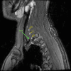
Rationale Knowledge of the relationship of the dorsal scapular artery (DSA) with the brachial plexus is limited. Objective We report a case of a variant DSA path, and revisit DSA origins and under-investigated relationship with the plexus in cadavers. Methods The DSA was examined in a male patient and 106 cadavers. Results In the case, we observed an unusual DSA compressing the lower plexus trunk, that resulted in intermittent radiating pain and paresthesia. In the cadavers, the DSA originated most commonly from the subclavian artery (71%), with 35% from the thyrocervical trunk. Nine sides of eight cadavers (seven females) had two DSA branches per side, with one branch from each origin. The most typical DSA path was a subclavian artery origin before passing between upper and middle brachial plexus trunks (40% of DSAs), versus between middle and lower trunks (23%), or inferior (4%) or superior to the plexus (1%). Following a thyrocervical trunk origin, the DSA passed most frequently superior to the plexus (23%), versus between middle and lower trunks (6%) or upper and middle trunks (4%). Bilateral symmetry in origin and path through the brachial plexus was observed in 13 of 35 females (37%) and 6 of 17 males (35%), with the most common bilateral finding of a subclavian artery origin and a path between upper and middle trunks (17%). Conclusion Variability in the relationship between DSA and trunks of the brachial plexus has surgical and clinical implications, such as diagnosis of thoracic outlet syndrome.
Keywords: anatomical variations; brachial plexus; dorsal scapular artery; subclavian artery; thoracic outlet syndrome; thyrocervical trunk.
Conflict of interest statement
Figures


References
-
- Huelke D F. A study of the transverse cervical and dorsal scapular arteries. Anat Rec. 1958;132(3):233–245. - PubMed
-
- Chaijaroonkhanarak W, Kunatippapong N, Ratanasuwan S. et al.Origin of the dorsal scapular artery and its relation to the brachial plexus in Thais. Anat Sci Int. 2014;89(2):65–70. - PubMed
-
- Murata H, Sakai A, Hadzic A, Sumikawa K. The presence of transverse cervical and dorsal scapular arteries at three ultrasound probe positions commonly used in supraclavicular brachial plexus blockade. Anesth Analg. 2012;115(2):470–473. - PubMed
-
- Read W T, Trotter M. The origins of transverse cervical and of transverse scapular arteries in American Whites and Negroes. Am J Phys Anthropol. 1941;28(2):239–247.
Grants and funding
LinkOut - more resources
Full Text Sources
Other Literature Sources
Molecular Biology Databases

