. 2016 Dec 15;8(12):5715-5722.
eCollection 2016.

Immunoglobulin E induces colon cancer cell apoptosis via enhancing cyp27b1 expression

Affiliations

Immunoglobulin E induces colon cancer cell apoptosis via enhancing cyp27b1 expression

Zhen-Shi Ye et al. Am J Transl Res. .

Abstract

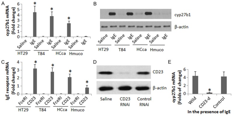
The pathogenesis of colon cancer (Cca) is to be further investigated. Vitamin D deficiency is associated with cancer growth; the underlying mechanism is unclear. Published data indicate that Cca cells express CD23. This study tests a hypothesis that exposure to IgE induces Cca cell apoptosis. In this study, the effect of ligation of CD23 by IgE on the expression of cyp27b1 was performed with Cca cells. The induction of apoptosis of Cca cells by IgE was assessed in a cell culture model. We observed that Cca cells express CD23; ligation of CD23 with IgE on Cca cells increased the expression of cyp27b1 in Cca, which promoted the conversion of VD3 to calcitriol, the latter increased the expression of FasL by Cca cells, and induced apoptosis of Cca cells. In conclusion, IgE is capable of inducing the cancer cell apoptosis via ligating CD23 and converting VD3 to calcitriol. The results suggest that IgE may have therapeutic potential in the treatment of Cca.

Keywords: Cyp27b1; Vitamin D; apoptosis; cancer; colon.


Figures

Figure 1
Figure 1
Ligation of CD23 increases cyp27b1 expression in HT29 cells. (A, B) The bars indicate the mRNA (A), the immune blots indicate the protein (B), of cyp27b1 in HT29 cells, T84 cells, human colon cancer cells (HCca) and human normal colon mucosa (Hmuco) after exposure to IgE (100 ng/ml; Antibodies Online) in the culture for 48 h. (C) The bars indicate the mRNA of FcεRI and CD23 in HT29 cells, T84 cells, HCca and Hmuco. (D) The immune blots show the results of CD23 RNAi in HT29 cells. (E) The bars indicate the mRNA of cyp27b1 in wild HT29 cells, or CD23-deficient (CD23-d) HT29 cells, or HT29 cells treated with control shRNA. The data are representatives of 3 independent experiments.
Figure 2
Figure 2
cyp27b1 modulates apoptosis in HT29 cells. The gated dot plots of (A-G) show the frequency of apoptotic cells. The treatment is denoted above each subpanel of the dot plots. IgE: HT29 cells were exposed to IgE (100 ng/ml) in the culture for 48 h. Cyp27b1-deficient: HT29 cells were treated with RNAi of cyp27b1. Control shRNA: HT29 cells were treated with control shRNA. (H) The bars show the summarized data of apoptotic HT29 cells in panels A-G. The data are summarized from 3 independent experiments. *, P<0.01, compared with group A.
Figure 3
Figure 3
IgE promotes VD3 metabolism in colon cancer cells. A. The immune blots indicate the protein levels of calcitriol in colon cancer cells after the treatment with IgE (100 ng/ml) or/and VD3 (50 nM) for 48 h. #, cyp27b1-deficient colon cancer cells. $, colon cancer cells were treated with control shRNA. B. The immune blots show the RNAi results of cyp27b1 in colon cancer cells (stimulated by IgE). C. The immune blots indicate the complex of calcitriol/VDR/RXR in HT29 cells after exposure to IgE/VD3 in the culture for 48 h. IgG: An isotype IgG using as a negative control. The data are a representative from 3 independent experiments.
Figure 4
Figure 4
cyp27b1 mediates IgE-induced apoptosis-related activities in HT29 cells. A, B. The bars indicate the mRNA levels of Fas and FasL in wild or cyp27b1-deficient (cyp27b1-d) HT29 cells after exposure to IgE (100 ng/ml) in the culture for 48 h. C. The bars indicate the binding to the FasL promoter in HT29 cells by the molecules denoted on the X axis after exposure to IgE or saline in the culture (by ChIP). D-G. The immune blots indicate the changes of FasL, Bax, caspase-3 and caspase-8 in HT29 cells after exposure to IgE or saline in the culture. Data of bars are summarized from 3 independent experiments and presented as mean ± SD. *, P<0.01, compared with the saline group. The immune blots are from one experiment that represents 3 independent experiments. Control HT29 cells: HT29 cells were treated with control shRNA.

References

    1. Smith RA, Andrews K, Brooks D, DeSantis CE, Fedewa SA, Lortet-Tieulent J, Manassaram-Baptiste D, Brawley OW, Wender RC. Cancer screening in the United States, 2016: a review of current American Cancer Society guidelines and current issues in cancer screening. CA Cancer J Clin. 2016;66:95–114. - PubMed
    1. Pai SG, Fuloria J. Novel therapeutic agents in the treatment of metastatic colorectal cancer. World J Gastrointest Oncol. 2016;8:99–104. - PMC - PubMed
    1. Arunachalam L, O’Grady H, Hunter IA, Killeen S. A systematic review of outcomes after transanal mesorectal resection for rectal cancer. Dis Colon Rectum. 2016;59:340–350. - PubMed
    1. Gulbake A, Jain A, Jain A, Jain A, Jain SK. Insight to drug delivery aspects for colorectal cancer. World J Gastroenterol. 2016;22:582–599. - PMC - PubMed
    1. Cho Y, Turner ND, Davidson LA, Chapkin RS, Carroll RJ, Lupton JR. Colon cancer cell apoptosis is induced by combined exposure to the n-3 fatty acid docosahexaenoic acid and butyrate through promoter methylation. Exp Biol Med (Maywood) 2014;239:302–310. - PMC - PubMed

LinkOut - more resources