. 2016 Nov 16;4(1):4-14.
doi: 10.1002/acn3.361. eCollection 2017 Jan.

POLG2 deficiency causes adult-onset syndromic sensory neuropathy, ataxia and parkinsonism

Affiliations

POLG2 deficiency causes adult-onset syndromic sensory neuropathy, ataxia and parkinsonism

Lionel Van Maldergem et al. Ann Clin Transl Neurol. .

Abstract

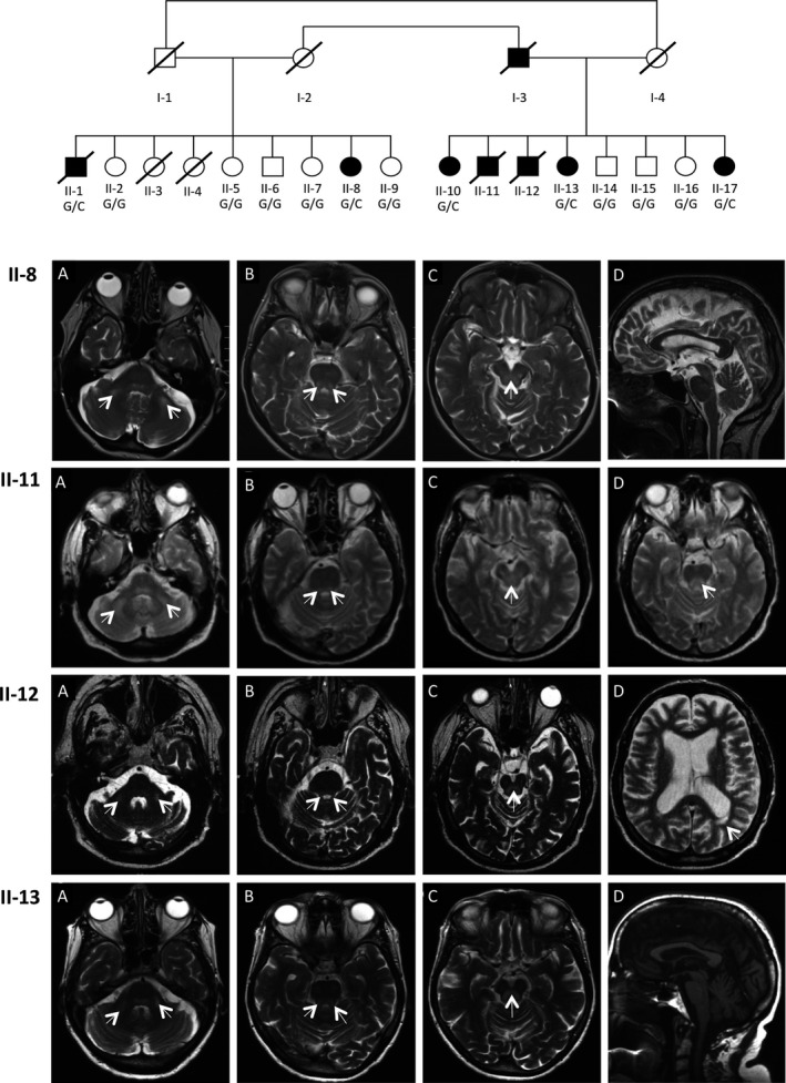
Objective: Mitochondrial dysfunction plays a key role in the pathophysiology of neurodegenerative disorders such as ataxia and Parkinson's disease. We describe an extended Belgian pedigree where seven individuals presented with adult-onset cerebellar ataxia, axonal peripheral ataxic neuropathy, and tremor, in variable combination with parkinsonism, seizures, cognitive decline, and ophthalmoplegia. We sought to identify the underlying molecular etiology and characterize the mitochondrial pathophysiology of this neurological syndrome.

Methods: Clinical, neurophysiological, and neuroradiological evaluations were conducted. Patient muscle and cultured fibroblasts underwent extensive analyses to assess mitochondrial function. Genetic studies including genome-wide sequencing were conducted.

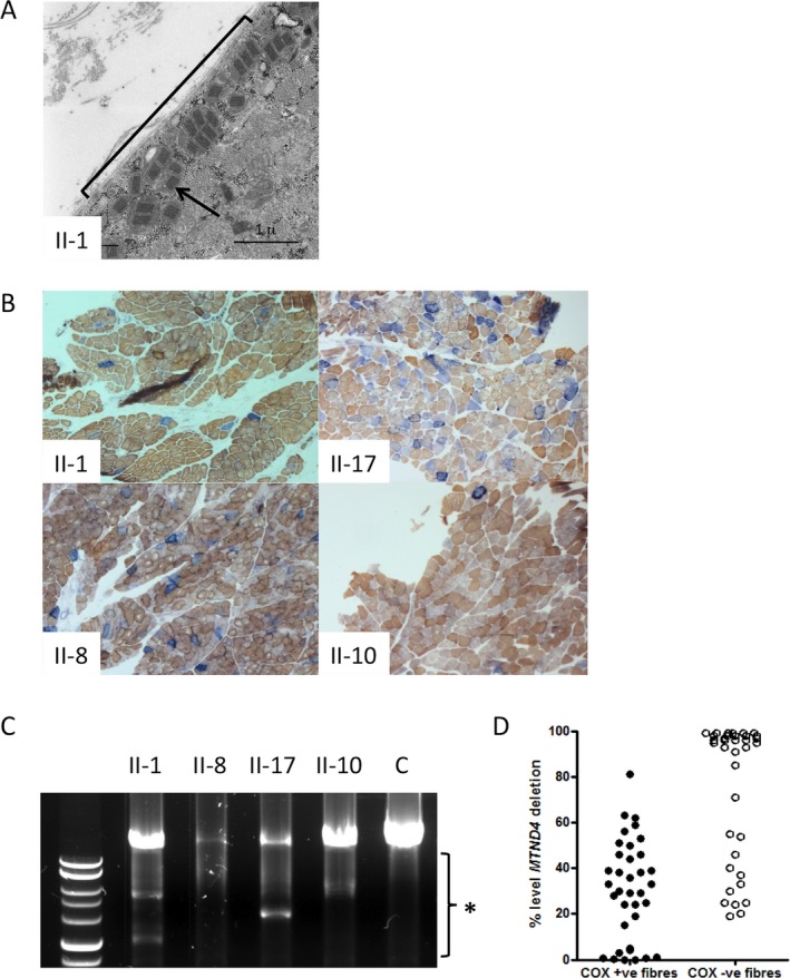
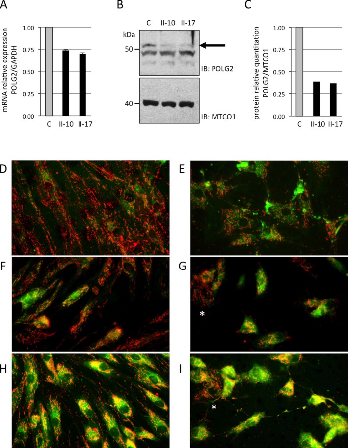
Results: Hallmarks of mitochondrial dysfunction were present in patients' tissues including ultrastructural anomalies of mitochondria, mosaic cytochrome c oxidase deficiency, and multiple mtDNA deletions. We identified a splice acceptor variant in POLG2, c.970-1G>C, segregating with disease in this family and associated with a concomitant decrease in levels of POLG2 protein in patient cells.

Interpretation: This work extends the clinical spectrum of POLG2 deficiency to include an overwhelming, adult-onset neurological syndrome that includes cerebellar syndrome, peripheral neuropathy, tremor, and parkinsonism. We therefore suggest to include POLG2 sequencing in the evaluation of ataxia and sensory neuropathy in adults, especially when it is accompanied by tremor or parkinsonism with white matter disease. The demonstration that deletions of mtDNA resulting from autosomal-dominant POLG2 variant lead to a monogenic neurodegenerative multicomponent syndrome provides further evidence for a major role of mitochondrial dysfunction in the pathomechanism of nonsyndromic forms of the component neurodegenerative disorders.


Figures

Figure 1
Figure 1
Pedigree and MRIs for family segregating POLG2 c.970‐1G>C variant. Affected individuals are represented as black symbols, whereas unaffected are open symbols. The genotype for the POLG2 c.970‐1G>C variant is indicated for individuals who were genotyped. Brain MRIs reveal abnormalities of the cerebellum and brain stem and white matter lesions. Axial T2‐weighted images revealed bilateral hyperintense lesions of (A) middle cerebellar peduncles, (B) upper cerebellar peduncles, and (C) periaqueductal grey matter in patients II‐8, II‐11, II‐12, and II‐13. Also shown are the following: II‐8 (D) Sagittal T2 image demonstrating mild supra and infratentorial atrophylenticular nuclei; II‐11 (D) Axial T2 image showing hypersignal of medial longitudinal fasciculus; II‐12 (D) Axial T2 image showing important hemispheric atrophy and periventricular white matter lesion; and II‐13 (D) Sagittal T1 image demonstrating moderate pontocerebellar and supratentorial atrophy.
Figure 2
Figure 2
Electron microscopy, histochemistry, and mtDNA analyses in patient muscle show signs of mitochondrial dysfunction. (A) Electron microscopy of Patient II‐1 skeletal muscle reveals subsarcolemmal accumulation of abnormal mitochondria with paracrystalline inclusions. The general region of subsarcolemnal accumulation is indicated with a black bracket and a black arrow points to one of the paracrystalline inclusions. (B) Sequential COX succinate dehydrogenase (SDH) histochemistry demonstrates a mosaic distribution of COX‐deficient muscle fibers (blue) among fibers exhibiting normal COX activity (brown). Illustrated are the images for Patients II‐1, II‐17, II‐8, and II‐10. (C) Long‐range PCR across the major mtDNA shows evidence of variable mtDNA deletions in muscle from patients II‐1, II‐17, II‐8, and II‐10. The black bracket indicates the region of the gel where deletion fragments segregate. (D) Quantitative, single‐fiber real‐time PCR reveals the majority—but not all—of COX‐deficient fibers contain high levels of a clonally expanded mtDNA deletion involving the MTND4 gene (36 COX‐positive and 36 COX‐deficient fibers laser captured from the biopsy material of patients II‐1, II‐8, and II‐17).
Figure 3
Figure 3
POLG2 mRNA and protein levels and mitochondrial membrane potential are decreased in patient fibroblasts. The level of POLG2 RNA and protein was measured in control (C) and patient cells (II‐10 and II‐17). (A) Quantitative real‐time PCR experiments were performed for human GAPDH and POLG2 and expressed as a relative quantitation of POLG2/GAPDH. (B) Mitochondrial lysates were analyzed by Western blotting using a rabbit polyclonal antisera raised against the recombinant human POLG2 protein. POLG2 is represented by the top band visible migrating at 55 kilodaltons (kDa), while two additional lower bands represent nonspecific binding. The black arrow points to the POLG2 band to help distinguish it from the nonspecific bands. (C) Relative quantitation of protein was measured by comparing the intensity of POLG2 and MTCO1 bands in controls and patients. D‐I. JC‐1 fluorescence studies of patient fibroblasts show reduced mitochondrial membrane potential. Imaging visualized mitochondria with inactive (green) or active (red) membrane potential. Membrane potential was assessed in untreated (D, F, H) and rotenone‐treated (E, G, I) fibroblast cells. Control fibroblasts are shown in D, E; Patient II‐1 fibroblast in F, G; Patient II‐17 fibroblast in H, I. A mosaic staining pattern of individual cells is present in patient fibroblasts, with some cells displaying predominantly red staining indicative of active mitochondria in these cells (these cells are marked with a white asterisk).

References

    1. Sommerville EW, Chinnery PF, Gorman GS, et al. Adult‐onset Mendelian PEO associated with mitochondrial disease. J Neuromusc Dis 2014;1:119–133. - PubMed
    1. Longley MJ, Clark S, Yu Wai Man C, et al. Mutant POLG2 disrupts DNA polymerase gamma subunits and causes progressive external ophthalmoplegia. Am J Hum Genet 2006;78:1026–1034. - PMC - PubMed
    1. Walter MC, Czermin B, Muller‐Ziermann S, et al. Late‐onset ptosis and myopathy in a patient with a heterozygous insertion in POLG2 . J Neurol 2010;257:1517–1523. - PubMed
    1. Young MJ, Longley MJ, Li FY, et al. Biochemical analysis of human POLG2 variants associated with mitochondrial disease. Hum Mol Genet 2011;20:3052–3066. - PMC - PubMed
    1. He L, Chinnery PF, Durham SE, et al. Detection and quantification of mitochondrial DNA deletions in individual cells by real‐time PCR. Nucleic Acids Res 2002;30:e68. - PMC - PubMed