. 2019 Jan 1;527(1):225-235.
doi: 10.1002/cne.24172. Epub 2017 Mar 10.

Expression of transcription factors divides retinal ganglion cells into distinct classes

Affiliations

Expression of transcription factors divides retinal ganglion cells into distinct classes

Neal T Sweeney et al. J Comp Neurol. .

Abstract

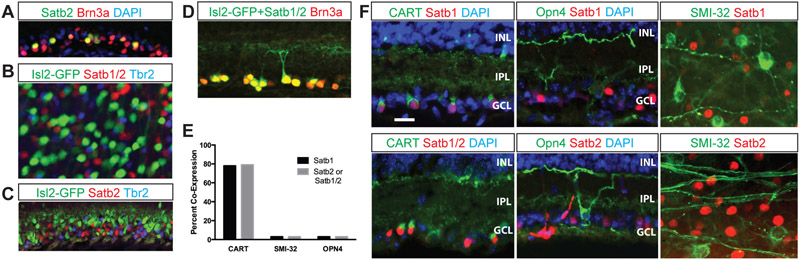
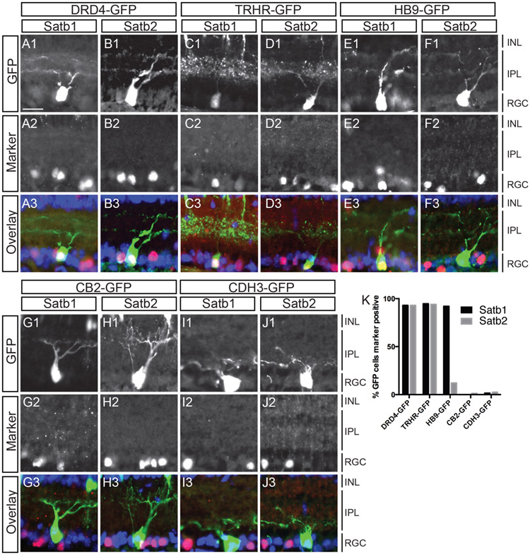
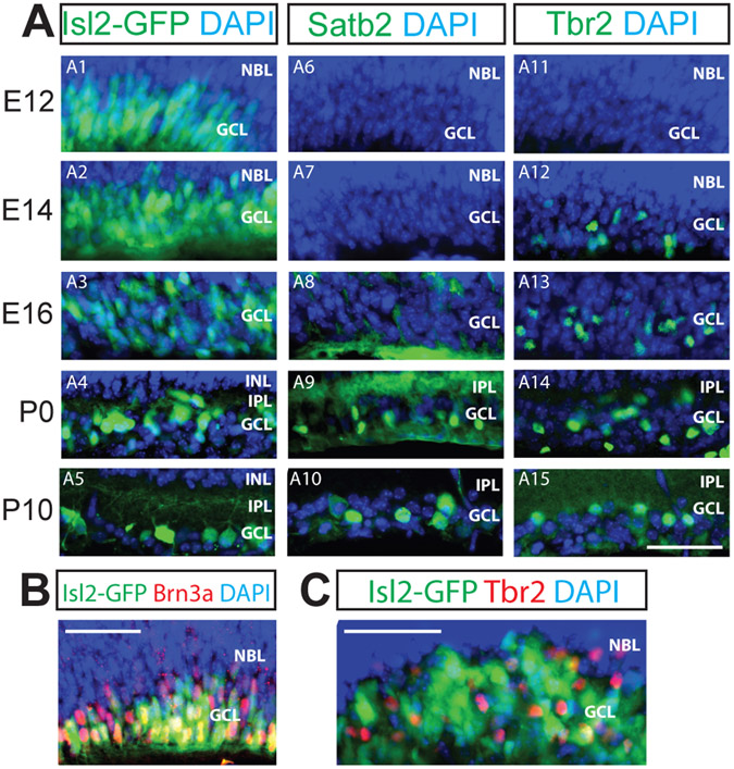
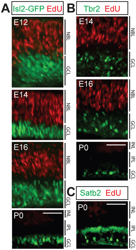
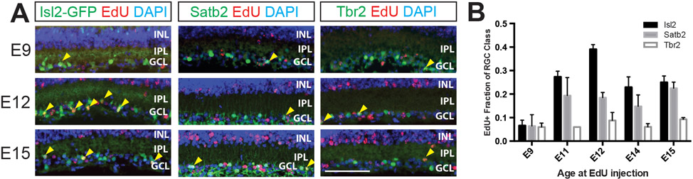
Retinal ganglion cells (RGCs) are tasked with transmitting all light information from the eye to the retinal recipient areas of the brain. RGCs can be classified into many different types by morphology, gene expression, axonal projections, and functional responses to different light stimuli. Ultimately, these classification systems should be unified into an all-encompassing taxonomy. Toward that end, we show here that nearly all RGCs express either Islet-2 (Isl2), Tbr2, or a combination of Satb1 and Satb2. We present gene expression data supporting the hypothesis that Satb1 and Satb2 are expressed in ON-OFF direction-selective (DS) RGCs, complementing our previous work demonstrating that RGCs that express Isl2 and Tbr2 are non-DS and non-image-forming, respectively. Expression of these transcription factors emerges at distinct embryonic ages and only in postmitotic cells. Finally, we demonstrate that these transcription factor-defined RGC classes are born throughout RGC genesis.

Keywords: RRID: AB_10615604; RRID: AB_11143446; RRID: AB_1608077; RRID: AB_2167511; RRID: AB_2301417; RRID: AB_2313614; RRID: AB_231491; RRID: AB_882455; cell fate; retinal ganglion cells; transcription factors.


Conflict of interest statement

Conflict of interest statement: The authors state that they have no conflict of interest to declare.

Figures

Figure 1.
Figure 1.. Isl2, Satb1, Satb2, and Tbr2 mark distinct RGC classes.
20 μm cryosections through adult retinas (unless otherwise indicated) were immunostained with the antibodies indicated; Isl2-GFP is native GFP from the transgenic line used. Satb1/2 refers to the use of Abcam ab51502 antibody that recognizes both Satb1 and Satb2. The color of the text matches the color of the fluorescent label or the nuclear DAPI staining. A: Section through adult ganglion cell layer of the retina showing Satb2-expressing RGCs also express Brn3a. B: Flat-mounted adult retina shows that Isl2, Satb1/2 and Tbr2 are expressed in largely non-overlapping populations of RGCs. C: 20 μm P0 cryosection stained with Isl2, Satb2 and Tbr2 show largely non-overlapping expression. D: Nearly all Brn3a-expressing cells in the RGC layer co-express either Isl2 or Satb1/2 (98%; 360 Brn3a-expressing cells counted) E: Quantification of staining data from F for either Satb2 staining (Abcam 34735), Satb1 staining (SC SC-5989) or Satb1/2 staining (Abcam 51502). F: Adult cryosections (CART, Opn4) or retinal flat mount (SMI-32) show that very few SMI-32-expressing or Opn4-expressing RGCs express Satb1 or Satb2 (SMI-32: 0% of Satb1-expressing, 103 Satb1-expressing cells counted; 0% of Satb2-expressing, 118 Satb2-expressing cells counted; Opn4: 0% of Satb1-expressing, 102 Satb1-expressing cells counted; 0% of Satb1/2, 97 Satb2-expressing cells counted), but a large percentage (79% of Satb1, 130 Satb1-expressing cells counted; 79% of Satb1/2, 92 Satb1/2-expressing cells counted) of CART-expressing RGCs also express Satb1/2 (and Satb2). IPL, inner plexiform layer; INL, inner nuclear layer; GCL, ganglion cell layer. Scale bar in F applies to all panels, 20 μm.
Figure 2.
Figure 2.. Satb1 and Satb2 are expressed in GFP-labeled On-Off DS RGCs but not in other defined RGC types.
(A-J) Sections through P8 eyes from BAC transgenic GFP lines were immunostained with antibodies against Satb1 and Satb2. Anti-GFP antibodies were used to increase signal and sections were treated with DAPI (blue) to visualize cell nuclei. Nearly all DRD4-GFP RGCs express Satb1 (92%; n=57) and Satb2 (96%; n=52). TRHR-GFP RGCs also express both Satb1 (95%; n=114) and Satb2 (94%; n=50). HB9-GFP RGCs express Satb1 (92%; n=51) but not Satb2 (3%; n=78) (E-F). Very few CB2-GFP RGCs were labeled by Satb1 (1%; n=85) or Satb2 (12%; n=74) and similarly, very few Cdh3-GFP RGCs expressed Satb1 (1.5%; n=66) or Satb2 (1.1%; n=85)(G-J). (K) Graphical representation of A-J. ONL, Outer nuclear layer; IPL, inner plexiform layer; INL, inner nuclear layer; GCL, ganglion cell layer. Scale bar: 20 uM in A for panels A-J.
Figure 3.
Figure 3.. Developmental onset of expression differs for Isl2, Satb2 and Tbr2.
Panels are labeled as in Figure 1. 20 μm (A, C) or 12 μm (B) retinal cryosections at the embryonic ages indicated on the left were immunostained with the antibodies indicated. A: Isl2 is widely expressed in the RGC layer at E12 and is gradually refined to its final expression pattern (A1-A5). Satb2 expression is first detected at E16 and becomes refined to the GCL at P0; Tbr2 expression is first detected at E14 (A11-A15). B: At E14, nearly all Brn3a-expressing RGCs also express Isl2 (97% of Brn3a-expressing cells; 347 Brn3a-expressing cells counted). C: At E16, few Tbr2-expressing RGCs colocalize with Isl2-expressing RGCs. NBL, neuroblast layer; IPL, inner plexiform layer; INL, inner nuclear layer; GCL, ganglion cell layer. Scale bars, 50 μm.
Figure 4.
Figure 4.. Isl2, Satb2 and Tbr2 expression is limited to post-mitotic RGCs.
20 μm retinal cryosections from mice injected with EdU at the ages indicated and sacrificed 4 hours later were immunostained with the antibodies indicated, following EdU visualization. A: EdU labeled RPCs at E12, E14, E16, and P0 do not express Isl2. B: Tbr2 is not expressed by progenitor cells at E14, E16 or P0. C: Satb2 is not expressed by progenitor cells at P0. NBL, neuroblast layer; INL, inner nuclear layer; IPL, inner plexiform layer; GCL, ganglion cell layer. For all three transcription factors, no cellular colocalization with Edu label was observed: n=3-5 animals/age, 5-7 sections/animal examined. Scale bars, 50 μm.
Figure 5.
Figure 5.. Isl2-, Satb2- and Tbr2-expressing RGCs are born over similar, broad windows of RGC differentiation.
20 μm P10 retinal cryosections from mice injected with EdU at the ages indicated were immunostained with the antibodies indicated after EdU visualization. A: RGCs expressing Isl2, Satb2, and Tbr2 can be EdU labeled at E9, E12 and E15. Yellow arrowheads indicate cells co-expressing EdU (red) and transgenic GFP (Isl2-GFP) or the transcription factor stained for (Satb2, Tbr2) in each column (green). Scale bar, 50 μm. B: Quantification of EdU birthdating experiment for five ages. The Y-axis measures the fraction of mature RGCs of each class that is EdU labeled, following mitotic labeling on the embryonic day indicated. INL, inner nuclear layer; IPL, inner plexiform layer; GCL, ganglion cell layer. N=3-5 animals, 5-6 images quantified per animal. Error bars represent S.E.M.

References

    1. Agrelo R, Souabni A, Novatchkova M, Haslinger C, Leeb M, Komnenovic V, Kishimoto H, Gresh L, Kohwi-Shigematsu T, Kenner L, et al. (2009). SATB1 defines the developmental context for gene silencing by Xist in lymphoma and embryonic cells. Dev Cell 16, 507–516. - PMC - PubMed
    1. Ahlfors H, Limaye A, Elo LL, Tuomela S, Burute M, Gottimukkala KV, Notani D, Rasool O, Galande S, and Lahesmaa R (2010). SATB1 dictates expression of multiple genes including IL-5 involved in human T helper cell differentiation. Blood 116, 1443–1453. - PMC - PubMed
    1. Anishchenko A, Greschner M, Elstrott J, Sher A, Litke AM, Feller MB, and Chichilnisky EJ (2010). Receptive field mosaics of retinal ganglion cells are established without visual experience. J Neurophysiol 103, 1856–1864. - PMC - PubMed
    1. Badea TC, Cahill H, Ecker J, Hattar S, and Nathans J (2009). Distinct roles of transcription factors brn3a and brn3b in controlling the development, morphology, and function of retinal ganglion cells. Neuron 61, 852–864. - PMC - PubMed
    1. Baden T, Berens P, Franke K, Roman Roson M, Bethge M, and Euler T (2016). The functional diversity of retinal ganglion cells in the mouse. Nature 529, 345–350. - PMC - PubMed

Publication types

Substances

LinkOut - more resources