. 2017 Feb;9(1):1759091416687871.
doi: 10.1177/1759091416687871.

C5a Increases the Injury to Primary Neurons Elicited by Fibrillar Amyloid Beta

Affiliations

C5a Increases the Injury to Primary Neurons Elicited by Fibrillar Amyloid Beta

Michael X Hernandez et al. ASN Neuro. 2017 Feb.

Abstract

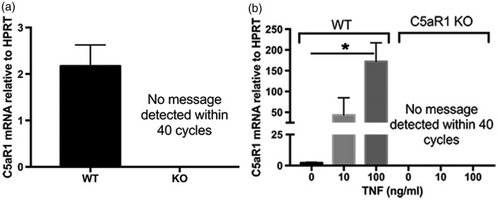
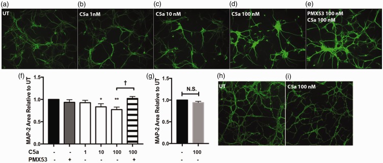
C5aR1, the proinflammatory receptor for C5a, is expressed in the central nervous system on microglia, endothelial cells, and neurons. Previous work demonstrated that the C5aR1 antagonist, PMX205, decreased amyloid pathology and suppressed cognitive deficits in two Alzheimer's Disease (AD) mouse models. However, the cellular mechanisms of this protection have not been definitively demonstrated. Here, primary cultured mouse neurons treated with exogenous C5a show reproducible loss of MAP-2 staining in a dose-dependent manner within 24 hr of treatment, indicative of injury to neurons. This injury is prevented by the C5aR1 antagonist PMX53, a close analog of PMX205. Furthermore, primary neurons derived from C5aR1 null mice exhibited no MAP-2 loss after exposure to the highest concentration of C5a tested. Primary mouse neurons treated with both 100 nM C5a and 5 µM fibrillar amyloid beta (fAβ), to model what occurs in the AD brain, showed increased MAP-2 loss relative to either C5a or fAβ alone. Blocking C5aR1 with PMX53 (100 nM) blocked the loss of MAP2 in these primary neurons to the level seen with fAβ alone. Similar experiments with primary neurons derived from C5aR1 null mice showed a loss of MAP-2 due to fAβ treatment. However, the addition of C5a to the cultures did not enhance the loss of MAP-2 and the addition of PMX53 to the cultures did not change the MAP-2 loss in response to fAβ. Thus, at least part of the beneficial effects of C5aR1 antagonist in AD mouse models may be due to protection of neurons from the toxic effects of C5a.

Keywords: C5a; C5aR1; amyloid; complement; neurodegeneration; primary neurons.


Figures

Figure 1.
Figure 1.
C5aR1 expression on primary neurons. Primary neurons from WT mice and C5aR1KO mice were generated using E15-E16 pups, cultured for 7 to 10 days, and RNA was collected from either (a) unstimulated cultures (WT n = 4; KO n = 2) or from (b) cultures treated with 0, 10, or 100 ng/ml of TNF-α for 24 hr (n = 2 for WT cells and n = 1 for C5aR1 knock out cells). Quantitative RT-PCR for C5aR1 expression relative to HPRT was performed with technical triplicates in each experiment. p values are calculated using unpaired two-tailed t test. Values of p < .05 were considered statistically significant.
Figure 2.
Figure 2.
C5a causes MAP-2 loss in a dose-dependent manner. Primary neurons from WT mice (a–e and f) or C5aR1KO mice (h–i and g) were generated using E15-E16 pups and cultured for 7 to 10 days. The cells were then stimulated with the indicated concentrations of hC5a, or 100 nM PMX53 for 24 hr. MAP-2 was visualized by immunocytochemistry (10× magnification) and quantified (f–g) using ImageJ software as described in M & M. Data are presented as percentage of MAP-2 area relative to untreated (UT) ± SEM. n = 4 independent experiments (f) and n = 3 independent experiments (g), each experiment having three coverslips per treatment, three to five images per coverslip. p < .05 *, p < .01 ** relative to untreated cultures using one-way ANOVA, uncorrected Fisher's LSD test (F), and p < .02 † unpaired two-tailed t test (f, g).
Figure 3.
Figure 3.
C5a increases toxicity in fAβ-treated neurons. Primary neurons from WT mice (a–e and k) or C5aR1KO mice (f–j and l) were generated using E15-E16 pups and cultured for 7 to 10 days. The cells were then stimulated or not with 5 µM fAβ, 100 nM hC5a, and 100 nM PMX53 for 24 hr. MAP-2 was visualized by immunocytochemistry (20 × magnification) and quantified using ImageJ software (k, l). Data are presented as mean MAP-2 area ± SEM of all image values from n = 3 independent experiments, each with three coverslips per treatment, five images per coverslip. p values are calculated using unpaired two-tailed t test. Values of p < .05 were considered statistically significant.
Figure 4.
Figure 4.
Primary neuronal cultures include GABAergic and glutamatergic neurons. Immunofluorescent staining of GAD67 (red, GABAergic neurons; left panel) or VGLUT1 (red, Glutamatergic neurons; right panel) in 7-day mouse neuron cultures. Nuclei are labeled with DAPI (blue; 10× magnification). Top right Inset in each panel is 20× magnification of region denoted by the arrow. Scale Bar: 50 µm.

Similar articles

Cited by

References

    1. Afagh A., Cummings B. J., Cribbs D. H., Cotman C. W., Tenner A. J. (1996) Localization and cell association of C1q in Alzheimer's disease brain. Experimental Neurology 138: 22–32. - PubMed
    1. Ager R. R., Fonseca M. I., Chu S. H., Sanderson S. D., Taylor S. M., Woodruff T. M., Tenner A. J. (2010) Microglial C5aR (CD88) expression correlates with amyloid-beta deposition in murine models of Alzheimer's disease. Journal of Neurochemistry 113: 389–401. - PMC - PubMed
    1. Aizenstein H. J., Nebes R. D., Saxton J. A., Price J. C., Mathis C. A., Tsopelas N. D., Ziolko S. K., James J. A., Snitz B. E., Houck P. R., Bi W., Cohen A. D., Lopresti B. J., DeKosky S. T., Halligan E. M., Klunk W. E. (2008) Frequent amyloid deposition without significant cognitive impairment among the elderly. Archives of Neurology 65: 1509–1517. - PMC - PubMed
    1. Akiyama H., et al. (2000) Inflammation and Alzheimer's disease. Neurobiology of Aging 21: 383–421. - PMC - PubMed
    1. Alexander J. J., Anderson A. J., Barnum S. R., Stevens B., Tenner A. J. (2008) The complement cascade: Yin-Yang in neuroinflammation-neuro-protection and -degeneration. Journal of Neurochemistry 107: 1169–1187. - PMC - PubMed

MeSH terms