. 2017 Jan 12:7:40476.
doi: 10.1038/srep40476.

Extracellular vesicle mediated intercellular communication at the porcine maternal-fetal interface: A new paradigm for conceptus-endometrial cross-talk

Affiliations

Extracellular vesicle mediated intercellular communication at the porcine maternal-fetal interface: A new paradigm for conceptus-endometrial cross-talk

Mallikarjun Bidarimath et al. Sci Rep. .

Abstract

Exosomes and microvesicles are extracellular vesicles released from cells and can contain lipids, miRNAs and proteins that affect cells at distant sites. Recently, microvesicles containing miRNA have been implicated in uterine microenvironment of pigs, a species with unique epitheliochorial (non-invasive) placentation. Here we report a novel role of conceptus-derived exosomes/microvesicles (hereafter referred to as extracellular vesicles; EVs) in embryo-endometrial cross-talk. We also demonstrate the stimulatory effects of EVs (PTr2-Exo) derived from porcine trophectoderm-cells on various biological processes including the proliferation of maternal endothelial cells (PAOEC), potentially promoting angiogenesis. Transmission immuno-electron microscopy confirmed the presence of EVs in tissue biopsies, PTr2-Exo and PAOEC-derived EVs (PAOEC-Exo). RT-PCR detected 14 select miRNAs in CD63 positive EVs in which miR-126-5P, miR-296-5P, miR-16, and miR-17-5P were the most abundant angiogenic miRNAs. Proteomic analysis revealed EV proteins that play a role in angiogenesis. In-vitro experiments, using two representative cell lines of maternal-fetal interface, demonstrated bidirectional EVs shuttling between PTr2 and PAOEC cells. Importantly, these studies support the idea that PTr2-Exo and PAOEC-Exo containing select miRNAs and proteins can be successfully delivered to recipient cells and that they may have a biological role in conceptus-endometrial cross-talk crucial for the pregnancy success.


Figures

Figure 1
Figure 1. Extracellular vesicles released by porcine endometrium and chorioallantoic membrane (CAM) were identified by transmission electron microscopy (TEM) on representative ultrathin sections.
(af) Endometrial and CAM biopsies were isolated from the conceptus attachment site at gestation day 20. TEM revealed vesicles of size in the range of approximately 50–150 nm, consistent with EVs in both the endometrium and CAM. Endometrial EVs (black arrows) appear to be localized in the extracellular space (a–c) while EVs (black arrows) in CAM are localized in the close proximity of cell membrane (d–f). Data is derived from three independent experiments. Scale bar: 500 nm.
Figure 2
Figure 2. Characterization of EVs isolated from culture supernatants of PTr2 and PAOEC cells.
(a) Transmission electron microscopy (TEM) of PTr2 derived EV pellets that are negatively stained with uranyl acetate and lead citrate. (b) Histogram of the number of isolated PTr2 derived EVs diameters. The Y axis shows the relative number of vesicles (%), and the × axis shows the vesicle diameter (nm). The size of EVs was approximately in the range of 26- to 125- nm (diameter [mean ± SD], 86 ± 21 nm). (c) TEM analysis of PAOEC derived EVs. (d) PAOEC derived EVs measured approximately in the range of 26- to 150- nm (diameter [mean ± SD], 99 ± 26 nm). (e) Western blotting detected CD63, exosomal marker, in the EV fraction as well as cellular fraction derived from both the PTr2 and PAOEC (f; cropped blots are displayed), respectively. (See also full-length blots in the Supplementary Figure S1). Calnexin (CANX) was only detected in cell lysates of PTr2 and PAOEC cells. (g) Characterization of EVs isolated from culture supernatant of PTr2 cells using transmission immunoelectron microscopy. Negatively stained EVs are labelled with 12-nm colloidal gold particles that recognize CD63 (black arrows) on the exosomal membrane. Data is derived from three independent experiments. Scale bar = 200 nm.
Figure 3
Figure 3. Porcine endometrium and CAM expresses CD63, a well characterized exosome marker.
CD63 immunoflourescence on formalin-fixed, paraffin-embedded porcine endometrial (a–c) and CAM (d–f) biopsies isolated at day 20 of gestation. Nuclei are stained with DAPI (blue; a,d,g), CD63 is stained with Anti-Rabbit CD63, reactive against pig (Red; b,e,h) followed by merge (c,f,i) to demonstrate its localization. *Endometrial stroma; LE: luminal epithelial layer of the uterus; Tr: Trophoblast, Magnification: 400x.
Figure 4
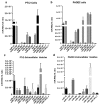
Figure 4. PTr2 and PAOEC derived EVs contain miRNAs.
(a) Real-time PCR confirmed the expression of literature curated 14 miRNAs that are involved in the angiogenesis regulation. In PTr2 cells, miR-16, miR-17-5P, miR-15b, let-7f, and miR-20a were relatively abundant. (b) PAOEC cells also expressed all miRNAs. Among these, miR-16, miR-17-5P, let-7f, miR-126-5P, and miR-296-5P were relatively abundant. (c) PTr2 derived EVs contain all 14 miRNAs; however, only miR-126-5P was relatively abundant compared to all other miRNAs. (d) PAOEC derived EVs only contained 10 out of 14 miRNAs while miR-126-5P being relatively abundant. miR-155-5P, miR-221-5P, let-7f, and miR-181c-1 were either absent or not detectable in the samples. Relative levels of miRNA expression normalized to RNU1A levels and data are presented as mean ± SEM. Data is derived from three independent experiments; n = 5.
Figure 5
Figure 5. Analysis of PTr2 and PAOEC derived-EV proteins identified by mass spectrometry using PANTHER software.
Extracellular vesicle proteins isolated from PTr2 and PAOEC cell supernatants were subjected to ontology and pathway analysis using PANTHER and Gene ontology algorithms and subsequently classified based on their (a) Biological Process and (b) Molecular function.
Figure 6
Figure 6. In vitro model of trophoblast-endothelial cell communication.
(a–l) In vitro transfer system using PTr2 cells are donors and PAOEC cells as recipients. Extracellular vesicles were isolated from culture supernatants of PTr2 cells. PTr2 derived fluorescently labelled EVs (20 μg/mL) were then added to PAOECs grown in a 6-well cell culture plate and allowed to incubate at 37 °C for 6 hrs (a–d) and 12 hrs (e–h) in a two set of experiments. PTr2 derived EVs were successfully internalized by the endothelial cells in a time dependent manner. Relative fluorescence emitted by the EVs was calculated in order to measure the concentration of EV uptake. There was a slight increase in the relative fluorescence indicating the increased uptake over a period of time (m). Nuclei are stained with DAPI (blue; a,e,i), cytoplasm is stained with CellTrackerTM Green BODIFY® dye (Green; b,f,j), EVs were labelled with CM-Dil (CellTracker, C7000; Red; c,g,k) and followed by merge (d,h,l) to demonstrate their localization in the cells. Control wells received the same preparation (PBS+DMSO+CM-Dil) except EVs in triplicate. Data is derived from three independent experiments.
Figure 7
Figure 7. In vitro model of endothelial-trophoblast cell communication.
(a–l) In vitro uptake of PAOEC derived EVs by PTr2 cells in a time dependent manner. PAOEC derived fluorescently labelled EVs (20 μg/mL) were then added to PTr2 cells grown in a 6-well cell culture plate and allowed to incubate at 37 °C for 6 hrs (a–d) and 12 hrs (e–h) in a two set of experiments. PTr2 cells were able to successfully uptake the PAOEC derived EVs in a time dependent manner. Concentration of EVs uptake was measured by calculating the relative fluorescence emitted by the PAOEC derived EVs. Slight decrease in the relative fluorescence was observed, indicating the disappearance of EVs in PTr2 cells over a period of time (m). Nuclei are stained with DAPI (blue; a,e,i), cytoplasm is stained with CellTrackerTM Green BODIFY® dye (Green; b,f,j), EVs were labelled with CM-Dil (CellTracker, C7000; Red; c,g,k) and followed by merge (d,h,l) to demonstrate their localization in the cells. Control wells received the same preparation (PBS+DMSO+CM-Dil) except EVs in triplicate. Data is derived from three independent experiments.
Figure 8
Figure 8. Effect of extracellular vesicles on cell proliferation.
(a) PTr2 derived EVs promote endothelial cell proliferation. PTr2 derived EVs in different concentrations (5, 10 or 20 μg protein/mL) were added to PAOECs grown in a 6-well cell culture plate and allowed to incubate at 37 °C for 24 hrs. PTr2 derived EVs significantly increased PAOEC proliferation in a dose-dependent manner (p < 0.05, n = 5). PAOECs treated with 10 μg/mL and 20 μg/mL of PTr2 derived EVs had significantly higher proliferation compared to other treatments. (b) PTr2 cells grown in cell culture plate were treated with PAOEC derived EVs in different concentrations (5, 10 or 20 μg protein/mL) and allowed to incubate at 37 °C for 24 hrs. PAOEC derived EVs had no significant effect on PTr2 cell proliferation. Similarly, (c) PAOEC derived EVs (5, 10 or 20 μg protein/mL) were added to PAOECs grown in a culture plate and allowed to incubate at 37 °C for 24 hrs. PAOEC derived EVs did not have significant effect on PAOEC cell proliferation. Finally, (d) PTr2 derived EVs did not have significant effect on PTr2 cell proliferation after 24 hrs of incubation at 37 °C. *p < 0.05, Data is presented as mean ± SEM and derived from three independent experiments.

References

    1. Valadi H. et al.. Exosome-mediated transfer of mRNAs and microRNAs is a novel mechanism of genetic exchange between cells. Nat. Cell Biol. 9, 654–659 (2007). - PubMed
    1. Simpson R. J., Jensen S. S. & Lim J. W. E. Proteomic profiling of exosomes: Current perspectives. Proteomics 8, 4083–4099 (2008). - PubMed
    1. Ng Y. H. et al.. Endometrial exosomes/microvesicles in the uterine microenvironment: a new paradigm for embryo-endometrial cross talk at implantation. PLoS One 8, e58502 (2013). - PMC - PubMed
    1. Luo S. S. et al.. Human villous trophoblasts express and secrete placenta-specific microRNAs into maternal circulation via exosomes. Biol. Reprod. 81, 717–729 (2009). - PubMed
    1. Donker R. B. et al.. The expression profile of C19MC microRNAs in primary human trophoblast cells and exosomes. Mol. Hum. Reprod. 18, 417–424 (2012). - PMC - PubMed

Publication types

MeSH terms