

Estimating 4D-CBCT from prior information and extremely limited angle projections using structural PCA and weighted free-form deformation for lung radiotherapy
- PMID: 28079267
- PMCID: PMC5508535
- DOI: 10.1002/mp.12102
Estimating 4D-CBCT from prior information and extremely limited angle projections using structural PCA and weighted free-form deformation for lung radiotherapy
Abstract
Purpose: To investigate the feasibility of using structural-based principal component analysis (PCA) motion-modeling and weighted free-form deformation to estimate on-board 4D-CBCT using prior information and extremely limited angle projections for potential 4D target verification of lung radiotherapy.
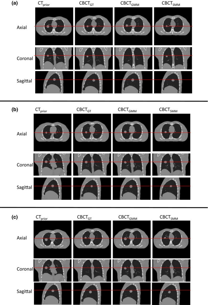
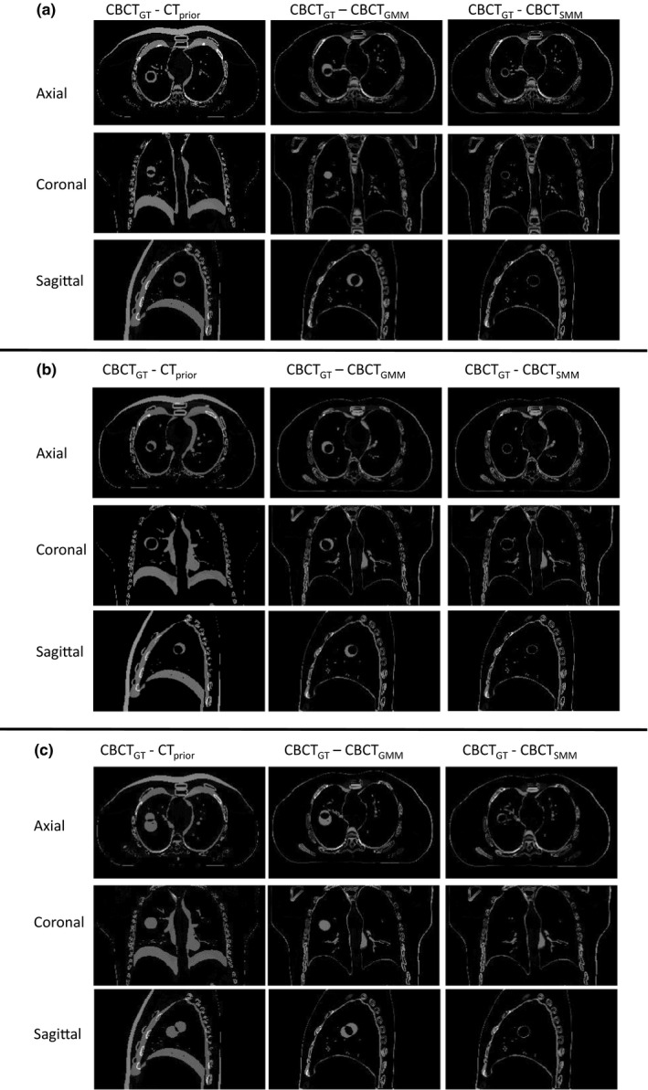
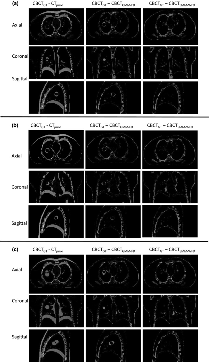
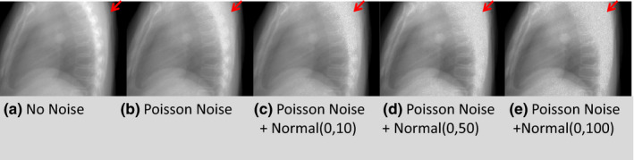
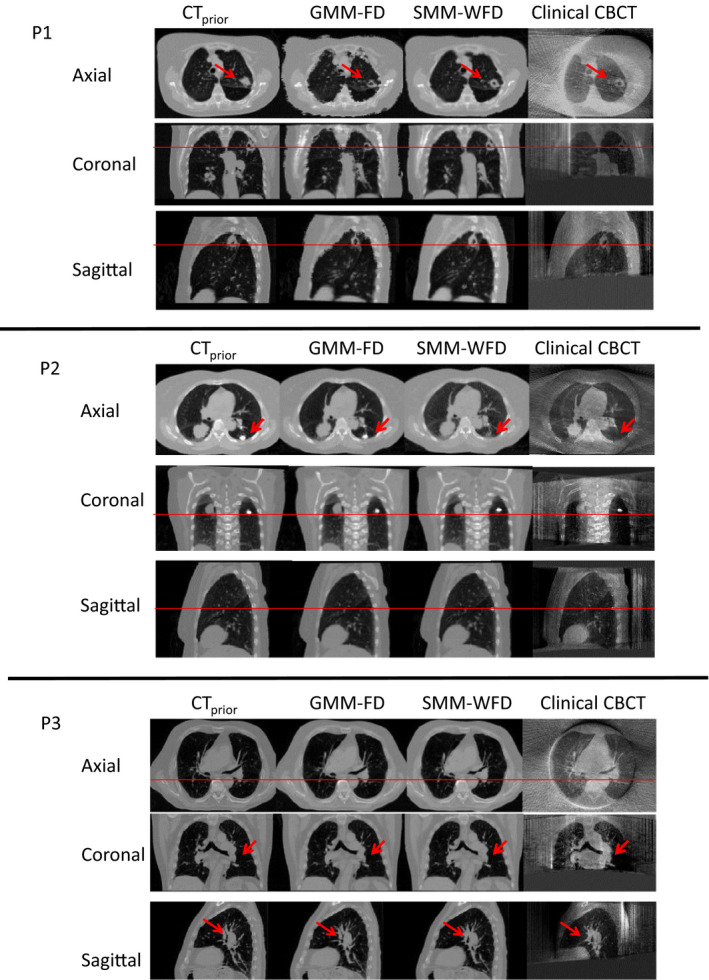
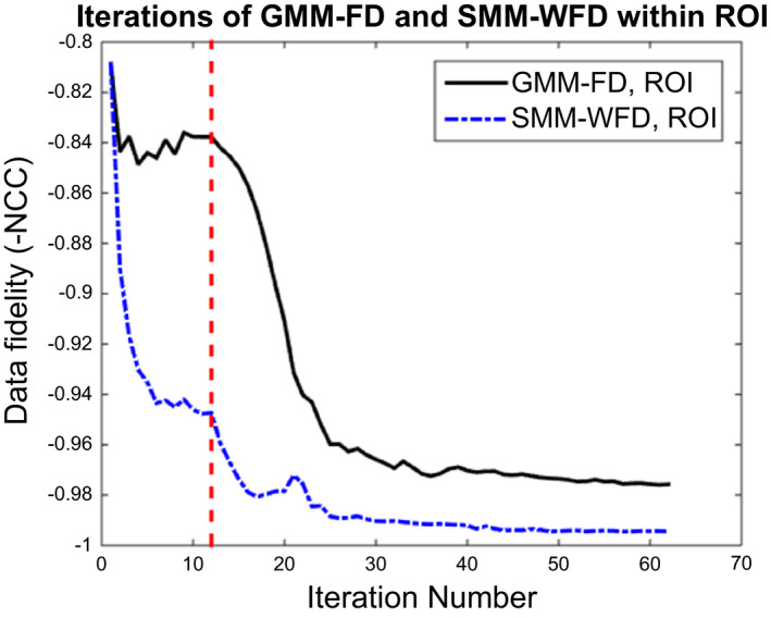
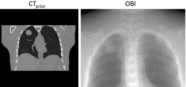
Methods: A technique for lung 4D-CBCT reconstruction has been previously developed using a deformation field map (DFM)-based strategy. In the previous method, each phase of the 4D-CBCT was generated by deforming a prior CT volume. The DFM was solved by a motion model extracted by a global PCA and free-form deformation (GMM-FD) technique, using a data fidelity constraint and deformation energy minimization. In this study, a new structural PCA method was developed to build a structural motion model (SMM) by accounting for potential relative motion pattern changes between different anatomical structures from simulation to treatment. The motion model extracted from planning 4DCT was divided into two structures: tumor and body excluding tumor, and the parameters of both structures were optimized together. Weighted free-form deformation (WFD) was employed afterwards to introduce flexibility in adjusting the weightings of different structures in the data fidelity constraint based on clinical interests. XCAT (computerized patient model) simulation with a 30 mm diameter lesion was simulated with various anatomical and respiratory changes from planning 4D-CT to on-board volume to evaluate the method. The estimation accuracy was evaluated by the volume percent difference (VPD)/center-of-mass-shift (COMS) between lesions in the estimated and "ground-truth" on-board 4D-CBCT. Different on-board projection acquisition scenarios and projection noise levels were simulated to investigate their effects on the estimation accuracy. The method was also evaluated against three lung patients.
Results: The SMM-WFD method achieved substantially better accuracy than the GMM-FD method for CBCT estimation using extremely small scan angles or projections. Using orthogonal 15° scanning angles, the VPD/COMS were 3.47 ± 2.94% and 0.23 ± 0.22 mm for SMM-WFD and 25.23 ± 19.01% and 2.58 ± 2.54 mm for GMM-FD among all eight XCAT scenarios. Compared to GMM-FD, SMM-WFD was more robust against reduction of the scanning angles down to orthogonal 10° with VPD/COMS of 6.21 ± 5.61% and 0.39 ± 0.49 mm, and more robust against reduction of projection numbers down to only 8 projections in total for both orthogonal-view 30° and orthogonal-view 15° scan angles. SMM-WFD method was also more robust than the GMM-FD method against increasing levels of noise in the projection images. Additionally, the SMM-WFD technique provided better tumor estimation for all three lung patients compared to the GMM-FD technique.
Conclusion: Compared to the GMM-FD technique, the SMM-WFD technique can substantially improve the 4D-CBCT estimation accuracy using extremely small scan angles and low number of projections to provide fast low dose 4D target verification.
Keywords: 4D CBCT; free-form deformation; limited angle; motion modeling.
© 2017 American Association of Physicists in Medicine.
Conflict of interest statement
None.
Figures

References
-
- Soike M, Kilburn JM, Lucas JT, et al. Image guided radiation therapy results in improved local control in lung cancer patients treated with fractionated radiation therapy for stage IIB‐IIIB disease. Int J Radiat Oncol Biol Phys. 2013;87:S547–S548.
-
- Zelefsky MJ, Kollmeier M, Cox B, et al. Improved clinical outcomes with high‐dose image guided radiotherapy compared with non‐IGRT for the treatment of clinically localized prostate cancer. Int J Radiat Oncol Biol Phys. 2012;84:125–129. - PubMed
-
- Cervino LI, Chao AK, Sandhu A, Jiang SB. The diaphragm as an anatomic surrogate for lung tumor motion. Phys Med Biol. 2009;54:3529–3541. - PubMed
-
- Keall PJ, Mageras GS, Balter JM, et al. The management of respiratory motion in radiation oncology report of AAPM Task Group 76. Med Phys. 2006;33:3874–3900. - PubMed
-
- Fakiris AJ, McGarry RC, Yiannoutsos CT, et al. Stereotactic body radiation therapy for early‐stage non‐small‐cell lung carcinoma: four‐year results of a prospective phase II study. Int J Radiat Oncol Biol Phys. 2009;75:677–682. - PubMed
Publication types
MeSH terms
Grants and funding
LinkOut - more resources
Full Text Sources
Other Literature Sources
Medical
