. 2017 Mar;37(3):1163-1178.
doi: 10.1177/0271678X16685105. Epub 2017 Jan 12.

PET imaging of cannabinoid type 2 receptors with [11C]A-836339 did not evidence changes following neuroinflammation in rats

Affiliations

PET imaging of cannabinoid type 2 receptors with [11C]A-836339 did not evidence changes following neuroinflammation in rats

Geraldine Pottier et al. J Cereb Blood Flow Metab. 2017 Mar.

Abstract

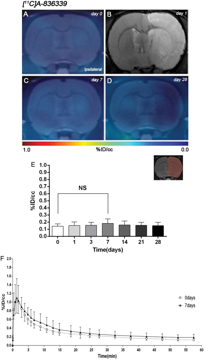
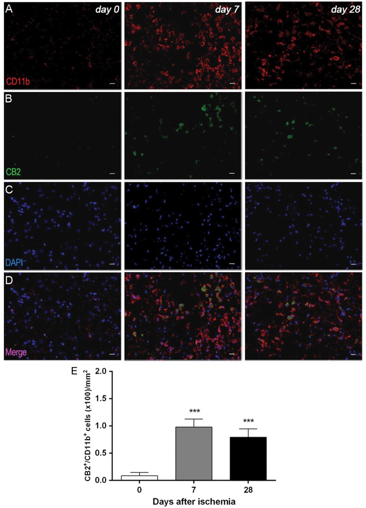
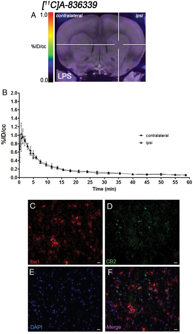
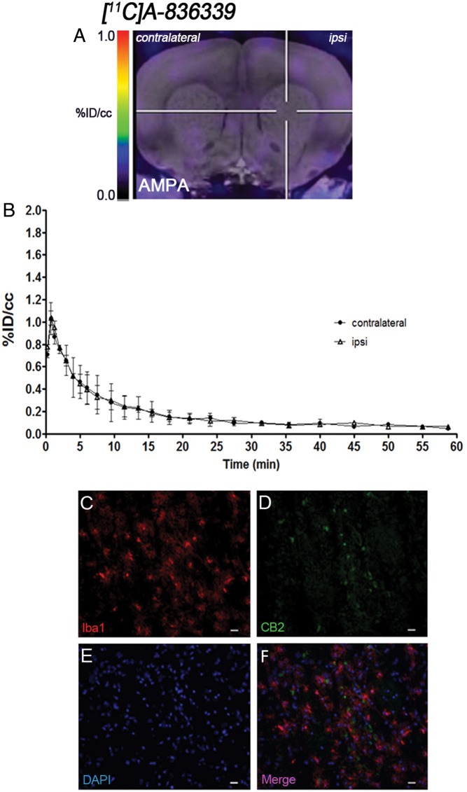
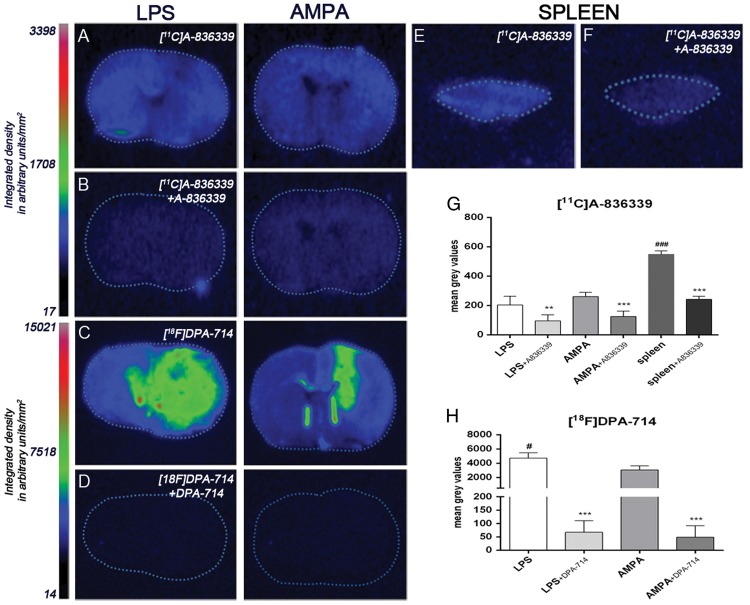
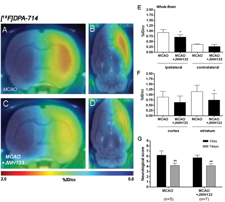
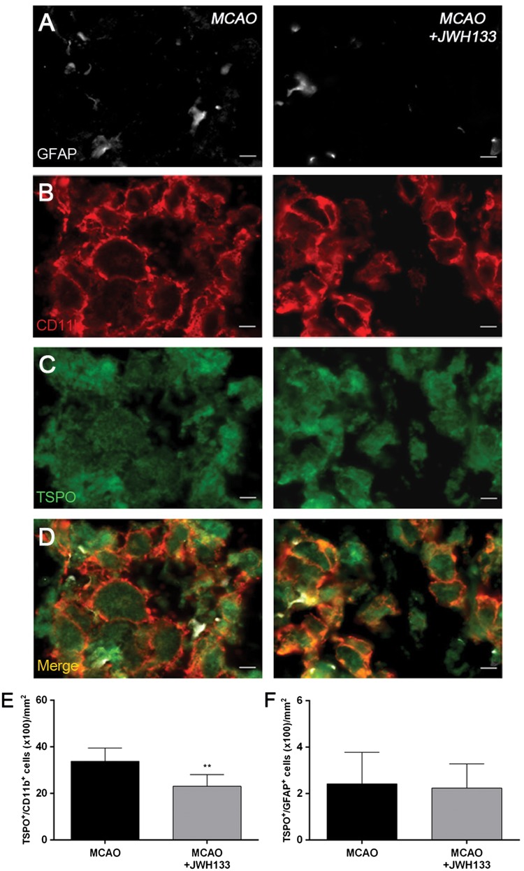
Cannabinoid type 2 receptors (CB2R) have emerged as promising targets for the diagnosis and therapy of brain pathologies. However, no suitable radiotracers for accurate CB2R mapping have been found to date, limiting the investigation of the CB2 receptor expression using positron emission tomography (PET) imaging. In this work, we report the evaluation of the in vivo expression of CB2R with [11C]A-836339 PET after cerebral ischemia and in two rat models of neuroinflammation, first by intrastriatal LPS and then by AMPA injection. PET images and in vitro autoradiography showed a lack of specific [11C]A-836339 uptake in these animal models demonstrating the limitation of this radiotracer to image CB2 receptor under neuroinflammatory conditions. Further, using immunohistochemistry, the CB2 receptor displayed a modest expression increase after cerebral ischemia, LPS and AMPA models. Finally, [18F]DPA-714-PET and immunohistochemistry demonstrated decreased neuroinflammation by a selective CB2R agonist, JWH133. Taken together, these findings suggest that [11C]A-836339 is not a suitable radiotracer to monitor in vivo CB2R expression by using PET imaging. Future studies will have to investigate alternative radiotracers that could provide an accurate binding to CB2 receptors following brain inflammation.

Keywords: TSPO; [11C]A-836339; [18F]DPA-714; cannabinoid type 2 receptors; cerebral ischemia; neuroinflammation; positron emission tomography.


Figures

Figure 1.
Figure 1.
Time course of the progression of [11C]A-836339 signal before and after middle cerebral artery occlusion (MCAO). The brain lesion at day 1 after reperfusion is shown with magnetic resonance imaging (MRI) alterations in T2-weighted (T2W) signal (b). Serial CB2 receptors PET binding images of coronal planes at control (day 0) (a), day 7 (c) and day 28 (d) after reperfusion. PET images are co-registered with a MRI (T2W) template to localize the PET signal. Images are generated by averaging all PET frames and correspond to the lesion evolution of the same animal over time. (e) %ID/cc (mean ± SD) of [11C]A-836339 was quantified in the entire ipsilateral cerebral hemisphere. The upper right panel shows the selected brain ROI for the quantification defined on a slice of a MRI (T2W) template. Rats (n = 6) were repeatedly examined by PET before (day 0) and at 1, 3, 7, 14, 21 and 28 after ischemia. (f) Time activity curves of [11C]A-836339 binding in the ipsilateral hemisphere at days 0 and 7 following cerebral ischemia.
Figure 2.
Figure 2.
Immunofluorescent labeling of CD11b (red), CB2 (green), and DAPI (blue) in the ischemic area, shown as three channels. The data show temporal evolution of microglia expressing CB2 at day 0 (control) (left column, n = 4), day 7 (middle column, n = 4) and day 28 (right column, n = 4) after ischemia. (a) CD11b-reactive microglia/macrophages increase at days 7 to 28 (b) corresponding to the temporal CB2 immunoreactivity after ischemia (b). (c) DAPI labeling evidence similar cellular density before and after MCAO. (d) Merged images of three immunofluorescent channels at different time points. The number of CD11b-reactive microglia/macrophages expressing CB2 receptor increases in the ischemic area following ischemia (e). Scale bars, 20 µm. ***p < 0.001 compared with day 0.
Figure 3.
Figure 3.
CB2 receptor binding with [11C]A-836339 PET at day 2 after LPS administration. (a) [11C]A-836339 PET image is co-registered with a MRI (T2W) template to localize the PET signal. The hairlines indicate the location of the injection site. PET image is generated by averaging all frames. (b) Average time activity curves of [11C]A-836339 binding at day 2 for VOIs placed in the ipsilateral and contralateral striatums after LPS injection. Immunofluorescent labeling of Iba1 (a), CB2 (b), DAPI (b) and the merge of the three channels (f). Scale bars, 20 µm.
Figure 4.
Figure 4.
CB2 receptor binding with [11C]A-836339 PET at day 7 after AMPA administration. (a) [11C]A-836339 PET image is co-registered with a MRI (T2W) template to localize the PET signal. The hairlines indicate the location of the injection site. PET image is generated by averaging all frames. (b) Average time activity curves of [11C]A-836339 binding at day 7 for VOIs placed in the ipsilateral and contralateral striatums after AMPA injection. Immunofluorescent labeling of Iba1 (a), CB2 (b), DAPI (b) and the merge of the three channels (f). Scale bars, 20 µm.
Figure 5.
Figure 5.
Autoradiograms of coronal brain sections after LPS and AMPA administration (a–d). The adjacent brain sections at the level of striatum show CB2 binding with [11C]A-836339 (a), [11C]A-836339 displacement with A-836339 (b) and the TSPO binding with [18F]DPA-714 (c) and [18F]DPA-714 displacement with DPA-714 (d) for both animal models. Autoradiograms of a section of rat spleen as positive control show CB2 binding with [11C]A-836339 (E) and [11C]A-836339 displacement with A-836339 (f). In vitro autoradiographic measures show [11C]A-836339 binging after LPS (n = 6), LPS + A-836339 (n = 6), AMPA (n = 6), AMPA + A-836339 (n = 6), spleen (n = 3), spleen + A-836339 (n = 3) (g) and [18F]DPA-714 binding after LPS (n = 3), LPS + DPA-714 (n = 3), AMPA (n = 3) and AMPA + DPA-714 (n = 3) (h). **p < 0.01 and ***p < 0.001 compared with no displacement; #p < 0.05 and ###p < 0.001 compared with AMPA, and AMPA/LPS, respectively.
Figure 6.
Figure 6.
Normalized coronal (a, c) and axial (b, d) PET images of [18F]DPA-714 at day 7 after middle cerebral artery occlusion (MCAO) in vehicle (a, b) and JWH133 (c, d) rats. PET images are co-registered with a MRI (T2W) rat template to localize anatomically the PET signal. [18F]DPA-714 uptake was quantified in vehicle (n = 5) and JWH133 (n = 7) at day 7 after ischemia as %ID/cc (mean ± SD) in the cerebral hemispheres, cortex and striatum (e–f). The neurologic score shows similar neurologic outcome at day 1 after ischemia (before the start of treatment) followed by a neurological outcome improvement at day 7 after cerebral ischemia (g). *p < 0.05 compared with vehicle; ##p < 0.01 compared with day 1.
Figure 7.
Figure 7.
Immunofluorescent labeling of GFAP (white), CD11b (red) and TSPO (green) in the ischemic area, shown as three channels. The data show temporal evolution of TSPO in microglial and astrocytic cells at day 7 after MCAO in vehicle (left column, n = 5) and JWH133-treated rats (right column, n = 7). GFAP-positive astrocytes do not change after treatments (a). CD11b-reactive microglia/macrophages (b) and TSPO receptor (c) decrease after MCAO in JWH133-treated rats. (d) Merged images of three immunofluorescent antibodies. The number of CD11b-reactive microglia/macrophages expressing TSPO decrease at day 7 after daily treatment with JWH133 (e). The number of GFAP-reactive astrocytes expressing TSPO shows similar values following treatment (f). **p < 0.01 compared with vehicle. Scale bars, 5 µm.

Similar articles

Cited by

References

    1. Manzanares J, Julian M, Carrascosa A. Role of the cannabinoid system in pain control and therapeutic implications for the management of acute and chronic pain episodes. Curr Neuropharmacol 2006; 4: 239–257. - PMC - PubMed
    1. Fernandez-Ruiz J, Moro MA, Martinez-Orgado J. Cannabinoids in neurodegenerative disorders and stroke/brain trauma: From preclinical models to clinical applications. Neurotherapeutics 2015; 12: 793–806. - PMC - PubMed
    1. Mackie K. Distribution of cannabinoid receptors in the central and peripheral nervous system. Handb Exp Pharmacol 2005; 168: 299–325. - PubMed
    1. Cabral GA, Raborn ES, Griffin L, et al. CB2 receptors in the brain: Role in central immune function. Br J Pharmacol 2008; 153: 240–251. - PMC - PubMed
    1. Miller AM, Stella N. CB2 receptor-mediated migration of immune cells: It can go either way. Br J Pharmacol 2008; 153: 299–308. - PMC - PubMed

LinkOut - more resources